. 2020 Nov 23;8(11):529.
doi: 10.3390/biomedicines8110529.

Adenosine A2A and A3 Receptors as Targets for the Treatment of Hypertensive-Diabetic Nephropathy

Affiliations

Adenosine A2A and A3 Receptors as Targets for the Treatment of Hypertensive-Diabetic Nephropathy

Daniela Patinha et al. Biomedicines. .

Abstract

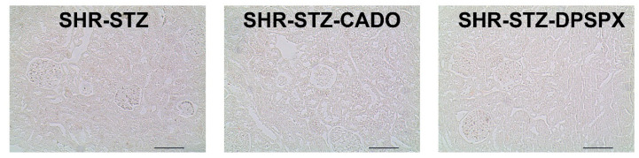
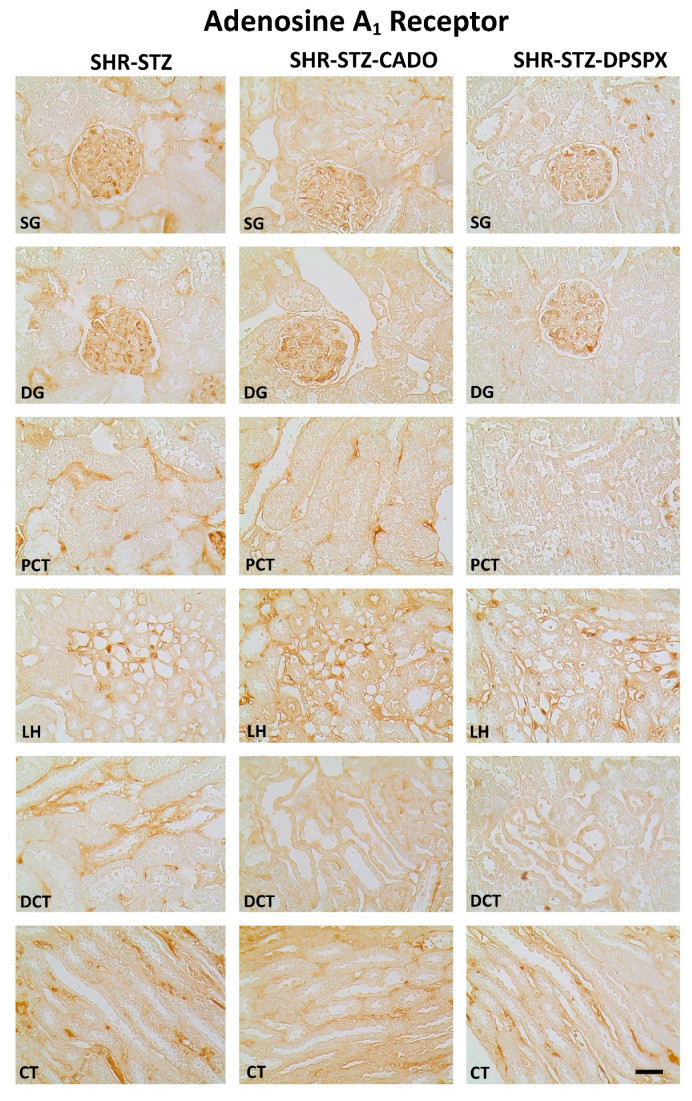
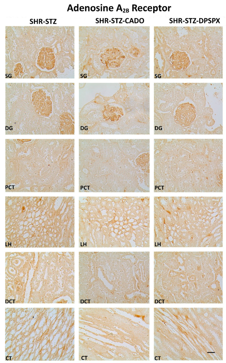
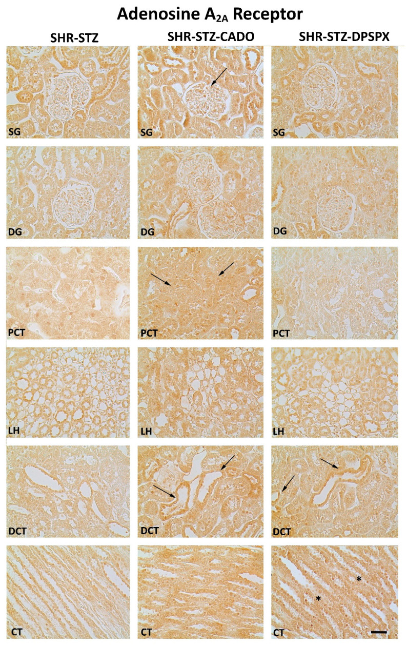
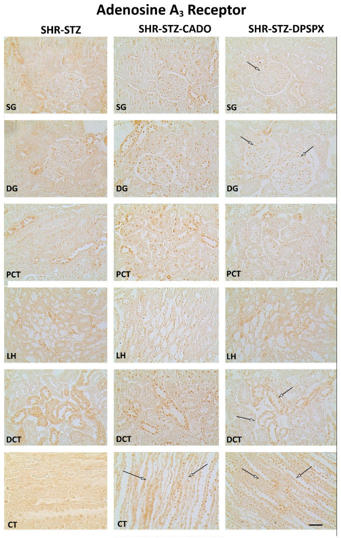
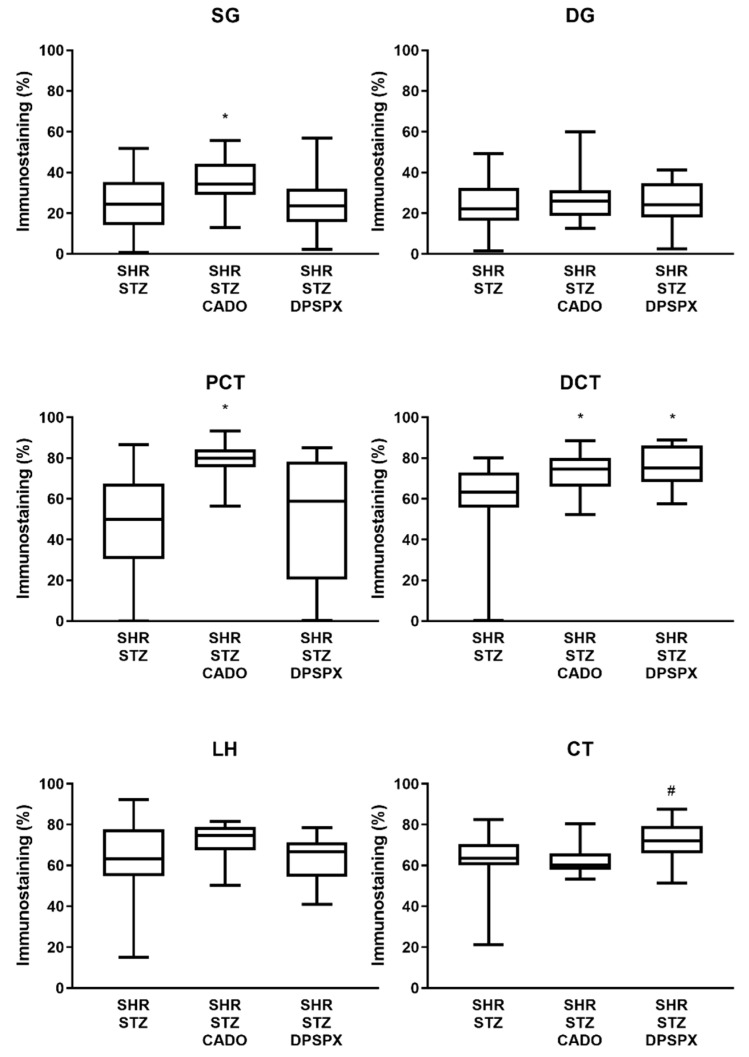
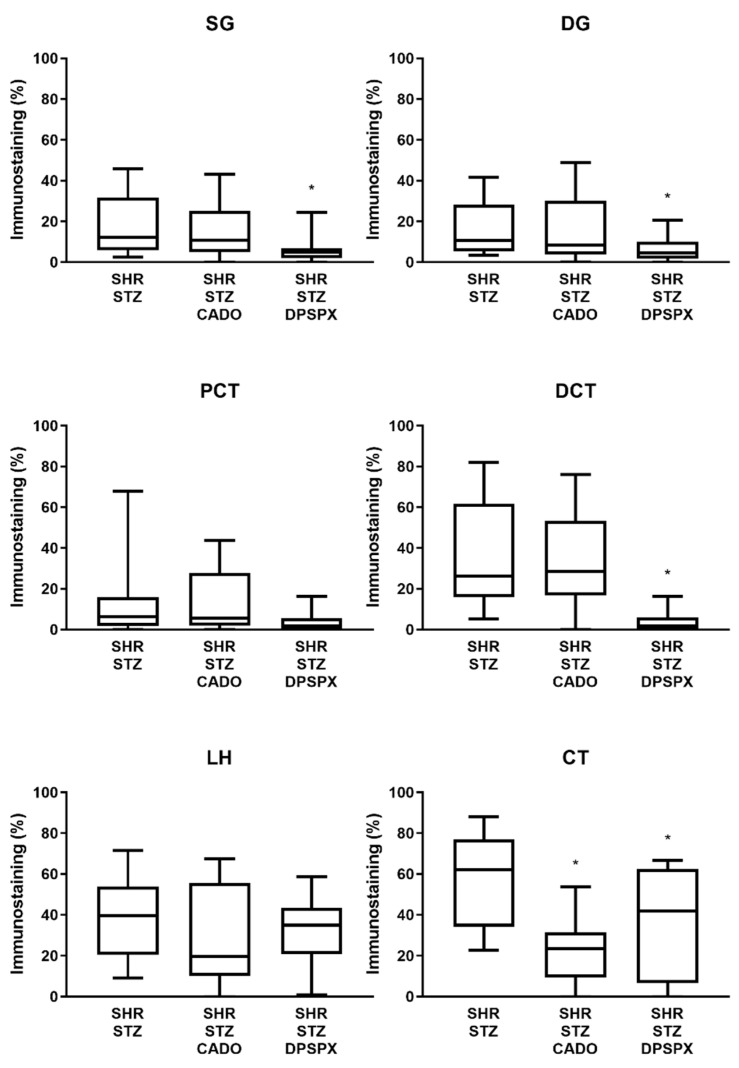
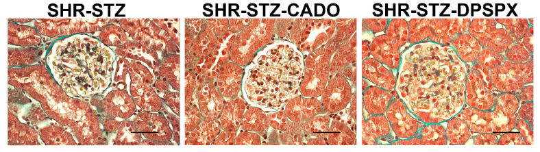
Diabetic nephropathy (DN) and hypertension are prime causes for end-stage renal disease (ESRD) that often coexist in patients, but are seldom studied in combination. Kidney adenosine levels are markedly increased in diabetes, and the expression and function of renal adenosine receptors are altered in experimental diabetes. The aim of this work is to explore the impact of endogenous and exogenous adenosine on the expression/distribution profile of its receptors along the nephron of hypertensive rats with experimentally-induced diabetes. Using spontaneously hypertensive (SHR) rats rendered diabetic with streptozotocin (STZ), we show that treatment of SHR-STZ rats with an agonist of adenosine receptors increases A2A immunoreactivity in superficial glomeruli (SG), proximal tubule (PCT), and distal tubule (DCT). Differently, treatment of SHR-STZ rats with a xanthinic antagonist of adenosine receptors decreases adenosine A3 immunoreactivity in SG, PCT, DCT, and collecting duct. There is no difference in the immunoreactivity against the adenosine A1 and A2B receptors between the experimental groups. The agonist of adenosine receptors ameliorates renal fibrosis, probably via A2A receptors, while the antagonist exacerbates it, most likely due to tonic activation of A3 receptors. The reduction in adenosine A3 immunoreactivity might be due to receptor downregulation in response to prolonged activation. Altogether, these results suggest an opposite regulation exerted by endogenous and exogenous adenosine upon the expression of its A2A and A3 receptors along the nephron of hypertensive diabetic rats, which has a functional impact and should be taken into account when considering novel therapeutic targets for hypertensive-diabetic nephropathy.

Keywords: adenosine receptors; diabetes; diabetic complications; diabetic nephropathy; hypertension.


Conflict of interest statement

The authors declare no conflict of interest.

Figures

Figure 1
Figure 1
Secondary antibody controls for immunohistochemistry experiments: Representative photomicrographs of kidney transversal sections from spontaneously hypertensive rats rendered diabetic with streptozotocin with no further pharmacological treatment (SHR-STZ) and continuously infused with 2-chloroadenosine (CADO) (non-selective agonist of adenosine receptors) or 1,3-dipropyl-8-sulfophenylxanthine (DPSPX) (non-selective antagonist of adenosine receptors), showing the absence of staining both in glomeruli and in tubular structures. Negative controls were incubated in parallel using 10% normal horse serum or blocking solution instead of the primary antibody in order to determine the background level due to nonspecific secondary antibody binding. Note the clean background obtained in all sections from the three groups of animals in the study. Bars: 50 µm.
Figure 2
Figure 2
Representative photomicrographs of the immunoreactivity against the adenosine A1 receptors in the superficial (SG) and deep glomeruli (DG), proximal convoluted tubule (PCT), distal convoluted tubule (DCT), loop of Henle (LH), and collecting tubule (CT) from kidney sections of SHR-STZ (left panel), SHR-STZ + CADO (middle panel), and SHR-STZ + DPSPX (right panel) animals. Bars: 20 µm.
Figure 3
Figure 3
Representative photomicrographs of the immunoreactivity against the adenosine A2B receptors in the superficial (SG) and deep glomeruli (DG), proximal convoluted tubule (PCT), distal convoluted tubule (DCT), loop of Henle (LH), and collecting tubule (CT) from kidney sections of SHR-STZ (left panel), SHR-STZ + CADO (middle panel), and SHR-STZ+DPSPX (right panel) animals. Bars: 20 µm.
Figure 4
Figure 4
Representative photomicrographs of the immunoreactivity against the adenosine A2A receptors in the superficial (SG) and deep glomeruli (DG), proximal convoluted tubule (PCT), distal convoluted tubule (DCT), loop of Henle (LH), and collecting tubule (CT) from kidney sections of SHR-STZ (left panel), SHR-STZ + CADO (middle panel), and SHR-STZ + DPSPX (right panel) animals. Filled arrows: evidence more pronounced immunoreactivity than that exhibited by the SHR-STZ control group; *: evidence more pronounced immunoreactivity than that exhibited by the SHR-STZ+CADO group. Bars: 20 µm.
Figure 5
Figure 5
Representative photomicrographs of the immunoreactivity against the adenosine A3 receptors in the superficial (SG) and deep glomeruli (DG), proximal convoluted tubule (PCT), distal convoluted tubule (DCT), loop of Henle (LH), and collecting tubule (CT) of SHR-STZ (left panel), SHR-STZ + CADO (middle panel), and SHR-STZ + DPSPX (right panel) animals. Open arrows: evidence less pronounced immunoreactivity than that exhibited by SHR-STZ. Bars: 20 µm.
Figure 6
Figure 6
Quantitative analysis of the immunostaining (staining fractional area in percentage of the tissue total area; using the semiautomated computer-assisted image analysis “SACAIA” method) for the adenosine A2A receptors in the six renal structures from SHR-STZ, SHR-STZ + CADO, and SHR-STZ + DPSPX rats. Superficial (SG) and deep (DG) glomeruli, proximal (PCT) and distal (DCT) convoluted tubules, loop of Henle (LH), and collecting tubule (CT). Values are the median and 25th–75th percentiles (P25-P75) from four rats. * p < 0.05 vs. the corresponding SHR-STZ group; # p < 0.05 vs. the corresponding SHR-STZ + CADO group.
Figure 7
Figure 7
Quantitative analysis of the immunostaining (staining fractional area in percentage of the tissue total area; using the SACAIA method) for the adenosine A3 receptors in the six renal structures from SHR-STZ, SHR-STZ+CADO, and SHR-STZ+DPSPX rats. Superficial (SG) and deep (DG) glomeruli, proximal (PCT) and distal (DCT) convoluted tubules, loop of Henle (LH), and collecting tubule (CT). Values are the median and 25th–75th percentiles (P25-P75) from four rats. * p < 0.05 vs. the corresponding SHR-STZ group.
Figure 8
Figure 8
Representative photomicrographs of Masson’s trichrome stain of glomeruli of SHR-STZ (left panel), SHR-STZ + CADO (middle panel), and SHR-STZ + DPSPX (right panel) animals. Bars: 20 µm.

References

    1. Oyarzun C., Garrido W., Alarcon S., Yanez A., Sobrevia L., Quezada C., San Martin R. Adenosine contribution to normal renal physiology and chronic kidney disease. Mol. Aspects Med. 2017;55:75–89. - PubMed
    1. Olivera A., Lamas S., Rodriguez-Puyol D., Lopez-Novoa J.M. Adenosine induces mesangial cell contraction by an a1-type receptor. Kidney Int. 1989;35:1300–1305. - PubMed
    1. Faulhaber-Walter R., Chen L., Oppermann M., Kim S.M., Huang Y., Hiramatsu N., Mizel D., Kajiyama H., Zerfas P., Briggs J.P., et al. Lack of a1 adenosine receptors augments diabetic hyperfiltration and glomerular injury. J. Am. Soc. Nephrol. 2008;19:722–730. - PMC - PubMed
    1. Truong L.D., Trostel J., McMahan R., Chen J.F., Garcia G.E. Macrophage a2a adenosine receptors are essential to protect from progressive kidney injury. Am. J. Pathol. 2016;186:2601–2613. - PMC - PubMed
    1. Min H.S., Cha J.J., Kim K., Kim J.E., Ghee J.Y., Kim H., Lee J.E., Han J.Y., Jeong L.S., Cha D.R., et al. Renoprotective effects of a highly selective a3 adenosine receptor antagonist in a mouse model of adriamycin-induced nephropathy. J. Korean Med. Sci. 2016;31:1403–1412. - PMC - PubMed

LinkOut - more resources