Review
. 2020 Nov 23;9(11):2523.
doi: 10.3390/cells9112523.

Role of Selective Autophagy in Spermatogenesis and Male Fertility

Affiliations
Review

Role of Selective Autophagy in Spermatogenesis and Male Fertility

Chunyu Lv et al. Cells. .

Abstract

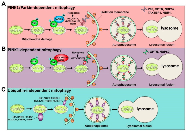
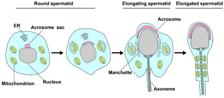
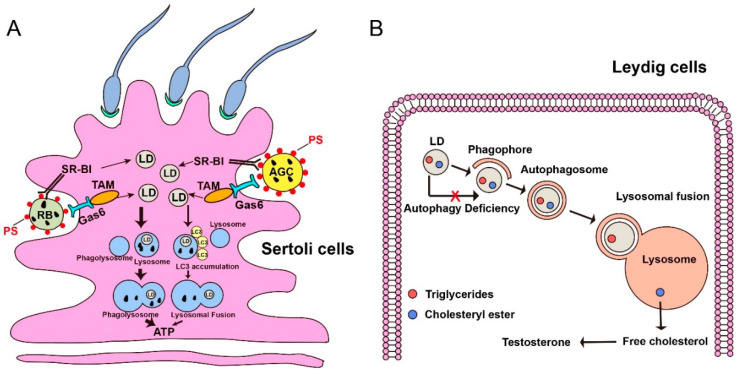
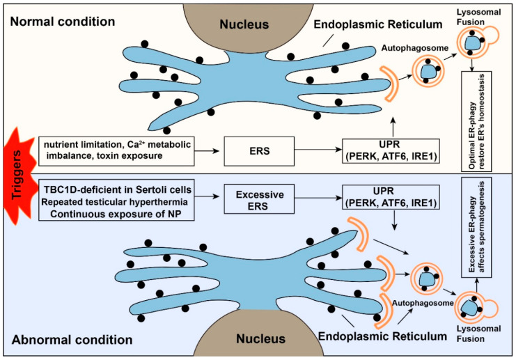
Autophagy is a "self-eating" process that engulfs cellular contents for their subsequent digestion in lysosomes to engage the metabolic need in response to starvation or environmental insults. According to the contents of degradation, autophagy can be divided into bulk autophagy (non-selective autophagy) and selective autophagy. Bulk autophagy degrades non-specific cytoplasmic materials in response to nutrient starvation while selective autophagy targets specific cargoes, such as damaged organelles, protein aggregates, and intracellular pathogens. Selective autophagy has been documented to relate to the reproductive processes, especially for the spermatogenesis, fertilization, and biosynthesis of testosterone. Although selective autophagy is vital in the field of reproduction, its role and the underlying mechanism have remained unclear. In this review, we focus on selective autophagy to discuss the recent advances in our understanding of the mechanism and role of selective autophagy on spermatogenesis and male fertility in mammals. Understanding the role of selective autophagy during spermatogenesis will promote the recognition of genetic regulation in male infertility, and shed light on therapies of infertile patients.

Keywords: autophagy; male fertility; selective autophagy; spermatogenesis.


Conflict of interest statement

The authors declared no potential conflicts of interest with respect to the research, authorship, and/or publication of this article.

Figures

Figure 1
Figure 1
Schematic illustration of the ubiquitin-dependent and ubiquitin-independent pathways of mitophagy is shown. The ubiquitin-dependent mitophagy can be divided into PINK1/Parkin-dependent mitophagy (A) and PINK1-dependent mitophagy (B). (A) Once mitochondria are damaged, PINK1 will accumulate on the OMM (outer mitochondrial membrane) and activate it by phosphorylation. The activated PINK1 recruits and activates PARKIN by phosphorylation. Then, the activated PARKIN produces the polyubiquitin chains recognized by receptors (P62, OPTN, NDP52, TAX1BP1, and NBR1). These receptors will bind with the LC3 adaptor to engulf mitochondria to complete autophagy. (B) The activated PINK1 recruits ULK1, DFCP1, and WIPI1, which can be recognized by receptors (OPTN and NDP52) to induce PINK1-dependent mitophagy. (C) Some proteins, including NIX, BNIP3, FUNDC1, BCL2L13, FKBP8, and NLRX1, can directly recognize the LC3 adaptor and initiate the mitophagy process.
Figure 2
Figure 2
Mitophagy is involved in spermatid differentiation. In the process of spermiogenesis, excessive mitochondrion in the residual bodies will be cleared by mitophagy, and the remaining mitochondrion will rearrange in elongated tubules and participate in the formation of spermatozoa.
Figure 3
Figure 3
Lipophagy promotes spermatogenesis in Sertoli cells and regulates the synthesis of testosterone in Leydig cells. (A) During spermatogenesis, apoptotic germ cells (AGCs) and residual bodies (RBs) are phagocytosed by Sertoli cells by the SR-BI/PS (Class B scavenger receptor type I / Phosphatide) system or TAM/Gas 6 (Translocation and assembly module/Growth arrest-specific 6) system. The lipid droplets can release from the breakdown of engulfed AGCs and RBs. In this condition, most LDs fused with the lysosome to form phagolysosome to generate energy for Sertoli cells, which further supports germ cell and spermatogenesis. While, minor LDs can have an LC3 accumulation signal and then fuse with the lysosome to produce ATP for energy support. PS, a type of phospholipid. LD, lipid droplet. Bigger arrow, a major pathway to degrade LDs in Sertoli cells. Smaller arrow, a minor pathway to degrade LDs in Sertoli cells. (B) LDs are composed of cholesteryl ester and triglycerides, which are localized in the Leydig cells. In normal conditions, lipophagy can degrade cholesterol esters into free cholesterol, an essential substrate for testosterone synthesis. When autophagy deficient, the substrate of testosterone will decrease due to the failure of lipophagy.
Figure 4
Figure 4
The dual role of endoplasmic reticulum autophagy. When cells face external stimuli, many protective mechanisms will be activated to restore the cells’ homeostasis. The upper panel shows endoplasmic reticulum stress (ERS) is a protective mechanism under normal conditions. The ubiquitin-proteasome system (UPS) and ER-phagy will be activated after the emergence of ERS. The normal function of ER and homeostasis of the cell will be restored. While the lower panel shows that under abnormal conditions, the intense or prolonged triggers (TBC1D20-deficient Sertoli cells of male mice, continuous exposure of nonylphenol, repeated testicular hyperthermia) will cause excessive ER-phagy, which affects the normal spermatogenic process.

Similar articles

Cited by

References

    1. Staub C., Johnson L. Review: Spermatogenesis in the bull. Animal. 2018;12:s27–s35. doi: 10.1017/S1751731118000435. - DOI - PubMed
    1. Satouh Y., Ikawa M. New Insights into the Molecular Events of Mammalian Fertilization. Trends Biochem. Sci. 2018;43:818–828. doi: 10.1016/j.tibs.2018.08.006. - DOI - PMC - PubMed
    1. Zhou R., Wu J., Liu B., Jiang Y., Chen W., Li J., He Q., He Z. The roles and mechanisms of Leydig cells and myoid cells in regulating spermatogenesis. Cell. Mol. Life Sci. 2019;76:2681–2695. doi: 10.1007/s00018-019-03101-9. - DOI - PMC - PubMed
    1. Mizushima N., Komatsu M. Autophagy: Renovation of cells and tissues. Cell. 2011;147:728–741. doi: 10.1016/j.cell.2011.10.026. - DOI - PubMed
    1. Stolz A., Ernst A., Dikic I. Cargo recognition and trafficking in selective autophagy. Nat. Cell Biol. 2014;16:495–501. doi: 10.1038/ncb2979. - DOI - PubMed

Publication types

LinkOut - more resources