Review
. 2020 Nov 23;9(11):2527.
doi: 10.3390/cells9112527.

Ex Vivo Culture Models to Indicate Therapy Response in Head and Neck Squamous Cell Carcinoma

Affiliations
Review

Ex Vivo Culture Models to Indicate Therapy Response in Head and Neck Squamous Cell Carcinoma

Imke Demers et al. Cells. .

Abstract

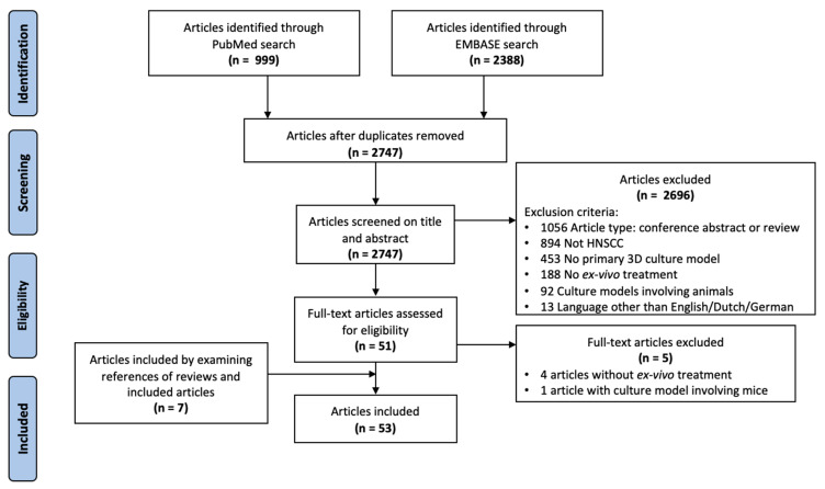
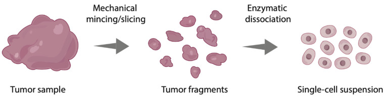
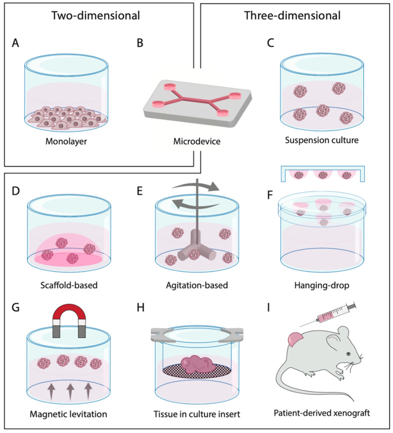
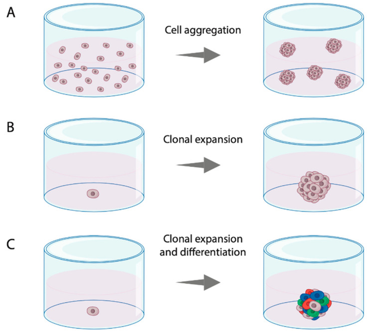
Head and neck squamous cell carcinoma (HNSCC) is characterized by a poor 5 year survival and varying response rates to both standard-of-care and new treatments. Despite advances in medicine and treatment methods, mortality rates have hardly decreased in recent decades. Reliable patient-derived tumor models offer the chance to predict therapy response in a personalized setting, thereby improving treatment efficacy by identifying the most appropriate treatment regimen for each patient. Furthermore, ex vivo tumor models enable testing of novel therapies before introduction in clinical practice. A literature search was performed to identify relevant literature describing three-dimensional ex vivo culture models of HNSCC to examine sensitivity to chemotherapy, radiotherapy, immunotherapy and targeted therapy. We provide a comprehensive overview of the currently used three-dimensional ex vivo culture models for HNSCC with their advantages and limitations, including culture success percentage and comparison to the original tumor. Furthermore, we evaluate the potential of these models to predict patient therapy response.

Keywords: 3D cell culture; drug response; ex vivo model; head and neck cancer; histoculture; organoid; personalized therapy; preclinical prediction model; primary cell culture; sensitivity testing.


Conflict of interest statement

Imke Demers and Johan Donkers declare no conflict of interest. Bernd Kremer reports grants from Pfizer and Novartis. Ernst Jan Speel reports grants from Pfizer and Novartis and honoraria from BMS. The funders had no role in the design of the study; in the collection, analyses, or interpretation of data; in the writing of the manuscript, or in the decision to publish the results.

Figures

Figure 1
Figure 1
Flow diagram of the systematic literature search performed.
Figure 2
Figure 2
Processing of the primary tumor sample into tumor fragments by mechanical modification and subsequently into a single-cell suspension by enzymatic dissociation.
Figure 3
Figure 3
Primary cell culture techniques divided into two-dimensional and three-dimensional models. (A) 2D monolayer from single-cell suspension; (B) micro(fluidic) device; (C) spheroids in suspension culture; (D) spheroids embedded in a scaffold-based system; (E) spheroids in an agitation-based system, e.g., a spinner flask; (F) spheroids in hanging-drop cultures; (G) spheroids formed by magnetic levitation; (H) histoculture in culture insert; (I) patient-derived xenograft mouse model with subcutaneous injection. Image of xenograft was modified from Servier Medical Art (http://smart.servier.com/), licensed under a Creative Common Attribution 3.0 Unported License (https://creativecommons.org/licences/by/3.0/).
Figure 4
Figure 4
Principles of sphere formation from primary tumor cells. (A) Sphere formed by aggregation of multiple cells in a single-cell suspension; (B) sphere formed by clonal expansion of a single cell with proliferating potential; (C) sphere formed by clonal expansion and lineage-dependent differentiation of a single cell with proliferating potential.

Similar articles

Cited by

References

    1. Bray F., Ferlay J., Soerjomataram I., Siegel R.L., Torre L.A., Jemal A. Global cancer statistics 2018: GLOBOCAN estimates of incidence and mortality worldwide for 36 cancers in 185 countries. CA Cancer J. Clin. 2018;68:394–424. doi: 10.3322/caac.21492. - DOI - PubMed
    1. Chow L.Q.M. Head and Neck Cancer. N. Engl. J. Med. 2020;382:60–72. doi: 10.1056/NEJMra1715715. - DOI - PubMed
    1. Gatta G., Botta L., Sanchez M.J., Anderson L.A., Pierannunzio D., Licitra L. Prognoses and improvement for head and neck cancers diagnosed in Europe in early 2000s: The EUROCARE-5 population-based study. Eur. J. Cancer. 2015;51:2130–2143. doi: 10.1016/j.ejca.2015.07.043. - DOI - PubMed
    1. Gillison M.L., Chaturvedi A.K., Anderson W.F., Fakhry C. Epidemiology of Human Papillomavirus-Positive Head and Neck Squamous Cell Carcinoma. J. Clin. Oncol. 2015;33:3235–3242. doi: 10.1200/JCO.2015.61.6995. - DOI - PMC - PubMed
    1. Pignon J.P., Bourhis J., Domenge C., Designé L. Chemotherapy added to locoregional treatment for head and neck squamous-cell carcinoma: Three meta-analyses of updated individual data. Lancet. 2000;355:949–955. doi: 10.1016/S0140-6736(00)90011-4. - DOI - PubMed

MeSH terms

LinkOut - more resources