

COVID-19-Induced Acute Bilateral Optic Neuritis
- PMID: 33238757
- PMCID: PMC7705770
- DOI: 10.1177/2324709620976018
COVID-19-Induced Acute Bilateral Optic Neuritis
Abstract
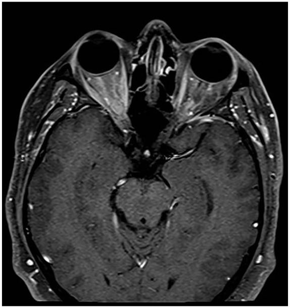
A 44-year-old male patient with no past medical history presented 2 weeks after seropositive coronavirus disease 2019 (COVID-19) infection with vision problems suggestive of optic neuritis. Radiological testing showed findings suspicious for acute bilateral optic neuritis. The patient had also anti-MOG antibodies. Whether this was an optic neuritis due to COVID-19, MOG antibody disease, or an activation of MOG antibody disease by COVID-19 is discussed in this case.
Keywords: COVID-19; bilateral optic neuritis.
Conflict of interest statement
Figures


References
-
- Balcer LJ. Optic neuritis. N Engl J Med. 2006;354:1273-1280. - PubMed
-
- Roed H, Frederiksen J, Langkilde A, Sørensen TL, Lauritzen M, Sellebjerg F. Systemic T-cell activation in acute clinically isolated optic neuritis. J Neuroimmunol. 2005;162:165-172. - PubMed
-
- Söderström M, Link H, Xu Z, Fredriksson S. Optic neuritis and multiple sclerosis: anti-MBP and anti-MBP peptide antibody-secreting cells are accumulated in CSF. Neurology. 1993;43:1215-1222. - PubMed
-
- Jacobs LD, Kaba SE, Miller CM, Priore RL, Brownscheidle CM. Correlation of clinical, magnetic resonance imaging, and cerebrospinal fluid findings in optic neuritis. Ann Neurol. 1997;41:392-398. - PubMed
Publication types
MeSH terms
Substances
LinkOut - more resources
Full Text Sources
Medical
Miscellaneous
