. 2021 Dec;27(6):668-684.
doi: 10.1177/1073858420954811. Epub 2020 Nov 25.

Cerebral Amyloid Angiopathy and Blood-Brain Barrier Dysfunction

Affiliations

Cerebral Amyloid Angiopathy and Blood-Brain Barrier Dysfunction

Monica Gireud-Goss et al. Neuroscientist. 2021 Dec.

Abstract

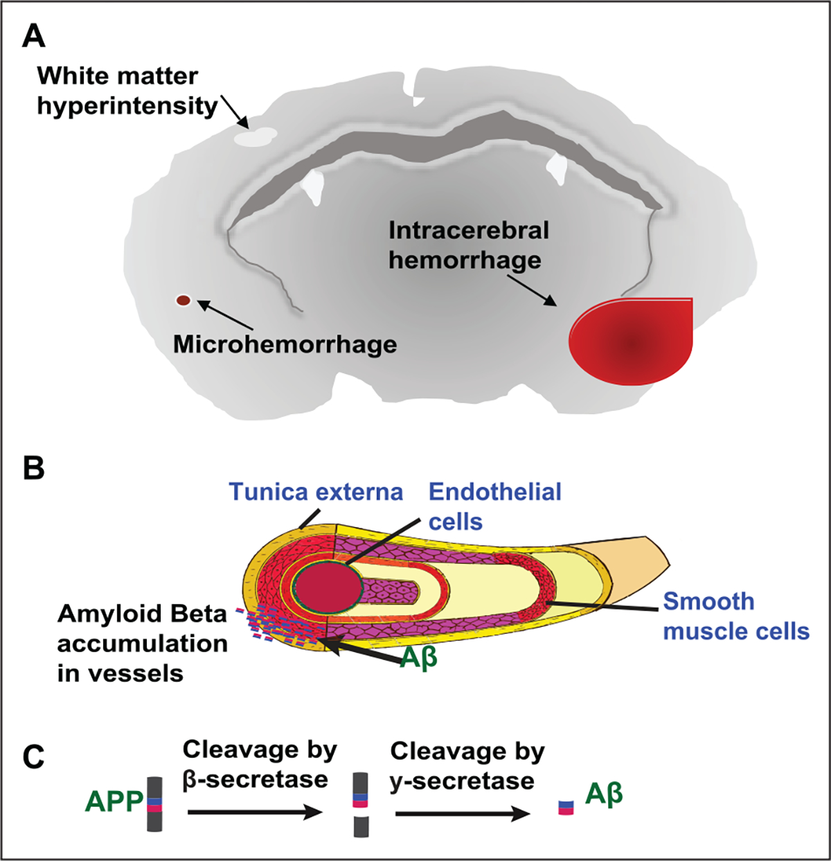
Cerebral hemorrhage, a devastating subtype of stroke, is often caused by hypertension and cerebral amyloid angiopathy (CAA). Pathological evidence of CAA is detected in approximately half of all individuals over the age of 70 and is associated with cortical microinfarcts and cognitive impairment. The underlying pathophysiology of CAA is characterized by accumulation of pathogenic amyloid β (Aβ) fragments of amyloid precursor protein in the cerebral vasculature. Vascular deposition of Aβ damages the vessel wall, results in blood-brain barrier (BBB) leakiness, vessel occlusion or rupture, and leads to hemorrhages and decreased cerebral blood flow that negatively affects vessel integrity and cognitive function. Currently, the main hypothesis surrounding the mechanism of CAA pathogenesis is that there is an impaired clearance of Aβ peptides, which includes compromised perivascular drainage as well as dysfunction of BBB transport. Also, the immune response in CAA pathogenesis plays an important role. Therefore, the mechanism by which Aβ vascular deposition occurs is crucial for our understanding of CAA pathogenesis and for the development of potential therapeutic options.

Keywords: BBB; CAA; amyloid beta; inflammation; intracerebral hemorrhage.


Conflict of interest statement

Declaration of Conflicting Interests

The author(s) declared no potential conflicts of interest with respect to the research, authorship, and/or publication of this article.

Figures

Figure 1.
Figure 1.
Pathological findings in cerebral amyloid angiopathy. (A) MRI showing CAA with microhemorrhage (red box). (B) Postmortem tissue from a stroke patient showing diffuse amyloid plaques (black) and CAA (white). (C) This image shows a lobar intracerebral hemorrhage as shown by a CT scan.
Figure 2.
Figure 2.
APP processing mechanisms and familial mutations. A protein sequence from amino acid residue 663–720 is presented, with the amino acid sequence for Aβ shown in red. β and γ secretases are responsible for the cleavage of Aβ from APP, whereas α-secretase leads to a non-amyloidogenic cleavage product. A number of known pathogenic point mutations within APP (with the listed mutation alias if known) identified in cases of familial CAA are shown.
Figure 3.
Figure 3.
Pathophysiology of CAA. (A) CAA pathology shows the presence of microhemorrhages, lobar cerebral hemorrhage, white matter hyperintensities, dilation of perivascular spaces, and has a preference for occipital lobe. (B) Aβ deposition occurs in small leptomeningeal or cortical vessels. (C) APP is cleaved by β-secretase and γ-secretase into Aβ fragments.
Figure 4.
Figure 4.
Ex vivo two-photon depiction of vascular amyloidosis using methoxy X04 amyloid dye. (A) A representative image showing a 12-month Tg2576 brain section with more punctate amyloids. (B) A representative image showing a 6-month TgSwDI brain section with vascular amyloidosis surrounding capillaries. (C) A representative image showing a 6-month TgSwDI brain section with large vessel amyloid accumulation following SMC pattern rings.
Figure 5.
Figure 5.
Clearance mechanisms of Aβ. (A) Aβ is produced by the cleavage of APP in neurons. (B) Aβ can be enzymatically degraded. (C) Aβ can be phagocytosed by glial cells. (D) Aβ can be cleared across the BBB by transendothelial waste clearance mechanisms. (E) Aβ can be cleared through perivascular drainage pathways. (F) Aβ accumulation in vessels leads to CAA. (G) Aβ accumulation in the brain parenchyma is observed in Alzheimer’s disease.
Figure 6.
Figure 6.
Depiction of CAA-laden vessel microenvironment and immunologic response. (A) Aβ deposits surrounded by innate immune cells such as astrocytes, activated microglia, and macrophages. (B) Phagocytosis of Aβ results in increased ROS production and increased pro-inflammatory cytokines expression (e.g., tumor necrosis factor-alpha, interleukins, and transforming growth factor beta-1) resulting in altered BBB integrity, leakage, and infiltration.

References

    1. Akiyama H, Kondoh T, Kokunai T, Nagashima T, Saito N, Tamaki N. 2000. Blood-brain barrier formation of grafted human umbilical vein endothelial cells in athymic mouse brain. Brain Res 858(1):172–6. - PubMed
    1. Albargothy NJ, Johnston DA, MacGregor-Sharp M, Weller RO, Verma A, Hawkes CA, and others. 2018. Convective influx/glymphatic system: tracers injected into the CSF enter and leave the brain along separate peri-arterial basement membrane pathways. Acta Neuropathol 136(1):139–52. - PMC - PubMed
    1. Attems J, Quass M, Jellinger KA, Lintner F. 2007. Topographical distribution of cerebral amyloid angiopathy and its effect on cognitive decline are influenced by Alzheimer disease pathology. J Neurol Sci 257(1–2):49–55. - PubMed
    1. Auriel E, Greenberg SM. 2012. The pathophysiology and clinical presentation of cerebral amyloid angiopathy. Curr Atheroscler Rep 14(4):343–50. - PubMed
    1. Banerjee G, Ambler G, Keshavan A, Paterson RW, Foiani MS, Toombs J, and others. 2020. Cerebrospinal fluid biomarkers in cerebral amyloid angiopathy. J Alzheimers Dis 74(4):1189–201. - PMC - PubMed

Publication types

MeSH terms

Substances