

Association of gestational age with MRI-based biometrics of brain development in fetuses
- PMID: 33238909
- PMCID: PMC7689975
- DOI: 10.1186/s12880-020-00525-9
Association of gestational age with MRI-based biometrics of brain development in fetuses
Abstract
Background: Reported date of last menstrual period and ultrasonography measurements are the most commonly used methods for determining gestational age in antenatal life. However, the mother cannot always determine the last menstrual period with certainty, and ultrasonography measurements are accurate only in the first trimester. We aimed to assess the ability of various biometric measurements on magnetic resonance imaging (MRI) in determining the accurate gestational age of an individual fetus in the second half of gestation.
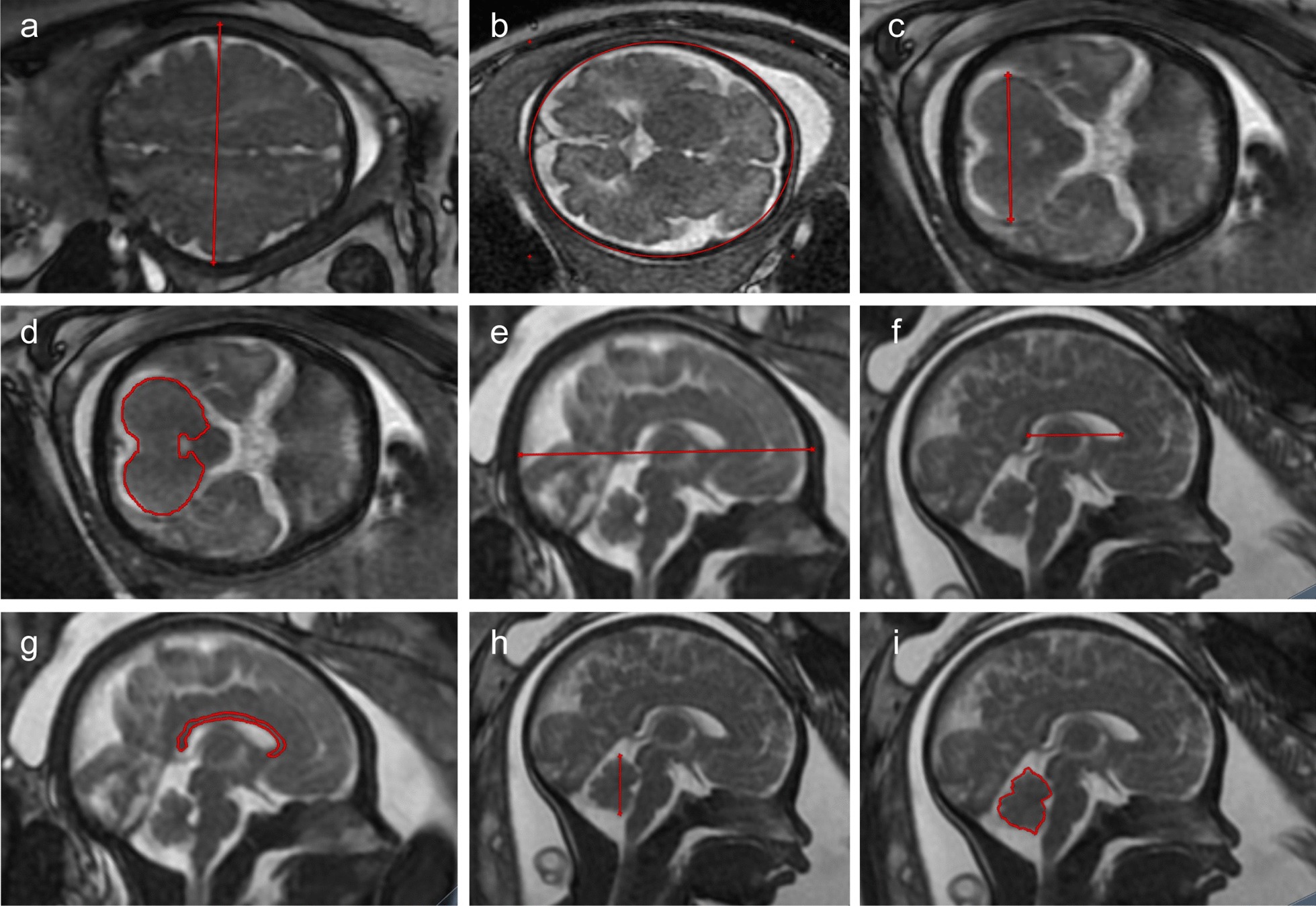
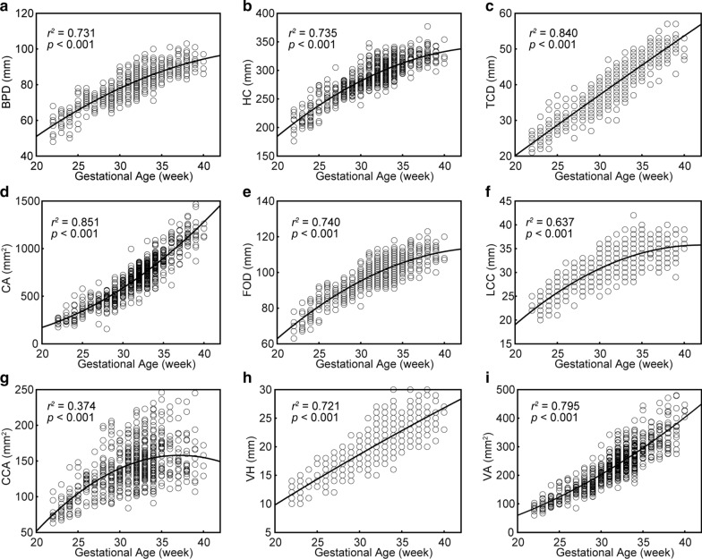
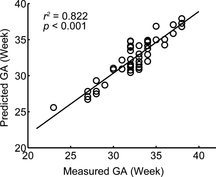
Methods: We used MRI to scan a total of 637 fetuses ranging in age from 22 to 40 gestational weeks. We evaluated 9 standard fetal 2D biometric parameters, and regression models were fitted to assess normal fetal brain development. A stepwise linear regression model was constructed to predict gestational age, and measurement accuracy was determined in a held-out, unseen test sample (n = 49).
Results: A second-order polynomial regression model was found to be the best descriptor of biometric measures including brain bi-parietal diameter, head circumference, and fronto-occipital diameter in relation to normal fetal growth. Normal fetuses showed divergent growth patterns for the cerebrum and cerebellum, where the cerebrum undergoes rapid growth in the second trimester, while the cerebellum undergoes rapid growth in the third trimester. Moreover, a linear model based on biometrics of brain bi-parietal diameter, length of the corpus callosum, vermis area, transverse cerebellar diameter, and cerebellar area accurately predicted gestational age in the second and third trimesters (cross-validation R2 = 0.822, p < 0.001).
Conclusions: These results support the use of MRI biometry charts to improve MRI evaluation of fetal growth and suggest that MRI biometry measurements offer a potential estimation model of fetal gestational age in the second half of gestation, which is vital to any assessment of pregnancy, fetal development, and neonatal care.
Keywords: Fetal brain maturity; Gestational age; Magnetic resonance imaging.
Conflict of interest statement
No potential conflict of interest was reported by the authors.
Figures
References
-
- Naidu K, Fredlund KL. Gestational age assessment. Treasure Island (FL); 2020.
Publication types
MeSH terms
Grants and funding
LinkOut - more resources
Full Text Sources
Other Literature Sources
Medical
