. 2020 Nov 25;11(1):496.
doi: 10.1186/s13287-020-02005-x.

Bone marrow mesenchymal stem cell-derived exosomes promote rotator cuff tendon-bone healing by promoting angiogenesis and regulating M1 macrophages in rats

Affiliations

Bone marrow mesenchymal stem cell-derived exosomes promote rotator cuff tendon-bone healing by promoting angiogenesis and regulating M1 macrophages in rats

Yao Huang et al. Stem Cell Res Ther. .

Abstract

Background: Rotator cuff tears (RCTs) often require reconstructive surgery. Tendon-bone healing is critical for the outcome of rotator cuff reconstruction, but the process of tendon-bone healing is complex and difficult. Mesenchymal stem cells (MSCs) are considered to be an effective method to promote tendon-bone healing. MSCs have strong paracrine, anti-inflammatory, immunoregulatory, and angiogenic potential. Recent studies have shown that MSCs achieve many regulatory functions through exosomes. The purpose of this study was to explore the role of bone marrow mesenchymal stem cell-derived exosomes (BMSC-Exos) in tendon-bone healing.

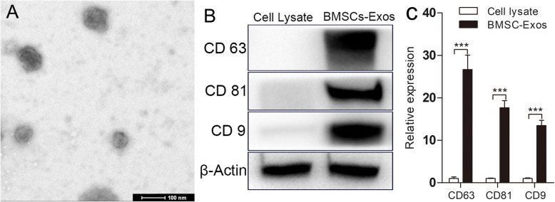
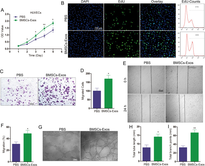
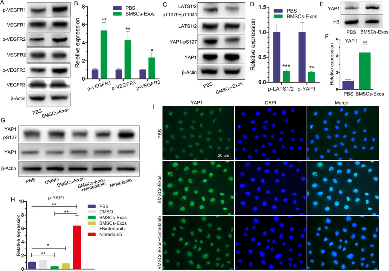
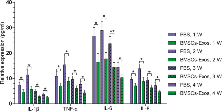
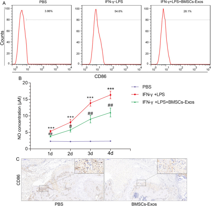
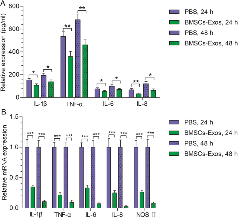
Methods: Our study found that BMSC-Exos promote the proliferation, migration, and angiogenic tube formation of human umbilical vein endothelial cells (HUVECs). The mechanism by which BMSC-Exos achieve this may be through the regulation of the angiogenic signaling pathway. In addition, BMSC-Exos can inhibit the polarization of M1 macrophages and inhibit the secretion of proinflammatory factors by M1 macrophages. After rotator cuff reconstruction in rats, BMSC-Exos were injected into the tail vein to analyze their effect on the rotator cuff tendon-bone interface healing.

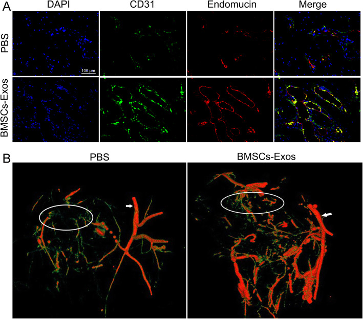
Results: It was confirmed that BMSC-Exos increased the breaking load and stiffness of the rotator cuff after reconstruction in rats, induced angiogenesis around the rotator cuff endpoint, and promoted growth of the tendon-bone interface.

Conclusion: BMSC-Exos promote tendon-bone healing after rotator cuff reconstruction in rats by promoting angiogenesis and inhibiting inflammation.

Keywords: Bone mesenchymal stem cells; Exosome; Rotator cuff; Shoulder; Tendinosis.


Conflict of interest statement

The authors declare that they have no competing interests.

Figures

Fig. 1
Fig. 1
BMSC-Exos were identified by TEM and WB. a Morphology observed by TEM. b Exosome surface markers detected by Western blot. c Quantitative analysis of b. Each experiment was independently repeated three times (*p < 0.05; **p < 0.01; ***p < 0.001)
Fig. 2
Fig. 2
Functional effects of BMSC-Exos on HUVECs. a, b BMSC-Exos significantly promoted the proliferation of HUVECs, as demonstrated by the Cell Counting Kit-8 (CCK-8) and EdU assays. Scar bar 100 μm. c BMSC-Exos significantly promoted the migration of HUVECs as determined by the transwell assay at 24 h. Scar bar 100 μm. d Quantitative analysis of the transwell assay. e BMSC-Exos significantly promoted the mobility of HUVECs as determined by the scratch wound assay. Scar bar 100 μm. f Quantitative analysis of the scratch wound assay. g BMSC-Exos significantly enhanced the tube formation ability at 6 h, as determined by the tube formation assay. Scar bar 250 μm. h, i Quantitative analysis of the tube formation assay. Each experiment was independently repeated three times (*p < 0.05; **p < 0.01; ***p < 0.001)
Fig. 3
Fig. 3
Functional effects of BMSC-Exos on angiogenesis-related signaling pathways in HUVECs. a BMSC-Exos promoted the phosphorylation of VEGFR at 12 h, as determined by WB in HUVECs. b Quantitative analysis of a. c BMSC-Exos inhibited the phosphorylation of LATS and YAP1 at 12 h, as determined by WB in HUVECs. d Quantitative analysis of c. e BMSC-Exos increased YAP1 expression in the nucleus at 12 h, as determined by WB in HUVECs. f Quantitative analysis of e. g Effects of BMSC-Exos and nintedanib on YAP1 phosphorylation at 12 h, as determined by WB in HUVECs. h Quantitative analysis of g. i Effects of BMSC-Exos and nintedanib on YAP1 expression at 12 h, as determined by immunofluorescence in HUVECs. Each experiment was independently repeated three times. Scar bar 20 μm
Fig. 4
Fig. 4
Effects of BMSC-Exos on the vessels around the rotator cuff insertion in rats. a BMSC-Exos promoted the expression of CD31 and endomucin around the tendon-bone interface 4 weeks after reconstruction, as determined by immunofluorescence (n = 9/group). Scar bar 100 μm. b BMSC-Exos promoted angiogenesis around the rotator cuff 4 weeks after reconstruction, as determined by angiography (n = 9/group) (white circle, rotator cuff insertion; white arrow, axillary artery)
Fig. 5
Fig. 5
BMSC-Exos reduced the expression of IL-1β, TNF-α, IL-6, and IL-8 in rat serum (each experiment was independently repeated three times)
Fig. 6
Fig. 6
Effects of BMSC-Exos on the polarization of M1 macrophages. a BMSC-Exos inhibited the expression of the surface marker CD86 in M1 macrophages (each experiment was independently repeated three times). b BMSC-Exos inhibited the release of NO by M1 macrophages (each experiment was independently repeated three times). c BMSC-Exos inhibited the expression and distribution of M1 macrophages around the tendon-bone interface 2 weeks after reconstruction (n = 9/group). *Compared with the PBS group. #Between the LPS and BMSC-Exos groups, #p < 0.05; ##p < 0.01
Fig. 7
Fig. 7
Effects of BMSC-Exos on M1 macrophage-related inflammatory factors. a BMSC-Exos inhibited the secretion of inflammatory factors by M1 macrophages. b BMSC-Exos inhibited mRNA expression of M1 macrophage-associated inflammatory factors. Each experiment was independently repeated three times
Fig. 8
Fig. 8
Mechanical and biological effects of BMSC-Exos on rotator cuff reconstruction in rats. a Biomechanical test of the reconstruction rotator cuff (supraspinatus). b, c BMSC-Exos promoted the maximum breaking load and stiffness at 4 and 8 weeks (n = 9/group). d BMSC-Exos promoted the Col I expression of the tendon-bone interface at 4 and 8 weeks (black arrows; n = 9/group). Scar bar 100 μm. e Quantitative analysis of d. f BMSC-Exos promoted Col II expression at the tendon-bone interface at 4 and 8 weeks (black arrows; n = 9/group). Scar bar 100 μm. g Quantitative analysis of f. h BMSC-Exos promoted the expression of Sharpey’s fibers and proteoglycan at the tendon-bone interface at 4 and 8 weeks (between two black lines; n = 9/group). Scar bar 200 μm. i Quantitative analysis of Sharpey’s fibers in h
Fig. 9
Fig. 9
BMSC-Exos promote tendon-bone healing after rotator cuff reconstruction by promoting angiogenesis and inhibiting M1 macrophage in rats

Similar articles

Cited by

References

    1. Frank JB, ElAttrache NS, Dines JS, Blackburn A, Crues J, Tibone JE. Repair site integrity after arthroscopic transosseous-equivalent suture-bridge rotator cuff repair. Am J Sports Med. 2008;36:1496–1503. doi: 10.1177/0363546507313574. - DOI - PubMed
    1. Gulotta LV, Nho SJ, Dodson CC, Adler RS, Altchek DW, MacGillivray JD, Registry HSSARC. Prospective evaluation of arthroscopic rotator cuff repairs at 5 years: part I--functional outcomes and radiographic healing rates. J Shoulder Elb Surg. 2011;20:934–940. doi: 10.1016/j.jse.2011.03.029. - DOI - PubMed
    1. Iyengar JJ, Samagh SP, Schairer W, Singh G, Valone FH, 3rd, Feeley BT. Current trends in rotator cuff repair: surgical technique, setting, and cost. Arthroscopy. 2014;30:284–288. doi: 10.1016/j.arthro.2013.11.018. - DOI - PubMed
    1. Atesok K, Fu FH, Wolf MR, Ochi M, Jazrawi LM, Doral MN, Lubowitz JH, Rodeo SA. Augmentation of tendon-to-bone healing. J Bone Joint Surg Am. 2014;96:513–521. doi: 10.2106/JBJS.M.00009. - DOI - PubMed
    1. Galanopoulos I, Ilias A, Karliaftis K, Papadopoulos D, Ashwood N. The impact of re-tear on the clinical outcome after rotator cuff repair using open or arthroscopic techniques - a systematic review. Open Orthop J. 2017;11:95–107. doi: 10.2174/1874325001711010095. - DOI - PMC - PubMed

Publication types

LinkOut - more resources