. 2021 Oct;70(10):1857-1871.
doi: 10.1136/gutjnl-2020-322980. Epub 2020 Nov 25.

NAIL: an evolutionarily conserved lncRNA essential for licensing coordinated activation of p38 and NFκB in colitis

Affiliations

NAIL: an evolutionarily conserved lncRNA essential for licensing coordinated activation of p38 and NFκB in colitis

Semih Can Akıncılar et al. Gut. 2021 Oct.

Abstract

Objective: NFκB is the key modulator in inflammatory disorders. However, the key regulators that activate, fine-tune or shut off NFκB activity in inflammatory conditions are poorly understood. In this study, we aim to investigate the roles that NFκB-specific long non-coding RNAs (lncRNAs) play in regulating inflammatory networks.

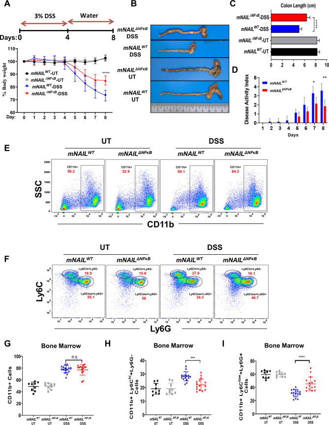
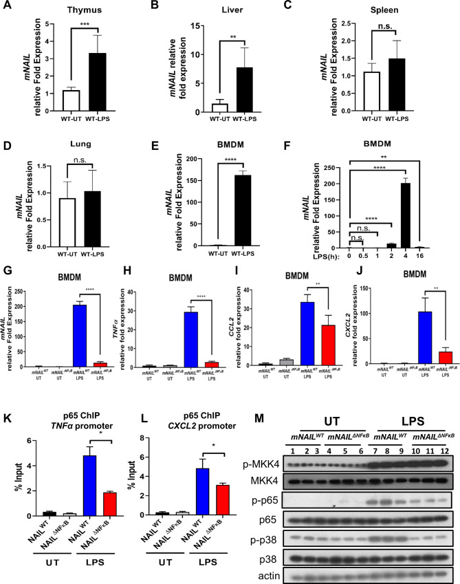
Design: Using the first genetic-screen to identify NFκB-specific lncRNAs, we performed RNA-seq from the p65-/- and Ikkβ-/- mouse embryonic fibroblasts and report the identification of an evolutionary conserved lncRNA designated mNAIL (mice) or hNAIL (human). hNAIL is upregulated in human inflammatory disorders, including UC. We generated mNAILΔNFκB mice, wherein deletion of two NFκB sites in the proximal promoter of mNAIL abolishes its induction, to study its function in colitis.

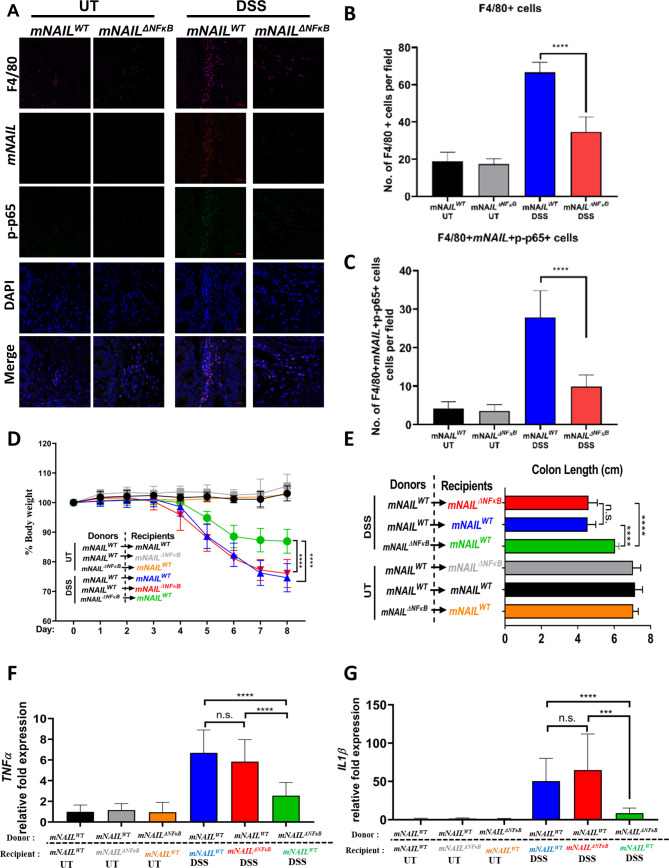
Results: NAIL regulates inflammation via sequestering and inactivating Wip1, a known negative regulator of proinflammatory p38 kinase and NFκB subunit p65. Wip1 inactivation leads to coordinated activation of p38 and covalent modifications of NFκB, essential for its genome-wide occupancy on specific targets. NAIL enables an orchestrated response for p38 and NFκB coactivation that leads to differentiation of precursor cells into immature myeloid cells in bone marrow, recruitment of macrophages to inflamed area and expression of inflammatory genes in colitis.

Conclusion: NAIL directly regulates initiation and progression of colitis and its expression is highly correlated with NFκB activity which makes it a perfect candidate to serve as a biomarker and a therapeutic target for IBD and other inflammation-associated diseases.

Keywords: chronic ulcerative colitis; inflammation; inflammatory bowel disease.


Conflict of interest statement

Competing interests: None declared.

Figures

Figure 1
Figure 1
Identification of lncRNAs regulated by NFκB signalling pathway. (A) NFκB signalling pathway and screening strategy used to identify NFκB-regulated lncRNAs. Crosses represent loss of the corresponding proteins (Ikkβ, Ikkγ, p65) using KO MEFs. (B) Heatmap of logCPM values of differentially expressed lncRNAs in WT, p65 -/- and Ikkβ -/- in immortalised MEFs exposed to TNFα for the indicated time points and analysed by paired-end RNA-sequencing. Gm16685 is shown with red arrow. (C) Schematic view of genetic loci of Gm16685 lncRNA (blue). Gm16685 is expressed from antisense direction to IL7 gene (red). NFκB binding motifs in the promoter region of Gm16685 shown as yellow highlighted sequences. (D) RT-qPCR analysis of Gm16685 transcript levels in immortalised WT MEFs exposed to TNFα for different time points as indicated. (E) RT-qPCR analysis of Gm16685 in WT (n=5), p65 -/- (n=8) and Ikkβ -/- (n=3) independent primary MEFs treated with TNFα for indicated time courses. P values were calculated using Student’s t-test method (***p<0.001). (F) Gene expression profile of 24 inflamed and patient match non-inflamed samples. Differentially expressed genes are shown in heatmap. LOC105375914 is shown with red arrow. (G) Box plot shows upregulation of hNAIL (LOC105375914) expression in inflamed colon tissue of UC patients. KO, knock out; MEFs, mouse embryonic fibroblasts; WT, wild type.
Figure 2
Figure 2
mNAILΔNFκB mice display decreased activation of NFκB and inflammation. (A) Schematic view of mNAIL lncRNA promoter targeting with CRISPR-Cas9 editing. NFκB binding motifs are shown in yellow in the promoter region of mNAIL. (B) mNAIL mice were generated using standard methods (refer to ‘Materials and methods’ section). mNAIL mice were born in normal Mendelian ratio (table below) and unchallenged mice appear normal. (C) Genotyping PCR results of mNAILWT and mNAILΔNFκB mice. PCR products from mNAILWT and mNAILΔNFκB alleles are shown. (D) Sanger sequencing results of mNAILWT and mNAILΔNFκB cells. (E–H) mNAILWT and mNAILΔNFκB MEFs (n=3) were treated with TNFα for the indicated time points. Expression analysis was performed by RT-qPCR for (E) mNAIL, (F) IL7, (G) TNFα and (H) IL1β genes. (I–K) WT MEFs were transfected with si-Control, si-NAIL #1 and si-NAIL #2 siRNAs. After 48 hours post-transfection, cells were treated with or without TNFα and harvested for gene expression analysis. Graphs show the gene expression analysis of (I) mNAIL and (J) TNFα by RT-qPCR. Actin was used as a control. Error bars indicate mean±SD of three independent experiments. P values were calculated using Student’s t-test method (*p<0.05; **p<0.01; n.s., not significant). (K) Cells lysate were analysed with western blot for the indicated proteins. MEFs, mouse embryonic fibroblasts; WT, wild type.
Figure 3
Figure 3
mNAILΔNFκB mice are resistant to dextran sulfate sodium (DSS)-induced colitis. (A) Scheme of DSS-induced colitis model is shown on the top panel. mNAILWT and mNAILΔNFκB mice were treated as shown in top panel. Body weight measurements from mice monitored for 8 days (bottom panel). (B and C) Colon length of DSS-treated mNAILWT and mNAILΔNFκB mice measured at day 8. (D) Disease activity index was recorded daily. (E) Bone marrow cells were isolated from the DSS-treated mNAILWT and mNAILΔNFκB mice at day 8 and were stained for CD11b (PE), Ly6c (BV711) and Ly6g (FITC) cell surface markers. Cells were analysed by FACS and gated as CD11b+cells. Representative FACS data were shown for mNAILWT and mNAILΔNFκB mice treated with or without DSS. (F) CD11b+cells were further gated for Ly6c and Ly6g markers. (G–I) Quantification of CD11b+, CD11b+Ly6Chi+Ly6G−, CD11b+Ly6Clow+Ly6G+cells gated in (E) and (F). Error bars indicate mean±SD of three independent experiments (UT: n=9, DSS: n=15). P values were calculated using Student’s t-test method (*p<0.05; **p<0.01; ***p<0.001; ****p<0.0001; n.s., not significant). DSS, dextran sulfate sodium; FACS, fluorescence-activated cell sorting; SSC, side scatter; side scatter; UT, untreated; WT, wild type.
Figure 4
Figure 4
mNAIL is expressed mainly in myeloid cells and its expression amplifies activation of NFκB and inflammation. (A–E) LPS (5 mg/kg) was injected intraperitoneally into C57BL/6N WT mice (n=6). (A) Thymus, (B) liver, (C) spleen and (D) lung tissues were collected after 4 hours and expression analysis was performed by RT-qPCR for mNAIL gene. (E, F) Bone marrow cells isolated from WT mice (n=6) and differentiated into bone marrow-derived macrophages (BMDM) for 7 days. BMDM cells were treated with or without LPS (200 ng/mL) for 4 hours. (E) Graph shows the mNAIL expression analysis by qPCR. (F) Time kinetics of mNAIL expression is shown in LPS-stimulated BMDM cells. Data were normalised to actin. (G–J) Bone marrow cells isolated from mNAILWT and mNAILΔNFκB mice (n=6) and differentiated into BMDM for 7 days. BMDM cells were treated with or without LPS (200 ng/mL) for 4 hours. Graph shows the gene expression analysis of (G) mNAIL, (H) TNFα, (I) CCL2 and (J) CXCL2 by qPCR. Data were normalised to actin. (K–L) mNAILWT and mNAILΔNFκB BMDM cells were treated with or without LPS, and ChIP analysis was performed for p65 (n=3). Graph shows the ChIP-qPCR analysis of p65 occupancy on the (K) TNFα and (L) CXCL2 promoters. Error bars indicate mean±SD of three biological replicates. P values were calculated using Student’s t-test method (*p<0.05; **p<0.01; ***p<0.001; ****p<0.0001; n.s., not significant). (M) Western blot shows the total and phosphorylated p65, p38 and MKK4 in BMDM cells treated with or without LPS. Each replicate is labelled as #1, #2 and #3. UT, untreated; WT, wild type.
Figure 5
Figure 5
mNAIL regulates expression of inflammatory genes in colitis. (A) Heat map shows the genes that are differentially expressed between mNAILWT and mNAILΔNFκB mice treated with DSS. Genes were first filtered for differential genes on DSS treatment between mNAILWT untreated (UT) and DSS to identify DSS-dependent genes. Next, this gene list was used to identify the differentially expressed genes between mNAILWT and mNAILΔNFκB mice treated with DSS. Differentially expressed genes were identified based on the criteria of 2 log2-fold difference with false discovery rate<0.05. (B–E) Expression analysis was performed by RT-qPCR for (B) mNAIL, (C) TNFα (D) IL1β and (E) CCL2 genes in the colon tissues of mNAILWT and mNAILΔNFκB mice treated with or without DSS at day 8 (n=15). (F–G) Protein levels of TNFα and IL1β cytokines were measured in colon samples by ELISA. Error bars indicate mean±SD of three independent experiments. P values were calculated using Student’s t-test method (**p<0.01; ***p<0.001; ****p<0.0001). (H) Total protein lysates isolated from the colon tissues of mNAILWT and mNAILΔNFκB mice treated with or without DSS at day 8 were analysed for phosphorylated and total p65 and p38. Actin was used for normalisation. DSS, dextran sulfate sodium; UT, untreated; WT, wild type.
Figure 6
Figure 6
mNAIL acts via immune cells to regulate inflammation. (A) Colon sections of the DSS-treated mNAILWT and mNAILΔNFκB mice at day 8 were stained with mNAIL specific FISH probe, F4/80 and p-p65 antibodies. Tissue slides were analysed for the indicated molecules by confocal microscopy. (B) Graph shows quantification of the number of F4/80+ cells from A. (C) Graph shows quantification of the number of F4/80+p-p65+cells from A. Error bars indicate mean±SD of three independent fields examined per mouse (n=3 per group). ***p<0.001; ****p<0.0001; n.s., not significant. P values were calculated by two-tailed Student’s t-test method. (D) mNAILWT or mNAILΔNFκB bone marrow reconstituted mNAILWT and mNAILΔNFκB mice were treated with DSS for 8 days. Graph shows the body weight measurements from mice monitored for 8 days (E) and the colon length at day 8. Graphs show gene expression analysis of (F) TNFα and (G) IL1β in the colon tissues of different bone marrow reconstituted groups treated with DSS for 8 days by qPCR. Data are normalised to actin. Error bars indicate mean±SD of two independent experiments (total n=12). P values were calculated using Student’s t-test method (***p<0.001; ****p<0.0001; n.s., not significant). DSS, dextran sulfate sodium; UT, untreated; WT, wild type.
Figure 7
Figure 7
mNAIL sequesters Wip1 phosphatase away from its substrate in vivo. (A) Colon tissues were collected from mNAILWT and mNAILΔNFκB mice (n=4) treated with and without DSS for 8 days. (A) p65 protein was immunoprecipitated and coprecipitated proteins were analysed by western blot. Actin was used as a loading control. Each replicates are indicated as #1, #2, #3 and #4. (B) Graph shows quantification of coprecipitated Wip1 from A) (total n=4). Error bars indicate mean±SD of four replicates. P values were calculated using Student’s t-test method (**p<0.01; ***p<0.001). (C) Summary of phenotypes of p65, p38 myeloid specific knock mice, Wip1 null mice and mNAILΔNFκB in DSS model. Model based on our studies. (D) In the colitis model, in the presence of NAIL, activation of p38 and p65 is coordinated in a timely manner. On intestinal damage and release of microbiota to the colon, myeloid progenitor cells differentiate into immature myeloid cells which give rise to macrophages that infiltrate inflamed colon and express inflammatory genes. In the absence of NAIL, Wip1 prevents phosphorylation of p65 and p38 which leads to defects in generation of immature myeloid cells, reduction of recruitment of macrophages and deregulated expression of inflammatory genes. In the absence of TNFα or other inflammatory stimuli, Wip1 phosphatase is bound to p65 and its other targets such as p38. On activation of NFκB by TNFα, p65 is released from IκB proteins and p65 accumulates in the nucleus to activate target genes such as the NAIL lncRNA. Phosphorylation of p65 (p–p65) by a number of kinases mainly (IKKs) in the cytoplasm is critical for it to effectively bind to a subset of target genes, and subsequently, recruit p300 histone acetyltransferase for activating chromatin remodelling and for effectively kick-starting NFκB target gene expression. Meanwhile, NAIL lncRNA stimulated due to activation of p65 and squelches Wip1 phosphatase away from p65 or other Wip1 targets such as p38 thereby temporally regulating the kinetics of p65 and p38 phosphorylation and hence activation of genes dependent downstream of these important pathways. In the absence of NAIL, p65 and other Wip1 targets (p38) are avidly bound by Wip1 even after stimulation and this leads to reduced p-p65 and p-p38 levels and also reduced p300 recruitment to target genes along with lesser interaction of p38 and NFκB targets, resulting in decreased expression of the inflammatory genes. DSS, dextran sulfate sodium; UT, untreated; WT, wild type.

Similar articles

Cited by

References

    1. Neurath MF. Cytokines in inflammatory bowel disease. Nat Rev Immunol 2014;14:329–42. 10.1038/nri3661 - DOI - PubMed
    1. Shen Z-H, Zhu C-X, Quan Y-S, et al. . Relationship between intestinal microbiota and ulcerative colitis: mechanisms and clinical application of probiotics and fecal microbiota transplantation. World J Gastroenterol 2018;24:5–14. 10.3748/wjg.v24.i1.5 - DOI - PMC - PubMed
    1. Atreya I, Atreya R, Neurath MF. NF-kappaB in inflammatory bowel disease. J Intern Med 2008;263:591–6. 10.1111/j.1365-2796.2008.01953.x - DOI - PubMed
    1. Cohn HM, Dave M, Loftus EV. Understanding the cautions and contraindications of immunomodulator and biologic therapies for use in inflammatory bowel disease. Inflamm Bowel Dis 2017;23:1301–15. 10.1097/MIB.0000000000001199 - DOI - PMC - PubMed
    1. Karin M, Greten FR. NF-kappaB: linking inflammation and immunity to cancer development and progression. Nat Rev Immunol 2005;5:749–59. 10.1038/nri1703 - DOI - PubMed

Publication types