. 2020 Nov 25;5(6):e00956-20.
doi: 10.1128/mSphere.00956-20.

Elucidation of DNA Repair Function of PfBlm and Potentiation of Artemisinin Action by a Small-Molecule Inhibitor of RecQ Helicase

Affiliations

Elucidation of DNA Repair Function of PfBlm and Potentiation of Artemisinin Action by a Small-Molecule Inhibitor of RecQ Helicase

Niranjan Suthram et al. mSphere. .

Abstract

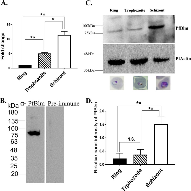
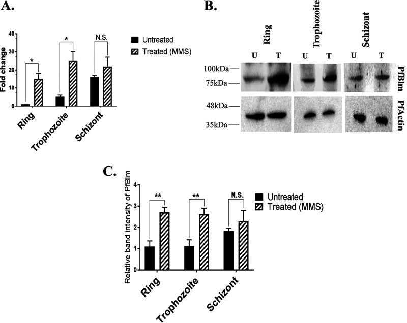
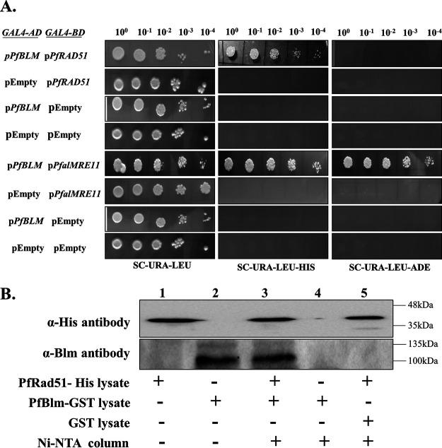
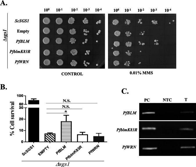
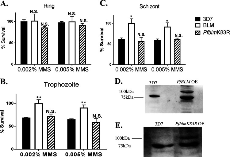
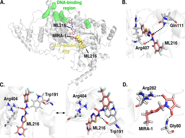
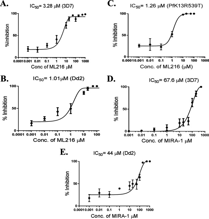
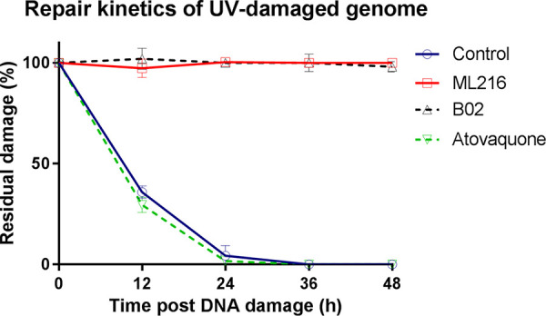
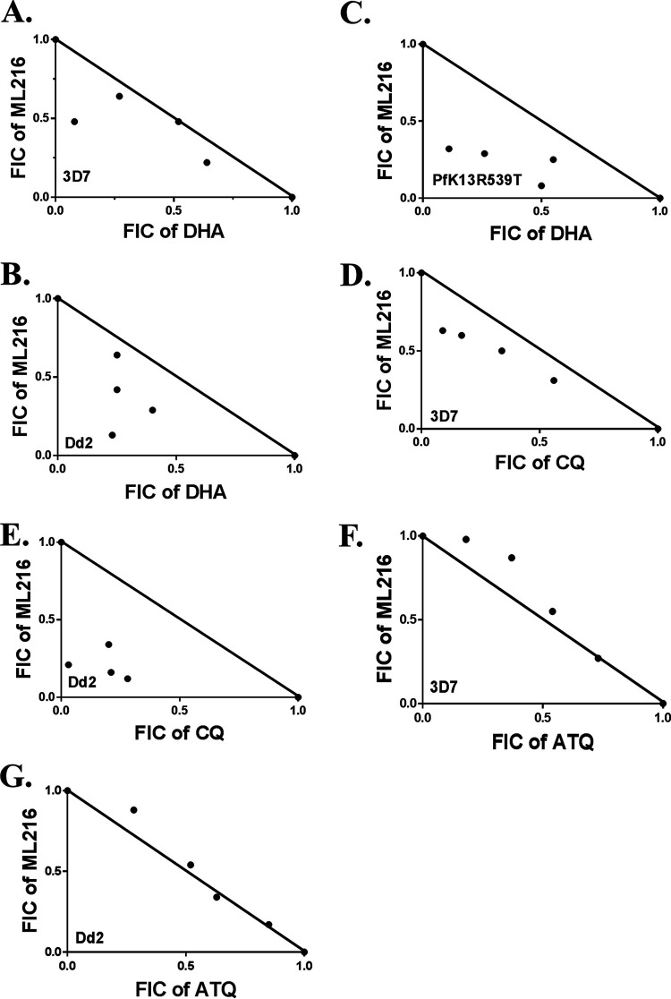
Artemisinin (ART)-based combination therapies are recommended as first- and second-line treatments for Plasmodium falciparum malaria. Here, we investigated the impact of the RecQ inhibitor ML216 on the repair of ART-mediated damage in the genome of P. falciparumPfBLM and PfWRN were identified as members of the RecQ helicase family in P. falciparum However, the role of these RecQ helicases in DNA double-strand break (DSB) repair in this parasite has not been explored. Here, we provide several lines of evidence to establish the involvement of PfBlm in DSB repair in P. falciparum First, we demonstrate that PfBlm interacts with two well-characterized DSB repair proteins of this parasite, namely, PfRad51 and PfalMre11. Second, we found that PfBLM expression was upregulated in response to DNA-damaging agents. Third, through yeast complementation studies, we demonstrated that PfBLM could complement the DNA damage sensitivity of a Δsgs1 mutant of Saccharomyces cerevisiae, in contrast to the helicase-dead mutant PfblmK83R Finally, we observe that the overexpression of PfBLM induces resistance to DNA-damaging agents and offers a survival advantage to the parasites. Most importantly, we found that the RecQ inhibitor ML216 inhibits the repair of DSBs and thereby renders parasites more sensitive to ART. Such synergism between ART and ML216 actions was observed for both drug-sensitive and multidrug-resistant strains of P. falciparum Taken together, these findings establish the implications of PfBlm in the Plasmodium DSB repair pathway and provide insights into the antiparasitic activity of the ART-ML216 combination.IMPORTANCE Malaria continues to be a serious threat to humankind not only because of the morbidity and mortality associated with the disease but also due to the huge economic burden that it imparts. Resistance to all available drugs and the unavailability of an effective vaccine cry for an urgent discovery of newer drug targets. Here, we uncovered a role of the PfBlm helicase in Plasmodium DNA double-strand break repair and established that the parasitic DNA repair mechanism can be targeted to curb malaria. The small-molecule inhibitor of PfBlm tested in this study acts synergistically with two first-line malaria drugs, artemisinin (ART) and chloroquine, in both drug-sensitive and multidrug-resistant strains of P. falciparum, thus qualifying this chemical as a potential partner in ART-based combination therapy. Additionally, the identification of this new specific inhibitor of the Plasmodium homologous recombination (HR) mechanism will now allow us to investigate the role of HR in Plasmodium biology.

Keywords: DNA repair; PfBlm; PfWrn; Plasmodium falciparum; homologous recombination; molecular docking; yeast complementation.


Figures

FIG 1
FIG 1
PfBLM expression is maximal at the mitotically active schizont stage. (A) Relative abundances of PfBLM transcripts during the ring (12 h postinvasion [hpi]), trophozoite (24 hpi), and schizont (38 hpi) stages quantified by real-time RT-PCR analysis. Data were normalized against PfARP. The mean values ± standard deviations (SD) from three independent experiments are plotted. (B) Specificity of anti-PfBlm antibody versus the preimmune sera. (C) Stage-specific expression of the PfBlm protein. PfActin was used as the loading control. The position of molecular markers is indicated on the left. Representative microscopic images of different blood stages are shown below the blot. (D) Quantification of Western blot data from three independent experiments. Data were normalized against loading control PfActin. Each bar represents mean density ± SD (n = 3). The P value was calculated using the two-tailed t test (* means a P value of <0.05, ** means a P value of <0.01, and N.S. means not significant).
FIG 2
FIG 2
DNA damage-induced upregulation of PfBLM during intraerythrocytic developmental stages. (A) Synchronized P. falciparum in vitro cultures at the ring, trophozoite, and schizont stages were either untreated (U) or treated (T) with 0.05% MMS for 6 h. Real-time RT-PCR on extracted RNA revealed the upregulation of PfBLM mRNA during the ring and trophozoite stages upon DNA damage, but its level was not changed during the schizont stage. PfARP transcripts were used as a loading control. Each bar represents the mean value ± SD (n = 3). (B) Western blots show that MMS induced the expression of the PfBlm protein at the ring and trophozoite stages, but in the case of the schizont stage, it remained unchanged. PfActin acted as a loading control. The position of molecular markers is indicated on the left. (C) Quantification of Western blot data from three independent experiments. Data were normalized against PfActin. Each bar represents the mean band intensity ± SD (n = 3). The P value was calculated using the two-tailed t test (* means a P value of <0.05, ** means a P value of <0.01, and N.S. means not significant).
FIG 3
FIG 3
PfBlm interacts with the key DNA repair proteins PfRad51 and PfalMre11. (A) Full-length PfBLM was fused to the GAL4 activation domain (GAL4-AD) of pGADC1. Similarly, full-length PfalMRE11 and PfRAD51 were fused to the GAL4 binding domain (GAL4-BD) of pGBDUC1. The interaction was tested in yeast strain PJ69-4A, which bears ADE2 and HIS as reporter genes. Starting with the same OD (1 OD/ml), 10-fold serially diluted cells were spotted onto plates lacking leucine and uracil to check for the presence of bait and prey plasmids. Simultaneously, cells were spotted onto plates lacking histidine and adenine to check the interaction. (B) Western blotting shows the interaction between PfRad51 and PfBlm in the Ni-NTA pulldown (lane 3). The cell lysate of histidine-tagged PfRad51 mixed with the empty GST cell lysate (lane 5) did not show a signal with anti-Blm antibody. The description of each lane is shown at the bottom. The position of molecular markers is indicated on the right.
FIG 4
FIG 4
PfBLM functionally complements the MMS sensitivity of the Δsgs1 mutant of S. cerevisiae. (A) Spotting assay on SC−His plates without (control) or with 0.01% MMS. The genotype of each strain is shown on the left. (B) A return-to-growth assay was performed, where all the complementation strains were either untreated or treated with 0.03% MMS for 2 h and allowed to grow on SC−His plates without MMS after washing. The percentage of cell survival for each strain was obtained by taking the ratio of the numbers of colonies formed between treated and untreated cultures. Each bar represents the mean number ± SD after normalization with untreated controls from three independent experiments (n = 3). The P value was calculated using the two-tailed t test (* means a P value of <0.05, and N.S. means not significant). (C) The expression of PfBLM, PfblmK83R, and PfWRN was confirmed by isolating RNA and performing semiquantitative RT-PCR from complementation strains. PC, positive control (Plasmodium genomic DNA used as the template); NTC, nontemplate control; T, test (cDNA from the respective complementation strains used as the template).
FIG 5
FIG 5
Overexpression of PfBLM provides a survival advantage to the parasites under DNA-damaging conditions. (A to C) Plasmodium falciparum 3D7 cells harboring PfBLM-GFP and PfblmK83R-GFP were either untreated or treated with 0.002% and 0.005% MMS for 2 h and subsequently allowed to grow for 48 h after washing. The experiment was also performed with 3D7 cultures, which acted as a control. After 48 h, smears were prepared, and infected erythrocytes were counted to obtain parasitemia data. The experiment was performed with three intraerythrocytic stages of parasites. Stages are indicated at the top. Percent survival was plotted by taking the ratio of the percentages of parasitemia between the treated and untreated cultures of the respective strains. Each bar represents the mean survival value ± SD (n = 3). Significance was calculated with respect to the wild-type strain (3D7). (D) Western blotting shows the expression of the PfBlm protein. The strains are indicated at the top. The expression of the PfBlm (80 kDa) protein in the 3D7 strain and the expression of GFP-tagged PfBlm (107 kDa) protein along with endogenous PfBlm (80 kDa) are shown. OE, overexpression. (E) Expression of the PfBlm (80 kDa) protein in the 3D7 strain (left) and expression of the GFP-tagged PfblmK83R (107 kDa) protein along with endogenous PfBlm (80 kDa) (right). The positions of molecular markers are indicated on the left. The P value was calculated using the two-tailed t test (* means a P value of <0.05, ** means a P value of <0.01, and N.S. means not significant).
FIG 6
FIG 6
In silico analysis of the binding poses and binding affinities of inhibitors against PfBlm. (A) Two binding poses for ML216 and a single binding pose for MIRA-1. ML216 can bind at either the ATP binding site or the DNA binding region, whereas MIRA-1 binds only in the vicinity of the ATP binding site. (B) ML216 interacts with Gln111 and Arg407 in the ATP binding site. (C) ML216 bound near the DNA binding region is seen to exist in two poses: one in which it interacts with Arg404 and one in which it interacts with Trp191. (D) MIRA-1 interacts with the residues Gly80 and Arg282 near the ATP binding site. Hydrogen bonds between the protein and the inhibitor are shown as dotted lines.
FIG 7
FIG 7
Compared to MIRA-1, ML216 is a potent inhibitor of the intraerythrocytic growth of P. falciparum. We employed a Giemsa staining method to evaluate percent parasitemia and deduced percent inhibition from the obtained parasitemia values. (A) Synchronous trophozoite-stage parasites (3D7) were grown for 48 h in the presence of various concentrations of ML216. Parasite growth inhibition at various concentrations of the drug was plotted to obtain the IC50 values. (B) Growth inhibition with ML216 in Dd2 parasites was plotted to obtain IC50 values. (C) Growth inhibition with ML216 in PfK13R539T parasites was plotted to obtain IC50 values. (D) The effect of MIRA-1 on 3D7 parasites was tested according to the same protocol as the one for ML216 treatment. Percent inhibition was plotted against various concentrations of the drug to obtain the IC50 values. (E) Growth inhibition with MIRA-1 in Dd2 parasites was plotted to obtain the IC50 values. Each assay was repeated three times for reproducibility (n = 3).
FIG 8
FIG 8
ML216 blocks the repair of UV-induced DNA damage in the nuclear genome of the parasite. ML216-, B02-, and atovaquone-pretreated and mock-treated cultures were UV irradiated at 100 J/m2 at the trophozoite stage. After UV treatment, cells were grown in the presence of sublethal doses of the respective drugs (ML216, B02, or atovaquone) for 48 h. The mock-treated culture was also grown in parallel in normal complete medium, which acted as the control. Genomic DNA was isolated before and after treatment (untreated and 0 h posttreatment) and at 12-h intervals until 48 h. The percentage of unrepaired damage was plotted at the indicated time points using GraphPad Prism software.
FIG 9
FIG 9
ML216 interacts synergistically with ART and CQ. (A to C) Isobolograms of dihydroartemisinin (DHA)-ML216 in the 3D7 (A), Dd2 (B), and PfK13R539T (C) strains. (D and E) Isobolograms of chloroquine (CQ)-ML216 in the 3D7 (D) and Dd2 (E) strains. (F and G) Isobolograms of atovaquone (ATQ)-ML216 in the 3D7 (F) and Dd2 (G) strains. Fixed-ratio drug combination assay. Each point represents the mean IC50 of the drug combination from three independent experiments. A solid line is plotted between the IC50 values of each drug when used alone. FIC, fractional inhibitory concentration.

Similar articles

Cited by

References

    1. Wongsrichanalai C, Sibley CH. 2013. Fighting drug-resistant Plasmodium falciparum: the challenge of artemisinin resistance. Clin Microbiol Infect 19:908–916. doi:10.1111/1469-0691.12316. - DOI - PubMed
    1. Frankenberg-Schwager M, Frankenberg D. 1990. DNA double-strand breaks: their repair and relationship to cell killing in yeast. Int J Radiat Biol 58:569–575. doi:10.1080/09553009014551931. - DOI - PubMed
    1. Roy N, Bhattacharyya S, Chakrabarty S, Laskar S, Babu SM, Bhattacharyya MK. 2014. Dominant negative mutant of Plasmodium Rad51 causes reduced parasite burden in host by abrogating DNA double-strand break repair. Mol Microbiol 94:353–366. doi:10.1111/mmi.12762. - DOI - PubMed
    1. Kirkman LA, Lawrence EA, Deitsch KW. 2014. Malaria parasites utilize both homologous recombination and alternative end joining pathways to maintain genome integrity. Nucleic Acids Res 42:370–379. doi:10.1093/nar/gkt881. - DOI - PMC - PubMed
    1. Bhattacharyya MK, Kumar N. 2003. Identification and molecular characterisation of DNA damaging agent induced expression of Plasmodium falciparum recombination protein PfRad51. Int J Parasitol 33:1385–1392. doi:10.1016/s0020-7519(03)00212-1. - DOI - PubMed

Publication types

MeSH terms

LinkOut - more resources