. 2020 Nov 25;5(6):e01109-20.
doi: 10.1128/mSphere.01109-20.

Pseudomonas Quinolone Signal-Induced Outer Membrane Vesicles Enhance Biofilm Dispersion in Pseudomonas aeruginosa

Affiliations

Pseudomonas Quinolone Signal-Induced Outer Membrane Vesicles Enhance Biofilm Dispersion in Pseudomonas aeruginosa

Adam C Cooke et al. mSphere. .

Abstract

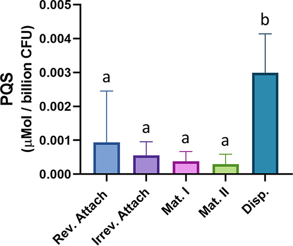
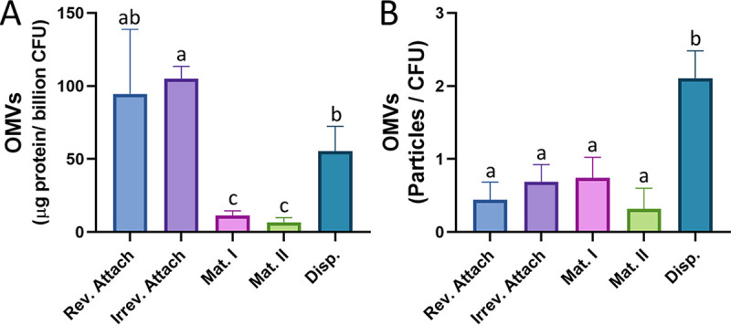
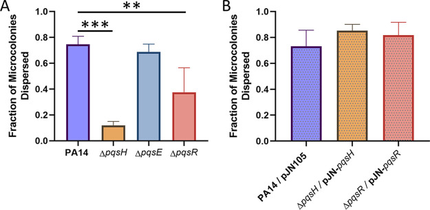
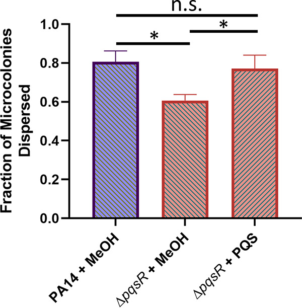
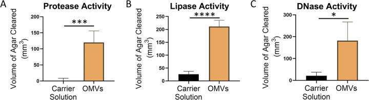
Bacterial biofilms are major contributors to chronic infections in humans. Because they are recalcitrant to conventional therapy, they present a particularly difficult treatment challenge. Identifying factors involved in biofilm development can help uncover novel targets and guide the development of antibiofilm strategies. Pseudomonas aeruginosa causes surgical site, burn wound, and hospital-acquired infections and is also associated with aggressive biofilm formation in the lungs of cystic fibrosis patients. A potent but poorly understood contributor to P. aeruginosa virulence is the ability to produce outer membrane vesicles (OMVs). OMV trafficking has been associated with cell-cell communication, virulence factor delivery, and transfer of antibiotic resistance genes. Because OMVs have almost exclusively been studied using planktonic cultures, little is known about their biogenesis and function in biofilms. Several groups have shown that Pseudomonas quinolone signal (PQS) induces OMV formation in P. aeruginosa Our group described a biophysical mechanism for this and recently showed it is operative in biofilms. Here, we demonstrate that PQS-induced OMV production is highly dynamic during biofilm development. Interestingly, PQS and OMV synthesis are significantly elevated during dispersion compared to attachment and maturation stages. PQS biosynthetic and receptor mutant biofilms were significantly impaired in their ability to disperse, but this phenotype was rescued by genetic complementation or exogenous addition of PQS. Finally, we show that purified OMVs can actively degrade extracellular protein, lipid, and DNA. We therefore propose that enhanced production of PQS-induced OMVs during biofilm dispersion facilitates cell escape by coordinating the controlled degradation of biofilm matrix components.IMPORTANCE Treatments that manipulate biofilm dispersion hold the potential to convert chronic drug-tolerant biofilm infections from protected sessile communities into released populations that are orders-of-magnitude more susceptible to antimicrobial treatment. However, dispersed cells often exhibit increased acute virulence and dissemination phenotypes. A thorough understanding of the dispersion process is therefore critical before this promising strategy can be effectively employed. Pseudomonas quinolone signal (PQS) has been implicated in early biofilm development, but we hypothesized that its function as an outer membrane vesicle (OMV) inducer may contribute at multiple stages. Here, we demonstrate that PQS and OMVs are differentially produced during Pseudomonas aeruginosa biofilm development and provide evidence that effective biofilm dispersion is dependent on the production of PQS-induced OMVs, which likely act as delivery vehicles for matrix-degrading enzymes. These findings lay the groundwork for understanding OMV contributions to biofilm development and suggest a model to explain the controlled matrix degradation that accompanies biofilm dispersion in many species.

Keywords: PQS; Pseudomonas aeruginosa; biofilms; dispersion; outer membrane vesicles; quorum sensing; secretion systems.


Figures

FIG 1
FIG 1
PQS production is elevated during dispersion. PQS was extracted from biofilm tube reactors grown to each of the five stages of development. Measured PQS production was normalized to micromoles per billion CFU. Error bars represent the standard deviations calculated from at least three biological replicates. Statistical significance was assessed by one-way ANOVA followed by Tukey’s post hoc test. Lowercase letters above the bars represent significance. Differences between bars that do not share a letter are statistically significant (P < 0.05).
FIG 2
FIG 2
OMV production varies across biofilm developmental stages. OMVs were harvested from each stage of biofilm development and quantified using two different methods. (A) Purified OMVs were quantified by the modified Lowry assay and normalized to micrograms protein per billion CFU. (B) Purified OMVs were also quantified using nanoparticle tracking and normalized to CFU. Error bars represent the standard deviations calculated from at least three biological replicates. Statistical significance was assessed by one-way ANOVA followed by Tukey’s post hoc test. Lowercase letters above the bars represent significance. Differences between bars that do not share a letter are statistically significant (P < 0.05).
FIG 3
FIG 3
PQS mutants are not deficient in reversible or irreversible attachment. Cultures were grown in 96-well plates, planktonic cells were removed, and attached biomass was quantified using crystal violet staining. (A) PA14 and ΔpqsA strains were grown for 2, 8, and 24 h. (B and C) PA14, ΔpqsH, ΔpqsE, and ΔpqsR strains were grown for 2 h (B) and 24 h (C). Error bars represent the standard deviations calculated from a minimum of three biological replicates. Statistical significance was determined using Student's two-tailed t tests for panel A and one-way ANOVA for panels B and C. *, P < 0.05.
FIG 4
FIG 4
P. aeruginosa dispersion is dependent on quinolone biosynthesis. Biofilms were grown in semibatch cultures in 24-well plates, and the fraction of microcolonies that had dispersed was determined. (A) PA14 wild type and pqsA mutant biofilms were assessed for dispersion after 4, 5, 6, and 7 days of growth. (B) Dispersion of the pqsA mutant overexpressing the pqsA gene was assessed after 6 days of growth and compared to that of the wild type and pqsA mutant. Representative images show microcolonies in PA14 wild-type (C), PA14 ΔpqsA (D), and PA14 ΔpqsA/pJN105-pqsA (E) biofilms after 6 days of growth. Central voids are clearly visible in panels C and E. Error bars represent the standard deviations calculated from at least three biological replicates. Scale bars, 100 μm. Statistical significance was determined using Student’s two-tailed t test for panel A and one-way ANOVA followed by Tukey’s post hoc test for panel B. n.s., P > 0.5; *, P < 0.05; **, P < 0.01.
FIG 5
FIG 5
Production of PQS specifically restores native biofilm dispersion. Biofilms were grown in semibatch cultures in 24-well plates for 6 days. (A) The fraction of microcolonies dispersed was found for PA14 wild-type biofilms as well as ΔpqsH, ΔpqsE, and ΔpqsR biofilms. (B) Overexpression of the missing genes in the mutant backgrounds restored the dispersion that was diminished in ΔpqsH and ΔpqsR biofilms. Bars represent the standard deviations calculated from at least three biological replicates. Statistical significance was analyzed by one-way ANOVA followed by Dunnett’s post hoc test. **, P < 0.01; ***, P < 0.001.
FIG 6
FIG 6
Exogenous PQS rescues ΔpqsR dispersion defect. PA14 wild-type and ΔpqsR biofilms were grown in semibatch cultures in 24-well plates for 4 days. For the following 2 days, the medium was exchanged every 12 h with fresh medium containing 40 μM PQS (+ PQS) or an equivalent amount of methanol (+ MeOH, vehicle control). Dispersion efficiency was then quantified for the strains under each condition. Error bars represent the standard deviations calculated from at least three biological replicates. Statistical significance was analyzed by ANOVA followed by Tukey’s post hoc test. n.s., P > 0.5; *, P < 0.05.
FIG 7
FIG 7
Purified OMVs display EPS-degrading activities. OMVs were harvested, washed with and resuspended in MV buffer, and added to wells punched into different types of agar. (A) Skim milk agar was used to assess protease activity. (B) Tributyrin agar was used to assess lipase activity. (C) DNase agar was used to assess DNase activity. Error bars represent the standard deviations calculated from three biological replicates. Significance was assessed using Student's two-tailed t tests. *, P < 0.05; ***, P < 0.001; ****, P < 0.0001.

Similar articles

Cited by

References

    1. Costerton JW, Stewart PS, Greenberg EP. 1999. Bacterial bioflims: a common cause of persistent infections. Science 284:1318–1322. doi:10.1126/science.284.5418.1318. - DOI - PubMed
    1. Potera C. 1999. Forging a link between biofilms and disease. Science 283:1837–1839. doi:10.1126/science.283.5409.1837. - DOI - PubMed
    1. Alhede M, Kragh KN, Qvortrup K, Allesen-Holm M, van Gennip M, Christensen LD, Jensen PØ, Nielsen AK, Parsek M, Wozniak D, Molin S, Tolker-Nielsen T, Høiby N, Givskov M, Bjarnsholt T. 2011. Phenotypes of non-attached Pseudomonas aeruginosa aggregates resemble surface attached biofilm. PLoS One 6:e27943. doi:10.1371/journal.pone.0027943. - DOI - PMC - PubMed
    1. Kragh KN, Hutchison JB, Melaugh G, Rodesney C, Roberts AEL, Irie Y, Jensen P, Diggle SP, Allen RJ, Gordon V, Bjarnsholt T. 2016. Role of multicellular aggregates in biofilm formation. mBio 7:e00237-16. doi:10.1128/mBio.00237-16. - DOI - PMC - PubMed
    1. Stoodley P, Sauer K, Davies DG, Costerton JW. 2002. Biofilms as complex differentiated communities. Annu Rev Microbiol 56:187–209. doi:10.1146/annurev.micro.56.012302.160705. - DOI - PubMed

Publication types

MeSH terms

LinkOut - more resources