. 2020 Nov 25;12(571):eaaw0285.
doi: 10.1126/scitranslmed.aaw0285.

Sampling interstitial fluid from human skin using a microneedle patch

Affiliations

Sampling interstitial fluid from human skin using a microneedle patch

Pradnya P Samant et al. Sci Transl Med. .

Abstract

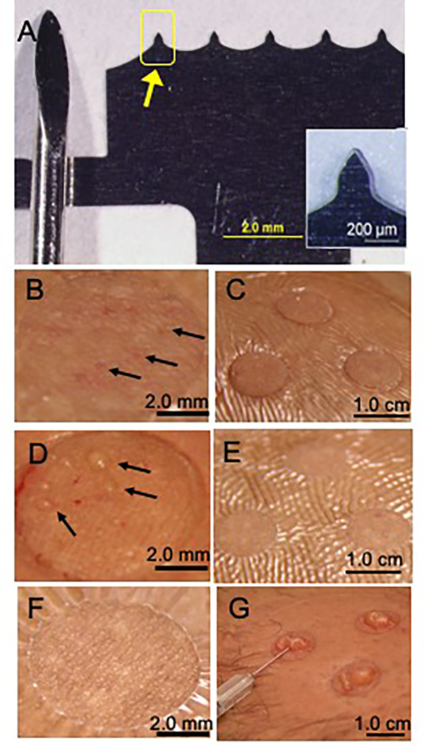
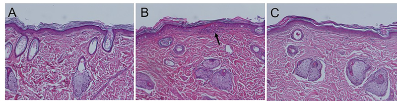
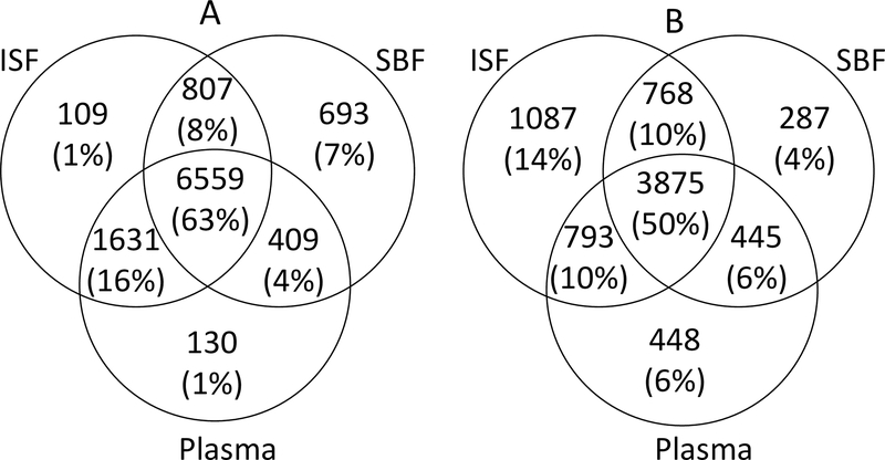
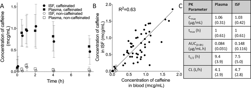
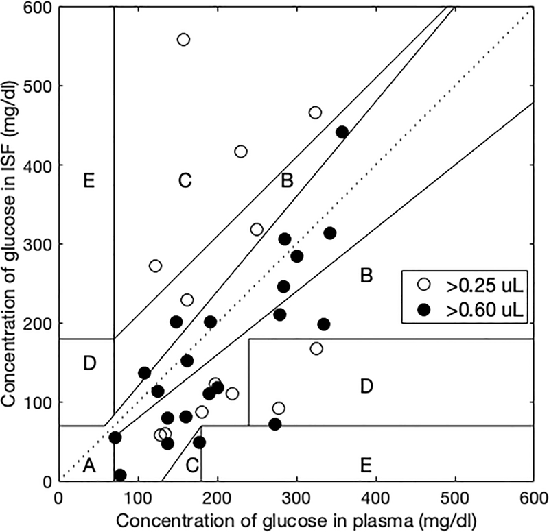
Tissue interstitial fluid (ISF) surrounds cells and is an underutilized source of biomarkers that complements conventional sources such as blood and urine. However, ISF has received limited attention due largely to lack of simple collection methods. Here, we developed a minimally invasive, microneedle-based method to sample ISF from human skin that was well tolerated by participants. Using a microneedle patch to create an array of micropores in skin coupled with mild suction, we sampled ISF from 21 human participants and identified clinically relevant and sometimes distinct biomarkers in ISF when compared to companion plasma samples based on mass spectrometry analysis. Many biomarkers used in research and current clinical practice were common to ISF and plasma. Because ISF does not clot, these biomarkers could be continuously monitored in ISF similar to current continuous glucose monitors but without requiring an indwelling subcutaneous sensor. Biomarkers distinct to ISF included molecules associated with systemic and dermatological physiology, as well as exogenous compounds from environmental exposures. We also determined that pharmacokinetics of caffeine in healthy adults and pharmacodynamics of glucose in children and young adults with diabetes were similar in ISF and plasma. Overall, these studies provide a minimally invasive method to sample dermal ISF using microneedles and demonstrate human ISF as a source of biomarkers that may enable research and translation for future clinical applications.


Conflict of interest statement

Competing interests: MRP is an inventor of patents licensed to companies developing microneedle-based products, is a paid advisor to companies developing microneedle-based products and is a founder/shareholder of companies developing microneedle-based products (Micron Biomedical). This potential conflict of interest has been disclosed and is managed by Georgia Tech and Emory University. PPS and MRP are inventors on a patent application (WO2019126735A1) submitted by Georgia Tech Research Corporation that covers ISF collection methods presented in this study.

Figures

Fig 1.
Fig 1.. Representative images of microneedle device and interstitial fluid collection by microneedle treatment compared to suction blister.
A. Photograph of stainless steel microneedle (MN) patch (right) shown next to a conventional medium-sized lancet (left). Each of the five MNs (arrow) is 250 μm in length, 200 μm in width at the base and tapering to 10 μm tip diameter. Inset shows a magnified view of a single MN. B. Magnified view of skin immediately after MN application. Micropores created from MN-punctured skin appear as faint red dots (arrows). C. Skin after MN application and vacuum administration (−50 kPa at room temperature for 20 min) to draw out interstitial fluid (ISF). Three treatment sites are shown, surrounded by Tegaderm skin covering, before ISF was removed from skin surface. D. Magnified view of skin immediately after MN treatment including vacuum administration. Droplets of ISF can be seen on the skin surface above micropores (arrows). E. Skin shown 24 h after MN treatment. F. Magnified view of skin 24 h after MN treatment. G. Photographs of suction blisters formed after extended vacuum application on human skin (−50 kPa to −70 kPa at 40°C for up to 1 h) being drained with a needle and syringe to collect suction blister fluid (SBF). Images (B-E,G) are all from the same subject and are representative of the study population (n=21).
Fig. 2.
Fig. 2.. Representative images of skin from the back of hairless rats before and after in vivo MN treatment.
A. H&E-stained skin section taken from the back before MN treatment. B. Histology of skin site taken from a biopsy 4 h after MN treatment. Black arrow shows a site of minor focal inflammation. C. Histology of skin 24 h after MN treatment.
Fig. 3.
Fig. 3.. Venn diagram showing the overlap of features in ISF from MN treatment, suction blister fluid and plasma from venipuncture.
Samples were analyzed using A. hydrophilic interaction chromatography (HILIC) and B. reverse-phase C18 liquid chromatography. After filtering, a total of 10,338 and 7,703 features were detected with HILIC and C18, respectively. A feature was considered “present” in a fluid if the feature was detected in that fluid in more than 10% of samples (≥ 3 of 20 samples). Figures not to scale. ISF, interstitial fluid; SBF, suction blister fluid.
Fig. 4.
Fig. 4.. Concentration of caffeine in ISF and plasma in human participants.
A. Concentration of caffeine in ISF and plasma for 8 h after consumption of caffeinated or caffeine-free soft drink (Diet Coke). Mean ± standard deviation, n = 9. B. Correlation between caffeine concentrations in ISF compared to plasma. Each point represents a single time point from a single participant. C. Pharmacokinetic parameters for caffeine concentrations in ISF and plasma. All data shown as mean (SD). Log-scale presentation of the data can be found in Fig S18. Cmax, highest caffeine concentration measured in ISF/plasma; tmax, time at which Cmax was measured; AUC(0–8h), Area under the curve; t1/2, half-life; CL, clearance; VD, volume of distribution
Fig. 5.
Fig. 5.. Clarke error grid showing correlation of glucose concentration in ISF and plasma.
Glucose concentration in ISF and plasma were measured in 15 children and young adults with type 1 diabetes before and for 3 h after eating a standard meal. Among ISF samples with volumes >0.6 μl (21 samples from 12 of the subjects) or >0.25 μl (34 samples from 14 of the subjects), 90% or 76% were in the A+B region of the Clarke error grid, respectively.

References

    1. Downing GJ, in Pharmaceutical Sciences Encyclopedia. (John Wiley & Sons, Inc, 2010).
    1. Heikenfeld J, Jajack A, Feldman B, Granger SW, Gaitonde S, Begtrup G, Katchman BA, Accessing analytes in biofluids for peripheral biochemical monitoring. Nature biotechnology 37, 407–419 (2019); published online EpubApr (10.1038/s41587-019-0040-3). - DOI - PubMed
    1. Deacon B, Abramowitz J, Fear of needles and vasovagal reactions among phlebotomy patients. Journal of Anxiety Disorders 20, 946–960 (2006); published online Epub2006/01/01/ (10.1016/j.janxdis.2006.01.004). - DOI - PubMed
    1. Fischbach FT, Dinning MB, A manual of laboratory and diagnostic tests. (Wolters Kluwer Health, ed. 8th edition, 2009), pp. 1283.
    1. Fogh-Andersen N, Altura BM, Altura BT, Siggaard-Andersen O, Composition of interstitial fluid. Clinical chemistry 41, 1522–1525 (1995); published online EpubOct ( - PubMed

Publication types