. 2020 Nov 25;12(571):eabc6659.
doi: 10.1126/scitranslmed.abc6659.

Systemic cancer therapy with engineered adenovirus that evades innate immunity

Affiliations

Systemic cancer therapy with engineered adenovirus that evades innate immunity

Svetlana Atasheva et al. Sci Transl Med. .

Abstract

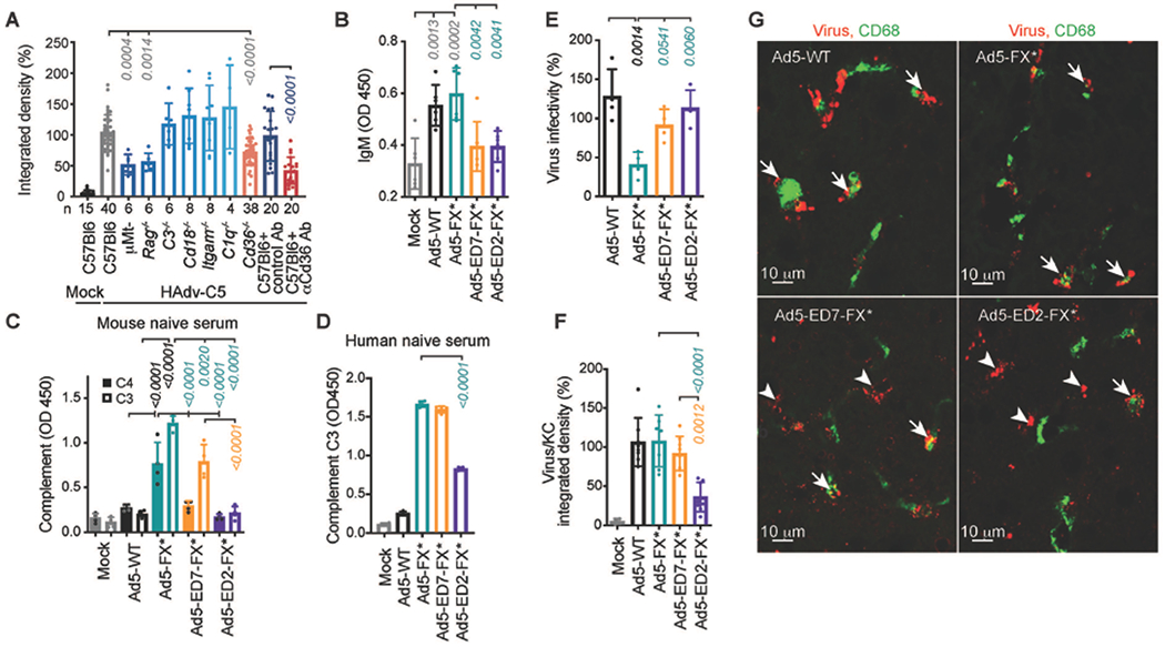
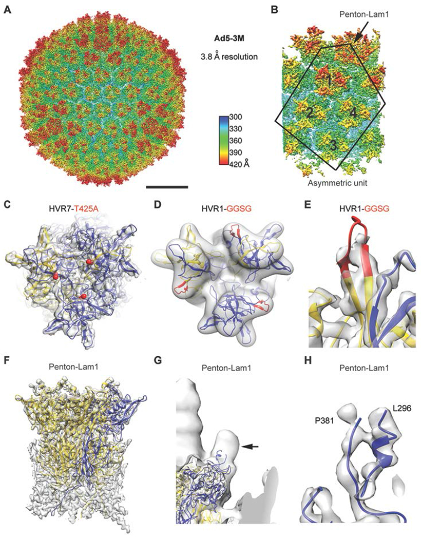
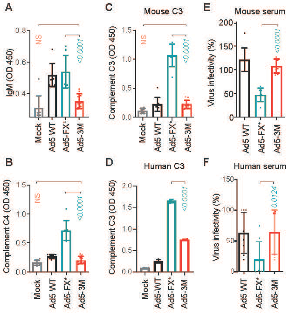
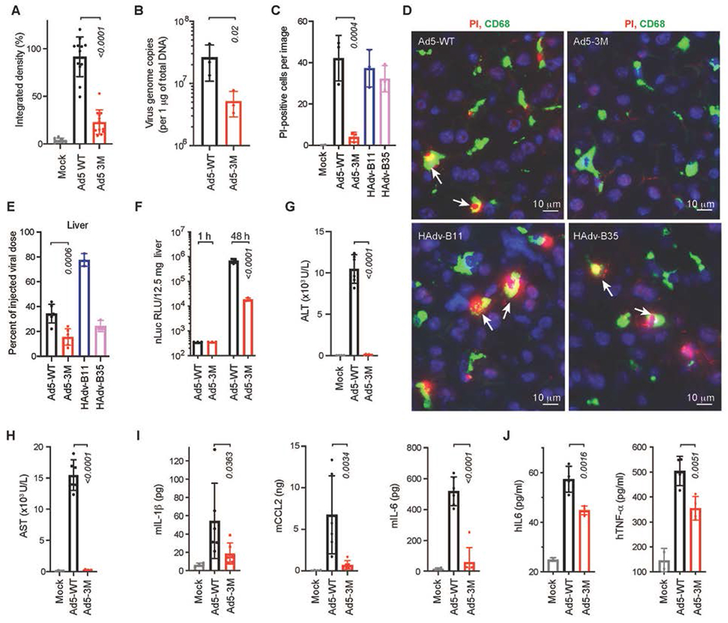
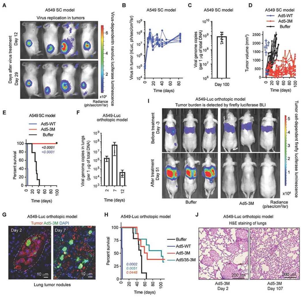
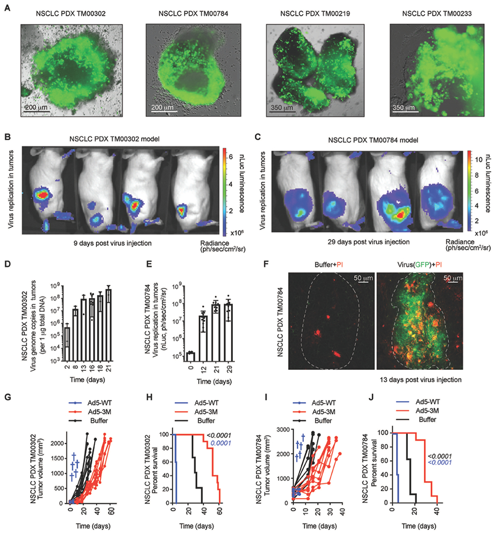
Oncolytic virus therapy is a cancer treatment modality that has the potential to improve outcomes for patients with currently incurable malignancies. Although intravascular delivery of therapeutic viruses provides access to disseminated tumors, this delivery route exposes the virus to opsonizing and inactivating factors in the blood, which limit the effective therapeutic virus dose and contribute to activation of systemic toxicities. When human species C adenovirus HAdv-C5 is delivered intravenously, natural immunoglobulin M (IgM) antibodies and coagulation factor X rapidly opsonize HAdv-C5, leading to virus sequestration in tissue macrophages and promoting infection of liver cells, triggering hepatotoxicity. Here, we showed that natural IgM antibody binds to the hypervariable region 1 (HVR1) of the main HAdv-C5 capsid protein hexon. Using compound targeted mutagenesis of hexon HVR1 loop and other functional sites that mediate virus-host interactions, we engineered and obtained a high-resolution cryo-electron microscopy structure of an adenovirus vector, Ad5-3M, which resisted inactivation by blood factors, avoided sequestration in liver macrophages, and failed to trigger hepatotoxicity after intravenous delivery. Systemic delivery of Ad5-3M to mice with localized or disseminated lung cancer led to viral replication in tumor cells, suppression of tumor growth, and prolonged survival. Thus, compound targeted mutagenesis of functional sites in the virus capsid represents a generalizable approach to tailor virus interactions with the humoral and cellular arms of the immune system, enabling generation of "designer" viruses with improved therapeutic properties.


Conflict of interest statement

Competing interests: D.M.S. has equity interest and is a chief scientific officer of AdCure Bio, which develops adenovirus technologies for therapeutic use. D.M.S. is an inventor on issued US patents No. 9,982,276, Penton-mutated, integrin-retargeted adenovirus vectors with reduced toxicity and their use; and No.10,376,549, Detargeted adenovirus variants and related methods; and pending US patent application 16/460,160 and European patent application 16740545.5, Detargeted adenovirus variants and related methods, submitted by AdCure Bio. All other authors declare that they have no competing interests.

Figures

Fig. 1.
Fig. 1.. Natural IgM bind HAdv-C5 through hexon HVR1 loop.
(A) Integrated density of HAdv-C5-positive staining within CD68- and F4/80-positive cells on liver sections from indicated gene-deficient mice or wild-type mice pre-treated with isotype control or anti-CD36 Ab, 30 minutes after intravenous HAdv-C5 administration. Number of replicates (n) are indicated under each bar. (B) ELISA measurement of natural IgM binding to indicated viruses in mouse plasma (n = 6). (C) Detection of complement components C4 and C3 deposition on the surface of the virions for indicated viruses in mouse serum (n = 4). (D) Detection of complement C3 deposition on the surface of the virions for indicated viruses in human naive serum, lacking HAdv-C5-specific neutralizing antibodies (n = 4). (E) Virus infectivity after incubation with 90% of the raw mouse serum normalized to virus infectivity with no serum treatment determined on HEK293 cells (n = 4). (F) Graphical representation and (G) representative images of immunofluorescent staining of sections of livers harvested from WT mice 30 minutes after intravenous injection with indicated viruses. Kupffer cell CD68-specific staining is in green. Staining with adenovirus-specific polyclonal antibodies is in red. Arrows point to virus staining co-localized with Kupffer cells and arrowheads show virus staining that does not co-localize with Kupffer cells (n = 4-8). The p-values of one-way ANOVA with multiple comparison adjustments are shown above the bars. Bars show mean ± SD. The color of the p-value number indicates the partner for multiple comparison tests. The methodology and statistical details are as described in Table S1 and Methods.
Fig. 2.
Fig. 2.. High-resolution cryo-EM structure of Ad5-3M virus, possessing compound mutations in hexon HVR1 and HVR7 loops and penton base RGD loop.
(A) Full Ad5-3M capsid structure at 3.8 Å resolution displayed with radial coloring. Scale bar, 250 Å. (B) Enlarged view of the icosahedral asymmetric unit (outlined), which includes one subunit of penton base with a laminin-α1 derived integrin interacting motif and 4 hexon trimers with mutations in HVR1 and HVR7 loops. (C) Top view of one Ad5-3M hexon trimer with the sites of the Thr-425-Ala (T425A) HVR7 loop point mutation depicted with red spheres. One hexon subunit is in gold, two hexon subunits are in blue, and the cryo-EM density is in transparent gray. (D) Ad5-3M hexon density filtered to 5-Å resolution superimposed with hexon coordinates including the modeled HVR1 loop containing the Gly-Gly-Ser-Gly (GGSG) sequence (red). (E) Enlarged view of Ad5-3M HVR1 loop model shown with unfiltered cryo-EM density. (F) Side view of the Ad5-3M penton base pentamer (one subunit in blue, four subunits in gold). (G) Ad5-3M penton base density filtered to 15-Å resolution reveals a protrusion at the base of integrin interacting loop (arrow). Density for the fiber, which protrudes from the center of the penton base, is observed at the left edge of the panel. (H) Enlarged view of ordered residues at either end of the Ad5-3M penton base integrin interacting loop shown with unfiltered cryo-EM density.
Fig. 3.
Fig. 3.. Ad5-3M virus is resistant to natural IgM- and complement-mediated opsonization and inactivation in mouse and human sera.
(A) ELISA measurement of natural IgM binding to the indicated viruses in mouse plasma. Detection of deposition of complement components C4 (B) and C3 (C) on the surface of the virions for indicated viruses in mouse serum using ELISA (n = 8-12). (D) Detection of deposition of human complement C3 on the surface of the virions for indicated viruses in human serum using ELISA (n = 4). (E and F) Virus inactivation in raw serum. Indicated viruses were incubated with 90% raw mouse serum (E) (n = 8), and human serum (F) containing low amounts of neutralizing antibodies (n = 8). Virus infectivity after incubation with serum was normalized to virus infectivity with no serum treatment. The p-values of one-way ANOVA with multiple comparison adjustments are shown above the bars. Bars show mean ± SD. The color of the p-value number indicates the partner for multiple comparison tests. N. S. – not significant; p > 0.05. The methodology and statistical details are as described in Table S1 and Methods.
Fig. 4.
Fig. 4.. Ad5-3M virus escapes sequestration in liver macrophages and fails to trigger hepatotoxicity and inflammatory cytokine activation after systemic delivery.
(A) Integrated density of virus-positive staining within CD68- and F4/80-positive cells for indicated viruses on liver sections at 1 h post virus injection, normalized to the median integrated density of Ad5-WT virus staining (n = 8-12). (B) Comparison of virus DNA copies in Kupffer cells by qPCR analysis for indicated viruses 30 min post intravenous virus injection (n = 3). (C) Graphical representation and (D) immunofluorescent staining of liver sections showing in vivo necrosis (PI permeability, red) of CD68-positive Kupffer cells (green) after administration of mice with indicated viruses. DAPI staining of the nuclei is in blue. Arrows point to the PI-positive necrotic Kupffer cells (n = 2-3). (E) Accumulation of viral DNA in livers of mice 1 hour after intravenous virus administration, when compared to the total injected dose for indicated viruses (n = 3-6). (F) Activity of virus-encoded nano-luciferase in liver lysates at the indicated time points (n = 3). Liver function was evaluated by measuring alanine aminotransferase (ALT) (G), and aspartate aminotransferase (AST) (H) in mouse serum 48 h post virus injection (n = 4-6). (I) Comparison of cytokine and chemokine concentration in the spleens of mice (pg or ng per 130mg of spleen tissue) at 1 h post intravenous virus injection (n = 4-6). (J) Amounts of human TNF-α and IL-6 released from primary human macrophages 72 h after their incubation with indicated viruses (n = 4). Bars show mean ± SD. The p-values of one-way ANOVA with multiple comparison tests are shown above the bars, the color of the p-value number indicates the comparison partner. The methodology for individual settings are as described in Table S1 and Methods.
Fig. 5.
Fig. 5.. Ad5-3M transduces tumors, suppresses tumor growth, and extends survival of mice with localized and disseminated tumors after systemic delivery.
(A) In vivo bioluminescence imaging (BLI) of subcutaneous tumor-bearing mice at days 12 and 29 post Ad5-3M treatment. The color indicates bioluminescence intensity of virus-encoded nano-luciferase. SC – subcutaneous. (B) Activity of virus-encoded nano-luciferase in tumor-bearing mice treated with Ad5-3M (n = 10). (C) The amounts of viral genomic DNA in the tumors on day 100 after Ad5-3M treatment (n = 10). (D) Kinetics of subcutaneous tumor growth in mice treated with Ad5-WT (n = 5), Ad5-3M (n = 15), or Buffer (n = 14). Blue crosses indicate deaths of animals treated with Ad5-WT. (E) Log-rank survival plot of subcutaneous tumor-bearing mice over time treated with Ad5-WT (n = 5), Ad5-3M (n = 15), or Buffer (n = 14). (F) Viral genome copies in the lungs of orthotopic tumor-bearing mice at the indicated time points post Ad5-3M treatment, measured by qPCR (n = 3-4). (G) Immunofluorescent staining of lung sections from disseminated orthotopic lung tumor-bearing mice at days 2 and 7 post Ad5-3M injection. Staining with human mitochondria-specific antibodies, recognizing human-derived tumor cells, is in red. Staining with adenovirus-specific polyclonal antibodies is in green. Nuclei-specific DAPI staining is in blue. The anatomical boundaries of tumor nodules are depicted with dotted lines. (H) Log-rank survival plot of mice with disseminated orthotopic lung tumors after treatment with Ad5-WT (n = 5), Ad5-3M (n = 8), Ad5/35-3M (n = 9), or Buffer (n = 9). The methodology and statistical details are as described in Table S1 and Methods. The color of the p-value number indicates the virus-treated comparison partner to the Buffer group. (I) In vivo whole body bioluminescent imaging of disseminated orthotopic lung tumor-bearing mice 3 days before (upper panels) and 51 days after (lower panels) indicated treatments. Representative images of tumor burden in mice on day 3 and on day 51 after treatment with buffer are shown. Selected images on day 51 after treatment with Ad5-3M are shown for 3 out of 8 mice that showed complete disappearance of tumor luminescence. The color indicates the intensity of luminescence, reflecting tumor burden in the lungs. (J) Hematoxylin and eosin staining of sections of lungs harvested from mice with disseminated lung tumors on days 2 and 107 after Ad5-3M treatment. Representative images are shown.
Fig. 6.
Fig. 6.. Ad5-3M transduces primary NSCLC tumors, suppresses tumor growth, and extends survival of PDX tumor-bearing mice after systemic administration.
(A) Fluorescent images of virus-driven GFP expression in primary NSCLC PDX tumor explants 3-5 days after Ad5-3M infection. Representative images of virus transduction for four individual PDX models are shown. In vivo BLI of NSCLC PDX models TM00302 (B) and TM00784 (C) subcutaneously grafted to NSG mice and treated with Ad5-3M intravenously. The color indicates intensity of luminescence of virus-encoded nano-luciferase. (D) Amounts of viral genomic DNA in TM00302-derived PDX tumors at the indicated times post intravenous Ad5-3M administration (n = 2-8). (E) Activity of virus-encoded nano-luciferase in subcutaneous TM00784 PDX tumors at the indicated time points after intravenous Ad5-3M administration (n = 4-12). (F) Fluorescent images of sections of PDX-tumors harvested from mice treated with Ad5-3M or buffer. Virus-driven GFP expression is in green and necrotic propidium iodide (PI)-positive cells are in red. The anatomical boundaries of tumor nodules are depicted with dotted lines. (G) Kinetics of tumor growth and (H) Log-rank survival plot of TM00302 PDX-tumor-bearing mice treated with Ad5-WT (n = 5), Ad5-3M (n = 10), or Buffer (n = 9). (I) Kinetics of tumor growth and (J) log-rank survival plot of TM00784 PDX-tumor-bearing mice treated with Ad5-WT (n = 5), Ad5-3M (n = 10), or buffer (n = 8). The blue crosses indicated death of the animals treated with Ad5-WT. The color of the p-value number indicates the comparison partner to Buffer group. The methodology and statistical details are as described in Table S1 and Methods.

References

    1. Senior M, Checkpoint inhibitors go viral. Nat Biotechnol 37, 12–17 (2019). - PubMed
    1. Andtbacka RH, Kaufman HL, Collichio F, Amatruda T, Senzer N, Chesney J, Delman KA, Spitler LE, Puzanov I, Agarwala SS, Milhem M, Cranmer L, Curti B, Lewis K, Ross M, Guthrie T, Linette GP, Daniels GA, Harrington K, Middleton MR, Miller WH Jr., Zager JS, Ye Y, Yao B, Li A, Doleman S, VanderWalde A, Gansert J, Coffin RS, Talimogene Laherparepvec Improves Durable Response Rate in Patients With Advanced Melanoma. Journal of clinical oncology : official journal of the American Society of Clinical Oncology 33, 2780–2788 (2015). - PubMed
    1. Andtbacka RH, Ross M, Puzanov I, Milhem M, Collichio F, Delman KA, Amatruda T, Zager JS, Cranmer L, Hsueh E, Chen L, Shilkrut M, Kaufman HL, Patterns of Clinical Response with Talimogene Laherparepvec (T-VEC) in Patients with Melanoma Treated in the OPTiM Phase III Clinical Trial. Annals of surgical oncology 23, 4169–4177 (2016). - PMC - PubMed
    1. Ochsenbein AF, Fehr T, Lutz C, Suter M, Brombacher F, Hengartner H, Zinkernagel RM, Control of early viral and bacterial distribution and disease by natural antibodies. Science 286, 2156–2159 (1999); published online EpubDec 10 (10.1126/science.286.5447.2156). - DOI - PubMed
    1. Maloney BE, Perera KD, Saunders DRD, Shadipeni N, Fleming SD, Interactions of viruses and the humoral innate immune response. Clin Immunol 212, 108351 (2020). - PMC - PubMed

Publication types