. 2021 Feb 1;81(3):698-712.
doi: 10.1158/0008-5472.CAN-20-2066. Epub 2020 Nov 25.

Axl and Mertk Receptors Cooperate to Promote Breast Cancer Progression by Combined Oncogenic Signaling and Evasion of Host Antitumor Immunity

Affiliations

Axl and Mertk Receptors Cooperate to Promote Breast Cancer Progression by Combined Oncogenic Signaling and Evasion of Host Antitumor Immunity

Viralkumar Davra et al. Cancer Res. .

Abstract

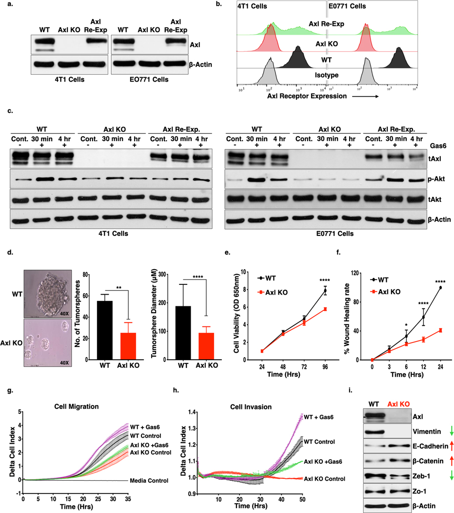
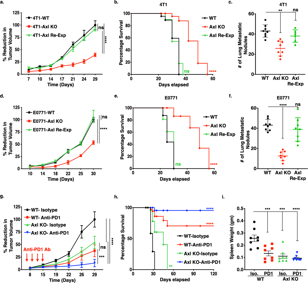
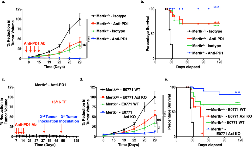
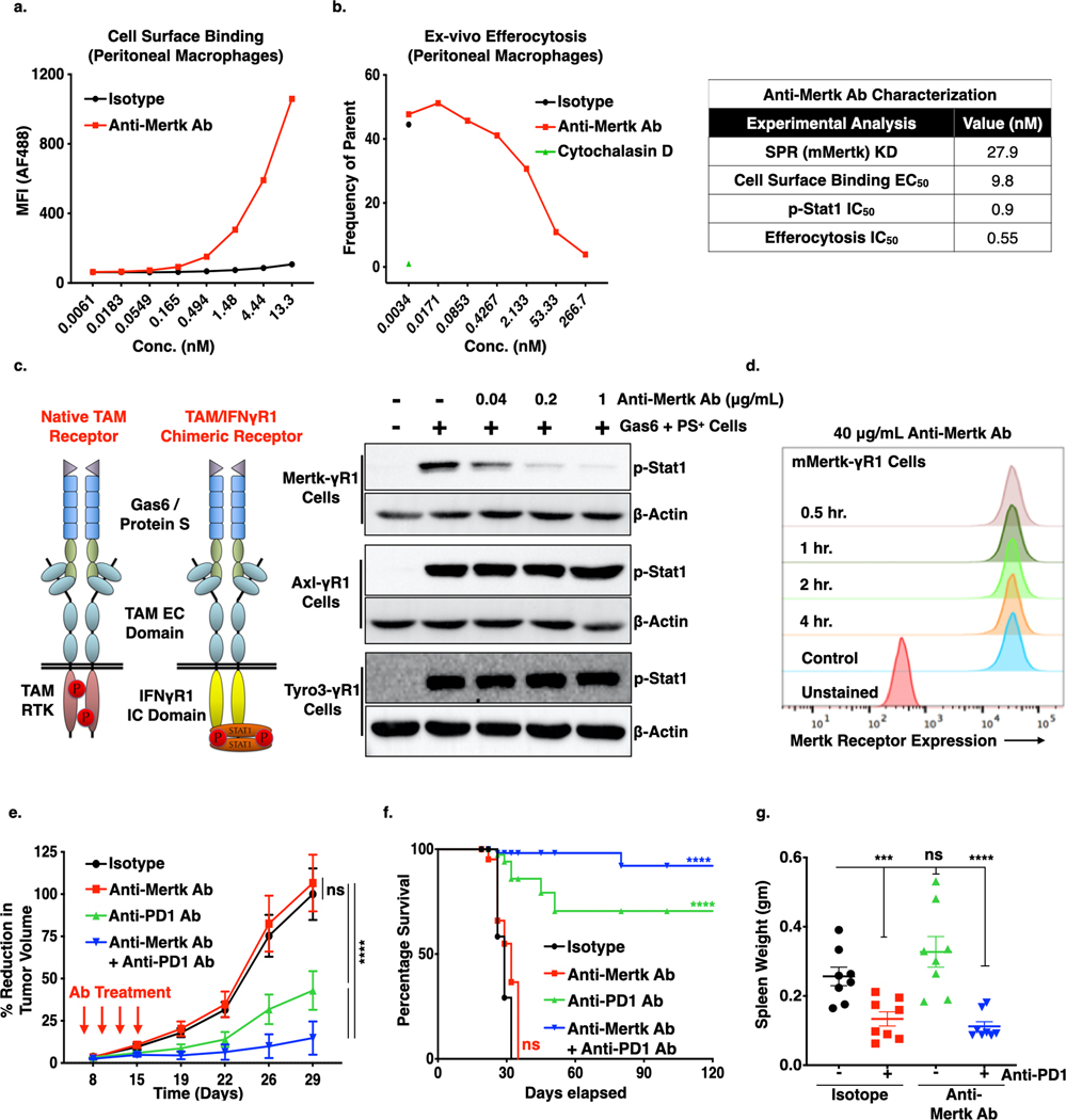
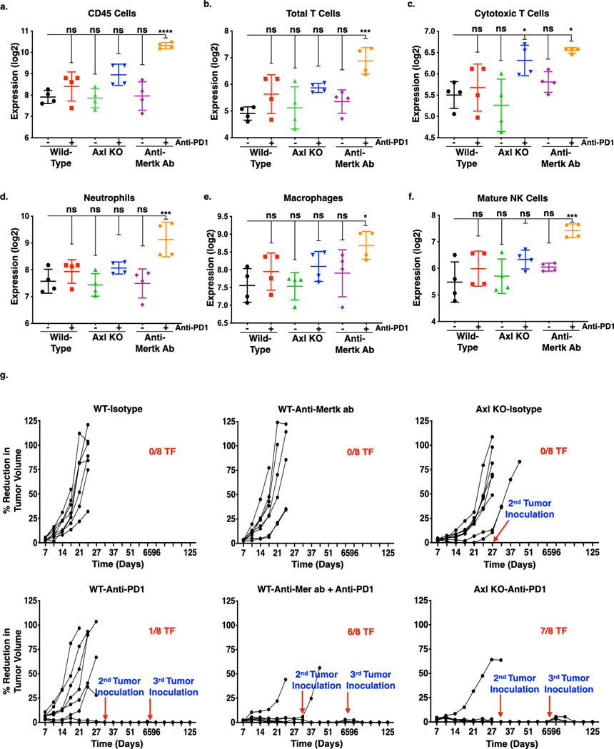
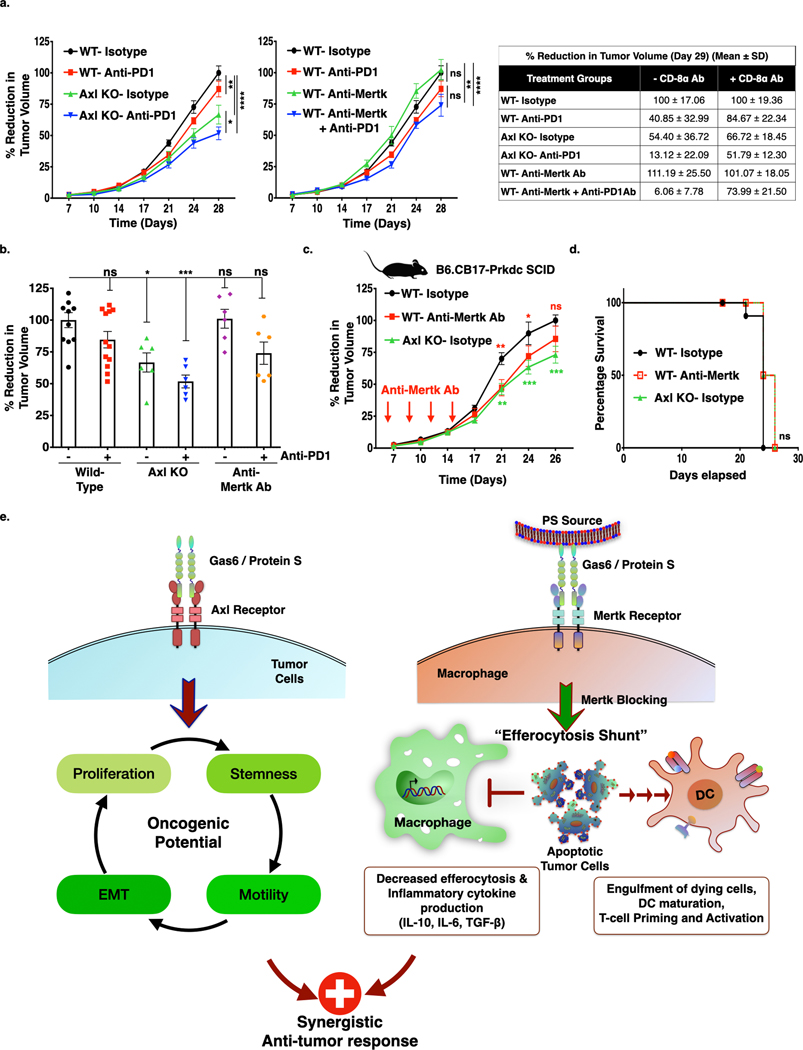
Despite the promising clinical benefit of targeted and immune checkpoint blocking therapeutics, current strategies have limited success in breast cancer, indicating that additional inhibitory pathways are required to complement existing therapeutics. TAM receptors (Tyro-3, Axl, and Mertk) are often correlated with poor prognosis because of their capacities to sustain an immunosuppressive environment. Here, we ablate Axl on tumor cells using CRISPR/Cas9 gene editing, and by targeting Mertk in the tumor microenvironment (TME), we observed distinct functions of TAM as oncogenic kinases, as well as inhibitory immune receptors. Depletion of Axl suppressed cell intrinsic oncogenic properties, decreased tumor growth, reduced the incidence of lung metastasis and increased overall survival of mice when injected into mammary fat pad of syngeneic mice, and demonstrated synergy when combined with anti-PD-1 therapy. Blockade of Mertk function on macrophages decreased efferocytosis, altered the cytokine milieu, and resulted in suppressed macrophage gene expression patterns. Mertk-knockout mice or treatment with anti-Mertk-neutralizing mAb also altered the cellular immune profile, resulting in a more inflamed tumor environment with enhanced T-cell infiltration into tumors and T-cell-mediated cytotoxicity. The antitumor activity from Mertk inhibition was abrogated by depletion of cytotoxic CD8α T cells by using anti-CD8α mAb or by transplantation of tumor cells into B6.CB17-Prkdc SCID mice. Our data indicate that targeting Axl expressed on tumor cells and Mertk in the TME is predicted to have a combinatorial benefit to enhance current immunotherapies and that Axl and Mertk have distinct functional activities that impair host antitumor response. SIGNIFICANCE: This study demonstrates how TAM receptors act both as oncogenic tyrosine kinases and as receptors that mediate immune evasion in cancer progression.


Figures

Figure 1.
Figure 1.. Genetic deletion of Axl receptor in murine breast cancer cells inhibit cell proliferation, formation of tumorspheres, cell motility and tumor growth in the preclinical murine breast cancer model.
(a-b.) Western blot (a.) and flow cytometry (b.) analysis of Axl receptor knockout (KO) using CRISPR/Cas9 technology and retroviral re-expression of Axl receptor in the Axl KO cells in the triple-negative (ER, PR or Her2/Neu) murine breast cancer 4T1-Luc-GFP and E0771 cells. c. Ligand, Gas6, mediated activation of Axl receptor was analyzed as phosphorylation of Akt (downstream signaling molecule of Axl) following Gas6 treatment for 30 mins and 4 hrs., in the 4T1 and E0771- WT, Axl KO and Axl Re-Exp. cells by immunoblot analysis. d. Tumorsphere formation from 4T1-WT and Axl KO cells on the ultra-low attachment plate. Phase-contrast micrographs representing tumorspheres (left panel) and column graphs showing number and size of tumorspheres formed from 1000– 4T1- WT and Axl KO cells. **P<0.01, ****P<0.001. Mean values ± SD are shown (n = 12). e. The effect of Axl receptor KO on cell proliferation as compared to 4T1-WT as analyzed MTT assay for 4 days. Mean values ± SD are shown (n = 8). f. Cell migration of the 4T1-WT and Axl KO as determined by wound healing assay. g-h. Real time cell migration (f) and invasion (g) of the 4T1-WT and Axl KO cells in the presence or absence of Gas6 through microporous membrane of CIM plates using Real-time xCELLigence system. Difference in the rate of migration and invasion was analyzed by two-way ANOVA. i. Axl deletion induces loss of mesenchymal markers, Vimentin and Zeb-1 and increase in epithelial markers, E-Cadherin and β-Catenin expression as analyzed by Western blot analysis of protein lysates from 4T1-WT and Axl KO cells.
Figure 2.
Figure 2.. Genetic Ablation of Axl receptor alone or in combination with anti-PD1 immunotherapy impairs tumor growth and metastasis in the murine breast cancer models.
(a-f.) In vivo tumor growth curves, corresponding Kaplan-Meier curves and number of lung metastatic nodules of 4T1 and E0771 tumor models. 5 X 10⁴ 4T1- (a.) and E0771-WT, Axl KO and Axl Re-Exp. (d.) cells were injected orthotopically into the mammary fat pad of 8-weeks BALB/c and c57BL/6 female mice respectively and tumor growth determined by means tumor volume measurement every 3 days over the period of 5 weeks. Kaplan-Meier curves depicting percentage survival of tumor bearing mice in corresponding 4T1 (b.) and E0771 (e.) tumors. Quantification of microscopic lung metastatic nodules from 4T1 (c.) and E0771 (f.) tumor bearing mice following lungs harvests at sacrifice on d35 and fixation bouin solution. g. Tumor growth analysis of E0771-WT and Axl KO tumor bearing mice (n=8/group) treated with mIgG1 Isotype control Ab or anti-PD1 Ab (5 mg/kg) on day 6, 9, 12, and 15. Arrows indicate antibody injections. h. Kaplan-Meier curve showing percentage survival of tumor bearing mice upon anti-PD1 immunotherapy. i. Analysis of splenomegaly, as a measure of extramedullary hematopoiesis, in the treated mice by means of spleen weight at sacrifice on d35. *P<0.05, ****P<0.001. Mean values ± SD are shown (n = 8).
Figure 3.
Figure 3.. Mertk deficiency decreases tumor malignancy and synergize with anti-PD1 immunotherapy in the E0771 murine breast cancer model.
a. 5X10⁴ E0771 WT cells were injected orthotopically in to the mammary fat pad of female c57BL/6- Mertk+/+ (WT) and Mertk−/− (Mertk KO) mice, and upon establishment of tumors, mice were treated with mIgG1 Isotype or anti-PD1 antibody (5 mg/kg) on day 6, 9, 12, and 15 and tumor growth determined by means tumor volume measurement every 3 days over the period of 5 weeks. (n=8/per group). Arrows indicate antibody injections. b. Kaplan-Meier curve showing percentage survival of tumor bearing mice upon anti-PD1 immunotherapy in the Mertk+/+ and Mertk−/− mice. c. Tumor free Mertk−/− mice treated with anti-PD1 immunotherapy were rechallenged with 1 X 10⁵ or 2 X 10⁵ E0771 WT cells d42 and d82 respectively, and tumor growth measurements were performed (n=16). d-e. Tumor growth curve (d.) and corresponding Kaplan-Meier curves (e.) showing percentage survival following injection of 5X10⁴ E0771 WT or Axl KO tumor cells in to the mammary fat pad of female c57BL/6- Mertk+/+ and Mertk−/− mice.
Figure 4.
Figure 4.. Mertk(−/−) macrophages decreases apoptotic cell efferocytosis and suppression of cytokine production in response to intraperitoneal LPS injection.
a. Mertk receptor expression in BMDM cultures upon stimulation with 0.1μM dexamethasone for 24 hrs. b. Flow cytometry analysis of efferocytosis in the dexamethasone treated Mertk(+/+) (WT) and Mertk(−/−) BMDMs. The efferocytosis was quantified by analyzing phRhodo positive macrophages after engulfment of stained irradiated CEM cells. The bar graphs showing normalized phRodo positive cells by mean fluorescence intensity. c. Peritoneal macrophages from Mertk(−/−) mice display decreased efferocytosis of phRodo stained irradiated CEM cells as compared to macrophages isolated from Mertk(+/+) mice. d. Efferocytosis in Mertk(+/+) and Mertk(−/−) peritoneal macrophages as analyzed by live imaging over the 4 hrs. e, f. Experimental mouse model depicting in vivo efferocytosis, where phRodo stained irradiated CEM cells injected in the peritoneal cavity of the mice for 1 hr, following by analysis for CD11b F4/80 positive macrophages with engulfed apoptotic cells. g. RNAseq analysis depicting signaling pathways affected by efferocytosis of apoptotic cells, when analzed by ingenuity pathway analysis. h. Gene expression analysis for genes modulated in inflammatory response and activation of macrophages pathways. i. The outline of LPS treatment in the c57BL/6- Mertk(+/+) and Mertk(−/−) mice. j. Mice were primed with 1 ml PBS injection into the peritoneal cavity. After 48 hrs, 20 μg LPS was injected intraperitoneally for 4 hrs, followed by isolation of peritoneal exudate, RNA isolation and qRT-PCR for cytokines including IL-6, IL-10 and TGF-β.
Figure 5.
Figure 5.. Effect of Anti-Mertk antibody on the Mertk receptor inhibition, efferocytosis, tumor growth and metastasis.
a. Cell surface binding analysis of Anti-Mertk monoclonal antibody (mAb) to the Mertk receptor on the peritoneal macrophages. b. In vivo efferocytosis showing decreased engulfment of apoptotic mouse thymocytes in the presence of anti-Mertk mAb as compared to isotype control. Cytochalasin D was used as a positive control (Left panel). Characterization of anti-Mertk antibody by surface plasma resonance (SPR) binding analysis, cell surface binding, p-Stat1 activation analysis in chimeric murine Mertk-γR1 cells and in vivo efferocytosis (Right tabel). d. Immunoblot analysis showing anti-Mertk antibody induced inhibition of Gas6 and apoptotic cells mediated activation Tyro3, Axl and Mertk receptors in the CHO cells expressing chimeric murine TAM-γR1 receptors. e. Flow-Cytometry based analysis showing effect of 40 μg/ml anti-Mertk ab on the Mertk receptor internalization in the CHO murine Mer-γR1 chimeric cells over the period of 4 hrs using Mertk specific flow-based antibody. f. Anti-tumor effect of anti-Mertk mAb in combination with Anti-PD1 immunotherapy. E0771 tumor bearing females C57/B16 (n=8/per group) were treated with mIgG1 Isotype control, anti-Mertk aAb (10mg/kg), anti-PD1 ab (5mg/Kg) alone and anti-Mertk ab in combination with anti-PD1 ab on day 6, 9,12, and15 and tumor growth was studied twice a week over the period of 4 weeks. g. Kaplan-Meier curve showing percentage survival of tumor bearing mice upon treatment with anti-Mertk ab and anti-PD1 immunotherapy. h. Upon sacrifice, spleens were also collected, and splenomegaly was quantified by means of spleen weight. *P<0.05, ****P<0.001. Mean values ± SD are shown (n = 8).
Figure 6.
Figure 6.. Effect of Axl receptor KO and anti-Mertk ab alone or in combination with anti-PD1 immunotherapy on innate immune cell infiltration and memory T-cell response in the primary mouse breast tumor.
The NanoString nCounter PanCancer immune profiling panel consisting of 770 genes for 24 different immune cell types and population characterization was uses to analyze immune cell infiltration in the primary tumors from each group (n = 4). a-g. The cell-type score comparison on the infiltrated immune cells in the primary tumors from each group were presented in log2 and graphically represented by GraphPad Prism. Expression profile of CD45 cells (CD45) (a), total T-cells (CD3D, CD3E, CD3G, CD6, SH2D1A) (b), cytotoxic T-cells (CD8A, CD8B, GZMA, GZMB, CD94, PRF1) (c), neutrophils (CSF3R, CD16, S100A12) (d), macrophages (CD163, CD68, CD84) (e), and mature NK (CD56dim) cells (NKp46, IL21R, KIR2DL3, KIR3DL1) (f). *P<0.05, **P<0.01, ****P<0.001. Mean values ± SD are shown (n = 4). g. Tumor free mice from E0771 WT and Axl KO tumor bearing mice treated with or without anti-PD1 mAb or E0771 WT tumor bearing mice treated with anti-Mertk mAb with or without anti-PD1 immunotherapy were rechallenged with 1 X 10⁵ or 2 X 10⁵ E0771 WT cells d42 and d82 respectively, and tumor growth measurements were performed up to d120 (n=8).
Figure 7.
Figure 7.. Anti-tumor effect of Axl receptor KO and anti-Mertk Antibody alone or in combination with anti-PD1 immunotherapy is T-cell dependent.
a. The Anti-CD8α antibody, which blocks CD8+ T-cell mediated immune response, has partially blocked anti-tumor effect of Axl receptor KO (Left panel), whereas completely blocked the anti-Mertk ab (Middle panel) mediated response in the combination with anti-PD1 immunotherapy. Comparision of decrease in tumor growth between mice treated with or without Anti-CD8α antibody at day 29. (Right table). b. Bar graph representing tumor growth at d28 from the individual mice from each group treated with Anti-CD8α antibody. (n=6–12) c. Graph representing tumor growth of Axl KO cells or WT tumor cells or WT tumor bearing mice treated with anti-Mertk antibody in the B6.CB17-Prkdc SCID mice. (n=6). d. Kaplan-Meier curve showing percentage survival in the SCID mice. e. Dual roles for Axl and Mertk contribute to tumorigenesis by distinct mechanisms. Axl, the expressed mainly on E0771 and 4T1 tumor cells, drives aggressive hallmarks of tumors by affecting stemness, migration, invasion, and EMT. Mertk, on the other hand, effects immunogenic signals in the TME, including efferocytosis and production of cytokines that impinge on the tumor milieu. We propose that anti-Mertk mAbs may block macrophage efferocytosis and the associated immunosuppressive effects associated with M2 macrophage activities.

Similar articles

Cited by

References

    1. Farkona S, Diamandis EP, Blasutig IM. Cancer immunotherapy: the beginning of the end of cancer? BMC Med 2016;14:73. - PMC - PubMed
    1. Sharma P, Allison JP. Immune checkpoint targeting in cancer therapy: toward combination strategies with curative potential. Cell 2015;161:205–14 - PMC - PubMed
    1. Birge RB, Boeltz S, Kumar S, Carlson J, Wanderley J, Calianese D, et al. Phosphatidylserine is a global immunosuppressive signal in efferocytosis, infectious disease, and cancer. Cell Death Differ 2016;23:962–78 - PMC - PubMed
    1. Dayoub AS, Brekken RA. TIMs, TAMs, and PS- antibody targeting: implications for cancer immunotherapy. Cell Commun Signal 2020;18:29. - PMC - PubMed
    1. Cheng X, Li L, Thorpe PE, Yopp AC, Brekken RA, Huang X. Antibody-Mediated Blockade of Phosphatidylserine Enhances the Antitumor Effect of Sorafenib in Hepatocellular Carcinomas Xenografts. Ann Surg Oncol 2016;23:583–91 - PubMed

MeSH terms

Substances