Review
. 2020 Nov 25;85(1):e00078-20.
doi: 10.1128/MMBR.00078-20. Print 2020 Nov 25.

Subdominance in Antibody Responses: Implications for Vaccine Development

Affiliations
Review

Subdominance in Antibody Responses: Implications for Vaccine Development

Gunnar Lindahl. Microbiol Mol Biol Rev. .

Abstract

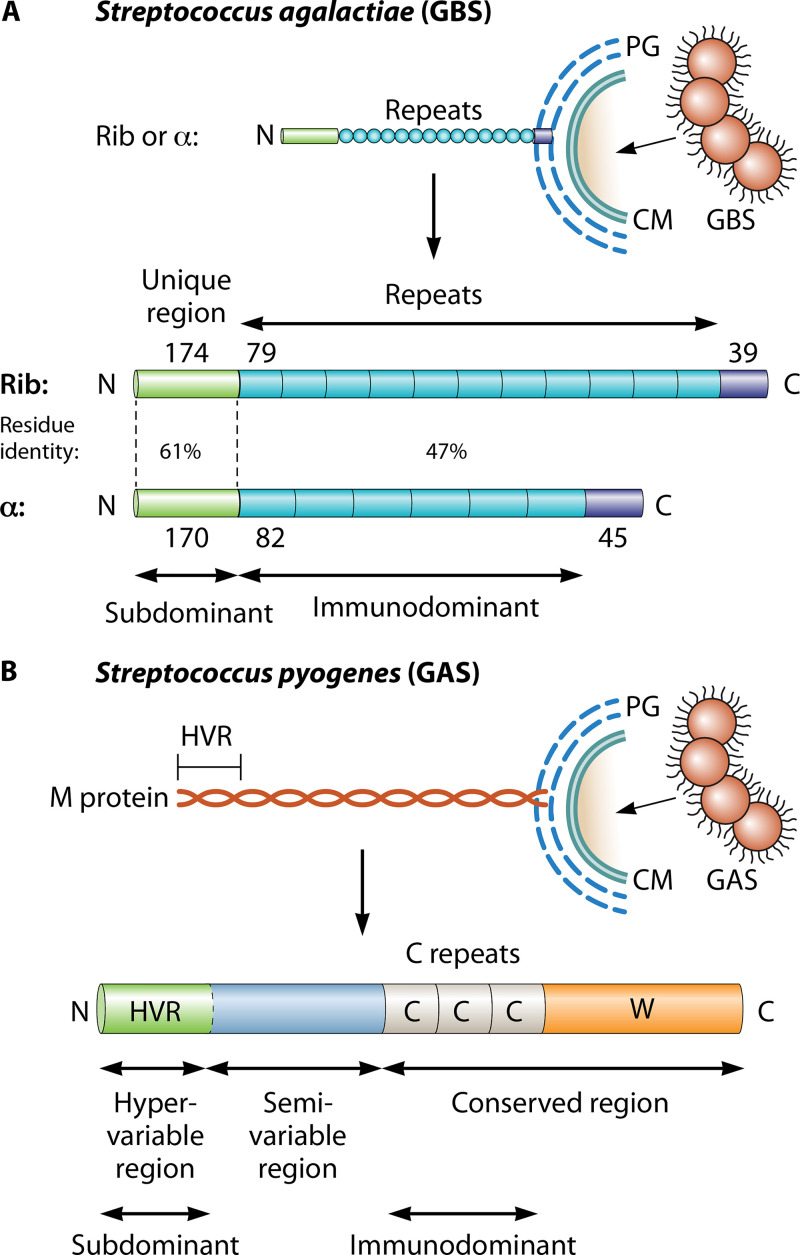
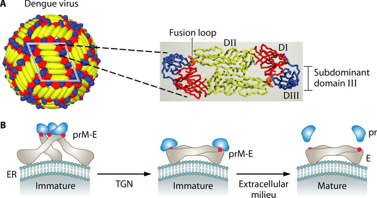
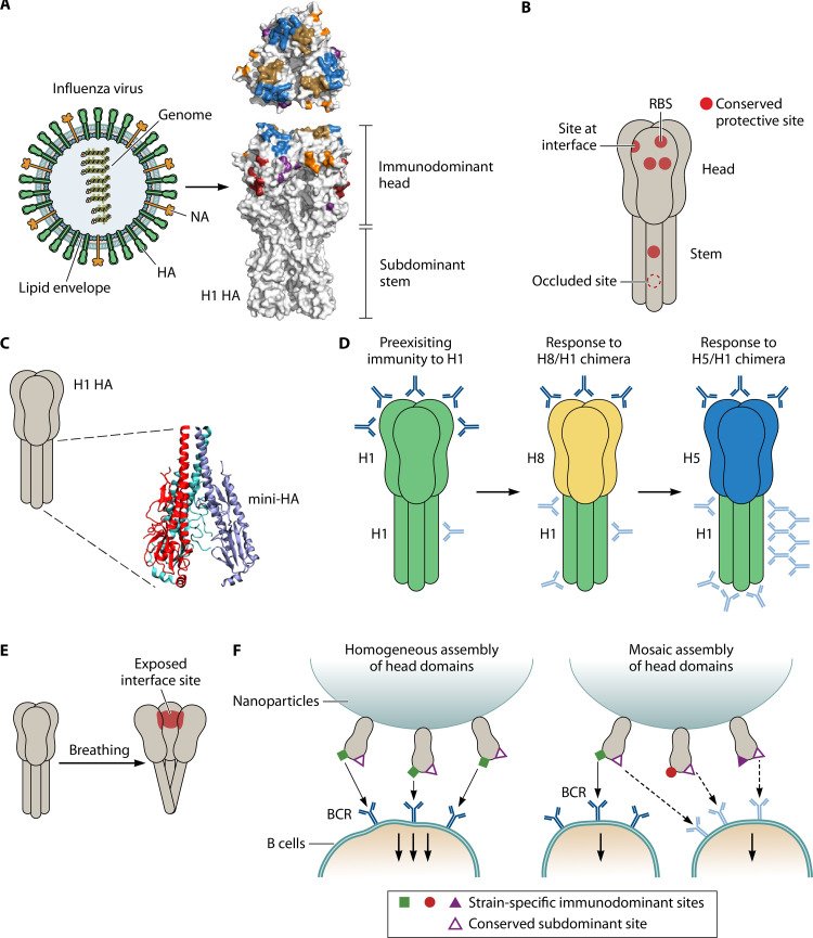
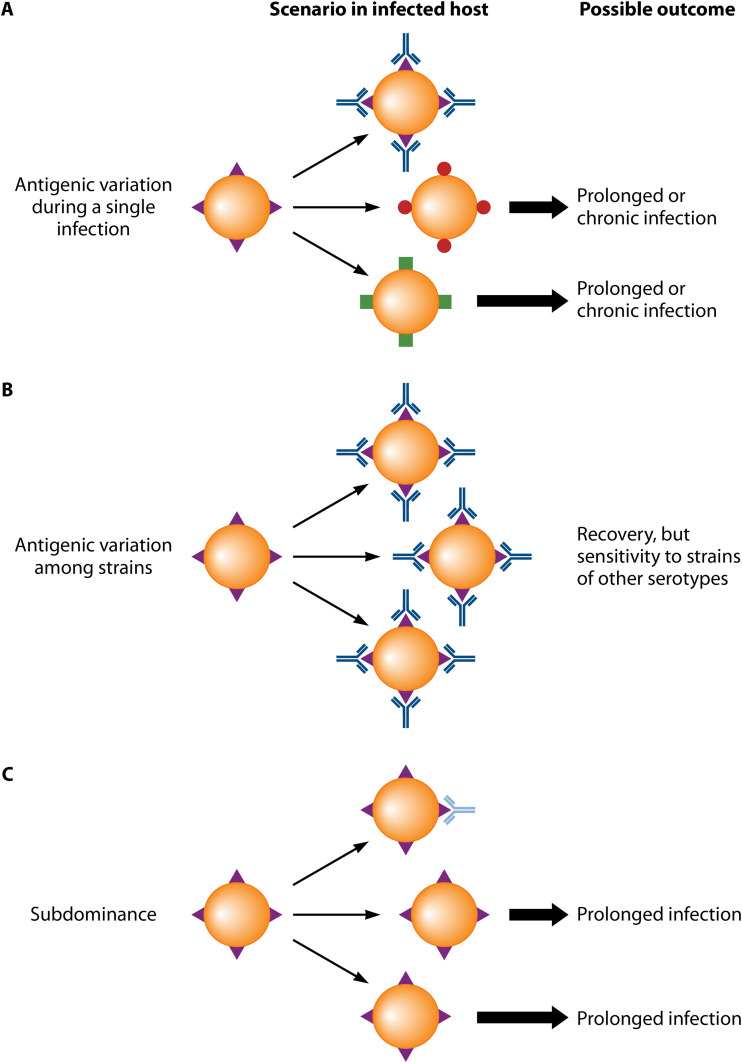
Vaccines work primarily by eliciting antibodies, even when recovery from natural infection depends on cellular immunity. Large efforts have therefore been made to identify microbial antigens that elicit protective antibodies, but these endeavors have encountered major difficulties, as witnessed by the lack of vaccines against many pathogens. This review summarizes accumulating evidence that subdominant protein regions, i.e., surface-exposed regions that elicit relatively weak antibody responses, are of particular interest for vaccine development. This concept may seem counterintuitive, but subdominance may represent an immune evasion mechanism, implying that the corresponding region potentially is a key target for protective immunity. Following a presentation of the concepts of immunodominance and subdominance, the review will present work on subdominant regions in several major human pathogens: the protozoan Plasmodium falciparum, two species of pathogenic streptococci, and the dengue and influenza viruses. Later sections are devoted to the molecular basis of subdominance, its potential role in immune evasion, and general implications for vaccine development. Special emphasis will be placed on the fact that a whole surface-exposed protein domain can be subdominant, as demonstrated for all of the pathogens described here. Overall, the available data indicate that subdominant protein regions are of much interest for vaccine development, not least in bacterial and protozoal systems, for which antibody subdominance remains largely unexplored.

Keywords: Plasmodium falciparum; Streptococcus agalactiae; Streptococcus pyogenes; antibodies; dengue virus; immune escape; immunodominance; influenza virus; subdominance; vaccine.


Figures

FIG 1
FIG 1
Fundamentals of immunodominance and subdominance in antibody responses. Panels A to C refer to antibody responses, and for comparison, panel D shows immunodominance in T cell responses. The antigen shown here is a protein, and for simplicity, it is represented schematically as a rod. However, it should be noted that most epitopes recognized by an antibody are conformational. Note that antibody immunodominance is a quantitative concept, not a qualitative one. (A) In the simplest case, one region of a protein is immunodominant, implying that it elicits the quantitatively dominating antibody response, while another region is subdominant. Antibodies are represented by Y-forms, with those directed against subdominant and immunodominant regions shown in light blue and dark blue, respectively. (B) In some cases, a hierarchy of immunodominance may be discerned among different parts of a protein. (C) An immunodominant region may include a subdominant site, a situation that has attracted particular interest in viruses. (D) Immunodominance in T cell responses. In the adaptive immune response to a protein, major histocompatibility complex (MHC) molecules form complexes with peptides derived from the protein, and these complexes are presented for recognition by T cell receptors (TCRs). However, peptides that are efficiently presented for recognition represent only a small minority of all peptides that potentially can be derived from the protein. These rare peptides correspond to T cell epitopes that are immunodominant in the responding individual (28, 234). In the figure, the peptide is enlarged compared to the epitope.
FIG 2
FIG 2
Circumsporozoite protein (CSP) of P. falciparum and its subdominant amino-terminal domain. (A) The sporozoite of P. falciparum is covered by CSP, in which a central repeat region is surrounded by distinct amino-terminal and carboxy-terminal domains. The repeat region is mainly composed of NANP tetrapeptide sequences (yellow) but starts with a single NPDP sequence (red) and also contains a few NVDP repeats (green) (47). While the repeat region is immunodominant, the amino-terminal region, which also is exposed on the sporozoite surface, is subdominant. In contrast, the carboxy-terminal region may be largely hidden in the intact CSP molecule. The immunodominant repeat region is the key component of the malaria vaccine RTS,S, as indicated. During an infection, CSP is cleaved at the RI site in the amino-terminal domain. Antibodies that bind close to this site (61) or to a junctional epitope located at the beginning of the repeat region (66–68) may protect against infection and may share the ability to block proteolysis at the RI site. Little is known about the structure of the subdominant amino-terminal domain, but it is noteworthy that the part located close to the repeats is charged (47), while the first 50 amino acid (aa) residues include 7 tyrosine residues. (B) Model for immunodominance and subdominance in CSP. The repeats of CSP may promote multipoint and high-avidity binding to cognate B cells, making the repeat region immunodominant by diverting the protein from B cells recognizing the nonrepeated domains, in particular, the surface-exposed amino-terminal domain (56, 57). The resulting subdominance of the amino-terminal domain may favor microbial virulence by allowing the microbe to evade potently protective antibodies directed against that domain. The limited protection conferred by antibodies to the repeats would be the price the microbe pays to achieve this result.
FIG 3
FIG 3
Subdominant domains in streptococcal surface proteins. (A) Schematic of the surface-exposed forms of the Rib and α proteins of Streptococcus agalactiae (GBS) (79, 80). These two proteins are the most common members of a family of elongated and highly repetitive streptococcal proteins. Each protein has a surface-distal amino-terminal domain, a region with long repeats, and a short carboxy-terminal region that promotes covalent linkage to the bacterial peptidoglycan (PG) layer, located outside the cell membrane (CM). The number of amino acid residues in a region (or repeat) is indicated. While all repeats are completely identical within Rib or α, they vary in number among clinical isolates and are different in Rib and α but show 47% residue identity. The amino-terminal domains are strikingly subdominant but are targets for protective antibodies (17). (B) Schematic of the surface-exposed form of Streptococcus pyogenes M protein. All strains of S. pyogenes express an M protein, encoded by the emm gene (95). This fibrillar coiled-coil protein has an amino-terminal hypervariable region (HVR) of ∼50 to 100 amino acid residues, a conserved carboxy-terminal region that includes C repeats (each with 35 or 42 residues), and a wall-spanning region (W). The HVR exhibits extreme sequence divergence among strains but is stable within a strain and represents a distinct domain that in many (but not all) M proteins specifically binds a human complement regulator. The central part of M protein is also variable among strains and typically includes domains that bind human plasma proteins, e.g., fibrinogen or IgA (95). While the HVR is the major target for protective antibodies, it is strikingly subdominant (18).
FIG 4
FIG 4
E protein of dengue virus and its subdominant domain III. (A) Schematic of dengue virus and its surface E protein. The mature dengue virion (left) is covered by 30 rafts, each of which contains three antiparallel E protein dimers. One raft is framed in black, and a single dimer is shown as ribbons (right). The positions of the three domains (DI to DIII) are indicated for one E protein monomer. While domain III is subdominant, the fusion loop in domain II is part of an immunodominant site. Adapted from reference . (B) Protein maturation during dengue virus replication (114). In the endoplasmic reticulum (ER), the surface of the immature virus particle is covered by trimeric prM-E spikes, in which prM prevents premature membrane fusion promoted by the fusion loop (red spot) in the E protein. During transport through the acidic trans-Golgi network (TGN), the complex is rearranged and prM is then cleaved into pr and M. The small M fragment is retained in the viral membrane, with negligible surface exposure, while the pr fragment remains bound to the FL. When the virus particle is released from the cell, pr dissociates from the complex and the FL becomes largely hidden within the E dimer (113–115). Adapted from reference with permission of AAAS.
FIG 5
FIG 5
Hemagglutinin (HA) of influenza virus: subdominant sites as targets for protective antibodies. (A) Models of influenza virus and hemagglutinin (HA). (Left) In a viral particle, the glycoproteins HA and neuraminidase (NA) are anchored in a lipid envelope surrounding the core, which contains the eight RNA segments of the genome. (Right) Top and side views of an HA trimer of subtype H1, showing the immunodominant head and the subdominant stem (149). The location of the five highly variable and immunodominant sites in the head are indicated (Sa, blue; Sb, gold; Ca1, purple; Ca2, orange; Cb, red). Right panel adapted from reference with permission from Springer Nature. (B) Cartoon showing the location of conserved sites (red) that are targets for protective antibodies in HA. These sites are of at least five types and include the RBS, a partially occluded site located at the monomer interface in the head, other conserved sites in the head, and two sites in the stem, one of which is occluded on native HA, i.e., not accessible to antibodies. Except for this occluded site in the stem, which has unique properties, the various sites may be described as subdominant. Of note, the whole head is variable and immunodominant compared to the stem, but it includes subdominant sites that are conserved and protective. In contrast, the entire stem is subdominant but contains protective sites. Panels C to F, and the corresponding legends, describe procedures that may allow an antibody response to be targeted to a subdominant site. (C) A stem-only construct (“mini-HA”), derived from the subdominant stem of H1 HA, elicits broadly protective antibodies (191). Figure based on PDB 5CJQ and produced with VMD (235). Of note, the structure of mini-HA differs slightly from that for the stem of intact HA, as it adopts a more open splayed conformation (191). (D) Use of chimeric HA proteins to focus responses on the subdominant stem (33, 196). (Left) Adults have preexisting antibodies to the head (top) and the stem (bottom) of H1 HA, although the response to the stem is very limited. (Middle) Immunization with a chimeric H8/1 protein may elicit a memory response to the H1 stem but only a primary response to the “exotic” H8 head. (Right) Boosting with an H5/1 chimera may further boost the response to the H1 stem while again eliciting a primary response to the “exotic” head. As in Fig. 1, antibodies directed against subdominant and immunodominant regions are shown in light blue and dark blue, respectively. (E) Schematic representation of “breathing” in an HA molecule, resulting in the exposure of a site in the head that is located at the monomer interface and is a target for protective antibodies (157, 159, 160). Formation of antibodies to this site may be favored by immunization with hyperglycosylated HA (197). (F) Use of mosaic nanoparticles to focus antibody responses on sites in the HA head that are subdominant and conserved (198). In this procedure, nanoparticles were covered with monomeric HA heads. (Left) A nanoparticle covered with heads from a single virus strain will elicit antibodies directed almost exclusively against strain-specific immunodominant sites, which promote very strong binding to cognate B cells, thereby diverting the antigen from B cells recognizing any other site(s). (Right) Use of a mosaic nanoparticle covered with several different heads will reduce the local concentration of strain-specific immunodominant sites, potentially favoring a response to a conserved and shared subdominant site, which may promote binding of relatively high avidity to cognate B cells.
FIG 6
FIG 6
Antibody escape through antigenic variation or subdominance: a comparison. Schematic comparing the well-known role of antigenic variation and the potential role of subdominance in immune escape. Of note, antigenic variation comes in two forms, as indicated below. (A) Antigenic variation during the course of a single infection. Protein variants are indicated in different colors. Microbes expressing new variants escape antibody attack and can grow (thick arrows) because they are not recognized by antibodies elicited by an earlier variant. This situation may result in the establishment of a prolonged infection, during which new antigenic variants are repeatedly selected for. (B) Antigenic variation among strains. In this case, an antibody response to strain-specific determinants typically results in recovery and immunity, but the host may subsequently be infected by a variant strain circulating in the population. The hemagglutinin (HA) of influenza virus offers a classical example. (C) Subdominance may promote immune escape by strongly limiting the formation of antibodies to a protective site. As a result, an infection may be prolonged.

Similar articles

Cited by

References

    1. Rodrigues CMC, Pinto MV, Sadarangani M, Plotkin SA. 2017. Whither vaccines? J Infect 74 Suppl 1:S2–S9. doi:10.1016/S0163-4453(17)30184-6. - DOI - PMC - PubMed
    1. Rappuoli R, Pizza M, Del Giudice G, De Gregorio E. 2014. Vaccines, new opportunities for a new society. Proc Natl Acad Sci U S A 111:12288–12293. doi:10.1073/pnas.1402981111. - DOI - PMC - PubMed
    1. Mariner JC, House JA, Mebus CA, Sollod AE, Chibeu D, Jones BA, Roeder PL, Admassu B, van 't Klooster GG. 2012. Rinderpest eradication: appropriate technology and social innovations. Science 337:1309–1312. doi:10.1126/science.1223805. - DOI - PubMed
    1. Koff WC, Burton DR, Johnson PR, Walker BD, King CR, Nabel GJ, Ahmed R, Bhan MK, Plotkin SA. 2013. Accelerating next-generation vaccine development for global disease prevention. Science 340:1232910. doi:10.1126/science.1232910. - DOI - PMC - PubMed
    1. Casadevall A, Pirofski LA. 2003. Exploiting the redundancy in the immune system: vaccines can mediate protection by eliciting 'unnatural' immunity. J Exp Med 197:1401–1404. doi:10.1084/jem.20030637. - DOI - PMC - PubMed

MeSH terms

LinkOut - more resources