. 2020 Dec 15;117(50):32017-32028.
doi: 10.1073/pnas.2016451117. Epub 2020 Nov 25.

Tolerogenic nanoparticles suppress central nervous system inflammation

Affiliations

Tolerogenic nanoparticles suppress central nervous system inflammation

Jessica E Kenison et al. Proc Natl Acad Sci U S A. .

Abstract

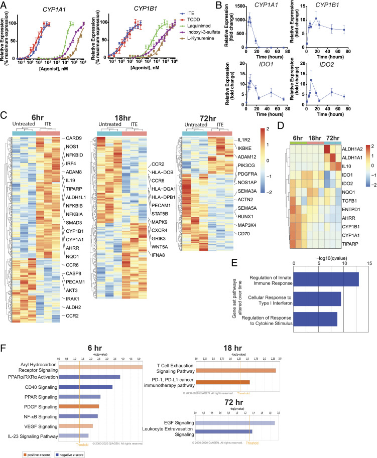
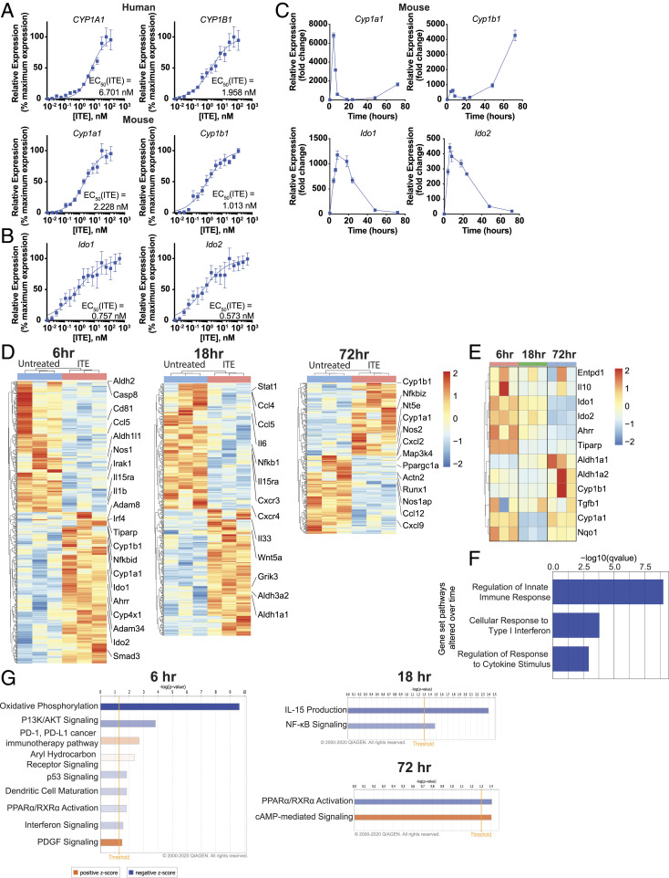
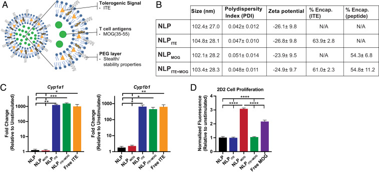
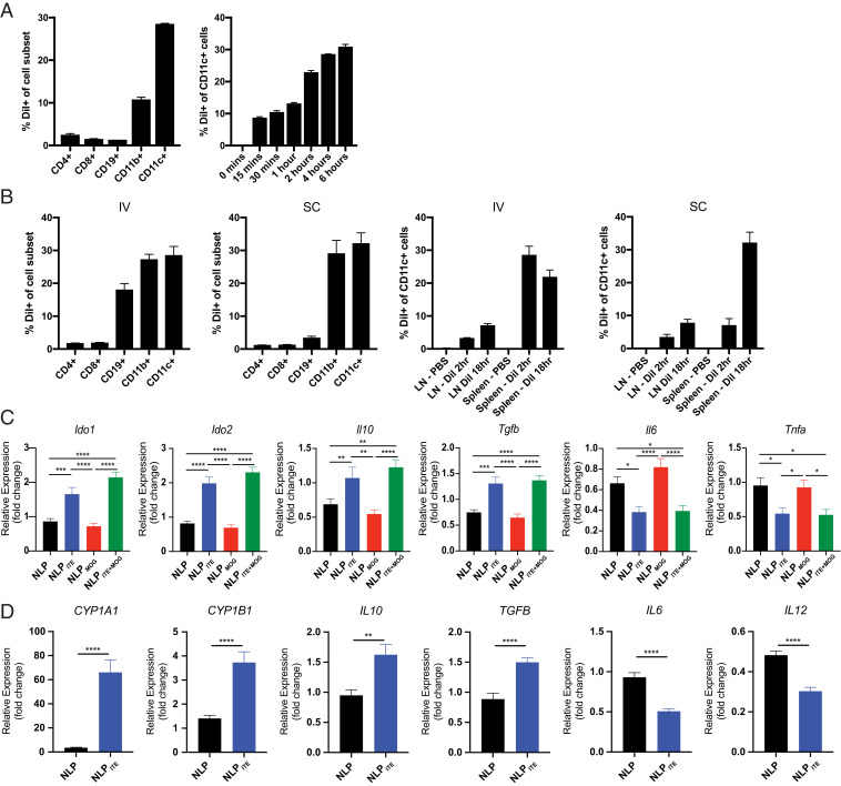
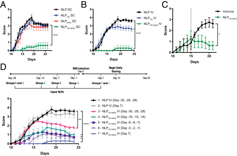
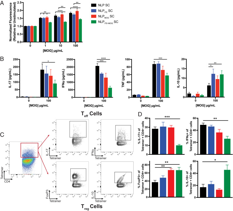
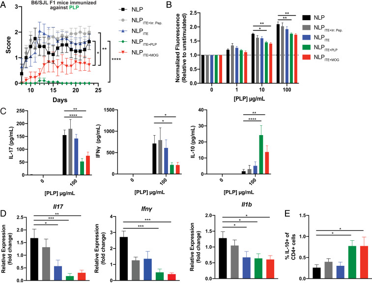
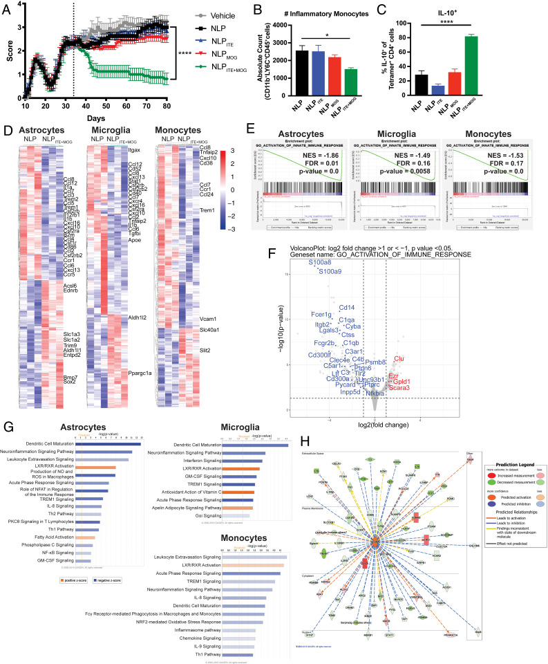
Therapeutic approaches for the induction of immune tolerance remain an unmet clinical need for the treatment of autoimmune diseases, including multiple sclerosis (MS). Based on its role in the control of the immune response, the ligand-activated transcription factor aryl hydrocarbon receptor (AhR) is a candidate target for novel immunotherapies. Here, we report the development of AhR-activating nanoliposomes (NLPs) to induce antigen-specific tolerance. NLPs loaded with the AhR agonist ITE and a T cell epitope from myelin oligodendrocyte glycoprotein (MOG)35-55 induced tolerogenic dendritic cells and suppressed the development of experimental autoimmune encephalomyelitis (EAE), a preclinical model of MS, in preventive and therapeutic setups. EAE suppression was associated with the expansion of MOG35-55-specific FoxP3+ regulatory T cells (Treg cells) and type 1 regulatory T cells (Tr1 cells), concomitant with a reduction in central nervous system-infiltrating effector T cells (Teff cells). Notably, NLPs induced bystander suppression in the EAE model established in C57BL/6 × SJL F1 mice. Moreover, NLPs ameliorated chronic progressive EAE in nonobese diabetic mice, a model which resembles some aspects of secondary progressive MS. In summary, these studies describe a platform for the therapeutic induction of antigen-specific tolerance in autoimmune diseases.

Keywords: EAE; MS; antigen-specific therapy; autoimmunity; nanoparticles.


Conflict of interest statement

Competing interest statement: J.E.K, A.J., N.K., S.T., D.N., A.P., V.P.S. are/were employees at AnTolRx; F.J.Q. is a consultant at AnTolRx. This work was partially funded by AnTolRx.

Figures

Fig. 1.
Fig. 1.
Temporal regulation of gene expression by AhR ligands in human DCs. (A) mRNA expression of the AhR target genes CYP1A1 and CYP1B1 in human peripheral blood DCs following incubation for 6 h with ITE, TCDD, laquinimod, indoxyl-3-sulfate, or l-kynurenine. Data were normalized to GAPDH, made relative to untreated samples, and presented here as a percentage of maximal induction. Data show the average of two independent experiments with DCs isolated from one independent healthy human donor per experiment. (B) mRNA expression of AhR target genes CYP1A1, CYP1B1, IDO1, and IDO2 in human peripheral blood DCs following incubation with 100 nM ITE for 0, 4, 6, 8, 18, 24, 48, and 72 h. Data are normalized to GAPDH and relative to time 0. Data show the average of three independent experiments with DCs isolated from one independent healthy human donor per experiment. (C) Heat map of differentially regulated genes determined by SMART-seq RNA-seq in DCs isolated from peripheral blood of three independent healthy human donors and treated for 6, 18, or 72 h with 100 nM ITE. Gene expression is row centered, log2 transformed, and saturated at −2 and +2 for visualization. (D) Heat map of expression of 12 genes known to be regulated by AhR activity as determined by SMART-seq RNA-seq in human peripheral DCs treated with 100 nM ITE for 6, 18, or 72 h. Gene expression is row centered, log2 transformed, and saturated at −2 and +2 for visualization. (E) Pathways identified by gene set enrichment analysis which were significantly altered over time following treatment in human peripheral blood DCs treated with ITE. (F) Ingenuity pathway analysis of the transcriptional profile of human peripheral blood DCs treated with ITE for 6, 18, or 72 h. Pathways associated with positive z-score are in orange; pathways associated with negative z-score are in blue; the relative strength of the z-score is represented by the intensity of the color.
Fig. 2.
Fig. 2.
Temporal regulation of gene expression by AhR ligands in murine DCs. (A) mRNA expression of AhR target genes Cyp1a1 and Cyp1b1 in human peripheral blood DCs or murine splenic DCs following incubation for 6 h with ITE. Data were normalized to Gapdh, made relative to untreated samples, and presented here as a percentage of maximal induction. Data show the average of three independent experiments with DCs isolated from one independent healthy human donor per experiment or a pool of DCs isolated from the spleens of 10 healthy B6 mice. (B) mRNA expression of AhR target genes Ido1 and Ido2 in murine splenic DCs following incubation for 6 h with ITE. Data were normalized to Gapdh, made relative to untreated samples, and presented here as a percentage of maximal induction. Data show the average of two independent experiments with DCs isolated from spleens of 10 healthy B6 mice per experiment. (C) mRNA expression of AhR target genes Cyp1a1, Cyp1b1, Ido1, and Ido2 in murine splenic DCs following incubation with 100 nM ITE for 0, 4, 6, 8, 18, 24, 48, and 72 h. Data are normalized to Gapdh and relative to time 0. Data are means ± SEM of one experiment representative of two independent experiments. (D) Heat map of differentially regulated genes determined by 3′ DGE-seq RNA-seq in murine splenic DCs isolated from three different pools of 10 mice each and treated for 6, 18, or 72 h with 100 nM ITE. Gene expression is row centered, log2 transformed, and saturated at −2 and +2 for visualization. (E) Heat map of the expression of 12 genes known to be regulated by AhR activity as determined by 3′ DGE-seq RNA-seq in murine splenic DCs treated with 100 nM ITE for 6, 18, or 72 h. Gene expression is row centered, log2 transformed, and saturated at −2 and +2 for visualization. (F) Pathways identified by gene set enrichment analysis which were significantly altered over time following treatment in murine splenic DCs treated with ITE. (G) Ingenuity pathway analysis of the transcriptional profile of murine splenic DCs treated with ITE for 6, 18, or 72 h. Pathways associated with positive z-score are in orange; pathways associated with negative z-score are in blue; the relative strength of the z-score is represented by the intensity of the color.
Fig. 3.
Fig. 3.
NLPs loaded with MOG35–55 and the tolerogenic AhR ligand ITE. (A) Schematic representation of NLPITE+MOG. (B) NLP characterization, including size, polydispersity index (PDI), zeta potential, % ITE, and % MOG35–55 encapsulation. Values represent the average of at least 17 different batches ± SD. (C) mRNA expression of AhR target genes cyp1a1 and cyp1b1 in murine splenic DCs following incubation for 6 h with NLP, NLPMOG, NLPITE (containing 10 nM ITE), NLPITE+MOG (containing 10 nM ITE), or free ITE (10 nM). (D) Proliferative response of 2D2 transgenic splenocytes activated for 72 h with NLP, NLPMOG (containing 10 μg/mL MOG), NLPITE (containing 1 μg/mL ITE), NLPITE+MOG (containing 10 μg/mL MOG and 1 μg/mL ITE), or free MOG (10 μg/mL). Data from C and D are means + SEM of one experiment representative of at least three independent experiments, with at least three biological replicates per experiment. *P < 0.05, **P < 0.01, ***P < 0.001, ****P < 0.0001, one-way ANOVA followed by Dunnett’s multiple comparison test.
Fig. 4.
Fig. 4.
NLPITE+MOG induces a tolerogenic phenotype in mouse and human DCs. (A) Murine splenocytes were isolated and cultured for 4 h in vitro with NLPDiI and examined by flow cytometry for NLPDiI uptake (Left). NLPDiI uptake by CD11c+ cells was measured at various timepoints (15 min to 6 h) (Right). (B) NLPDiI uptake by splenocytes as evaluated by flow cytometry 2 h after IV or SC NLP injection (Left two). NLPDiI uptake in spleen or LN CD11c+ cells measured 2 h or 18 h after IV or SC NLP injection (Right two). (C) Ido1, Ido2, Il10, TGFβ, Il6, and Tnfα expression in murine splenic DCs after culture for 6 h in vitro with NLP, NLPMOG, NLPITE, or NLPITE+MOG. For evaluation of Il6 and Tnfα, murine splenic DCs were stimulated with LPS during culture with NLPs. Data are depicted as the mean + SEM of three independent experiments with three biological replicates per experiment. (D) Human DCs were isolated from three independent healthy donor PBMCs, cultured for 6 h in vitro with NLP or NLPITE and CYP1A1, CYP1B1, IL10, TGFβ, IL6, and IL12 expression was analyzed by RT-PCR. For evaluation of IL6 and IL12, DCs were stimulated with LPS during culture with NLPs. Data are depicted as the mean + SEM of three independent healthy donors. *P < 0.05, **P < 0.01, ***P < 0.001, ****P < 0.0001, one-way ANOVA followed by Dunnett’s multiple comparison test, or Student’s t test.
Fig. 5.
Fig. 5.
NLPITE+MOG suppresses acute EAE in C57BL/6J mice. EAE was induced in B6 mice with MOG35–55, and NLPs were administered following various treatment regimens. (A) Clinical scores of mice treated SC with NLPs once a week beginning the day after disease induction. (B) Clinical scores of mice treated IV with NLPs administered once on day 7 after disease. (C) Clinical scores of mice treated IV with NLPs administered once on day 15 after disease. (D) Clinical scores of mice treated daily IV with NLPs for 3 d 1 mo prior, 2 wk prior, 1 wk prior, 1 d prior, or a single injection 1 wk after EAE induction. Data are mean ± SEM (n = 5 mice per group). *P < 0.05, **P < 0.01, ****P < 0.0001, two-way repeated measures ANOVA followed by Tukey’s multiple comparison test.
Fig. 6.
Fig. 6.
NLPITE+MOG suppresses the encephalitogenic T cell response. (A) Proliferative response and (B) cytokine secretion following restimulation with MOG35–55 of splenocytes from SC NLP-treated B6 mice, 26 d after EAE induction. (C) Gating strategy for the identification of MOG38–49 tetramer-positive IL-17+, IFNγ+, FoxP3+, and IL-10+ CD4+ T cells. (D) Frequency of MOG38–49 tetramer-positive IL-17+, IFNγ+, FoxP3+, and IL-10+ CD4+ T cells in the brain and spinal cord of B6 EAE SC NLP-treated mice. Data are mean + SEM (n = 5 mice per group). *P < 0.05, **P < 0.01, ***P < 0.001, ****P < 0.0001, one-way ANOVA followed by Dunnett’s multiple comparison test.
Fig. 7.
Fig. 7.
NLPITE+MOG ameliorates EAE in B6 × SJL F1 mice induced using PLP. SC NLPs were administered once a week beginning 1 d after disease induction using PLP139–151 in C57BL/6 × SJL F1 mice. (A) Clinical scores of NLP-treated mice (graph shows the average ±SEM of two independent experiments with n = 5 mice per group, per experiment. *P < 0.05, **P < 0.01, ***P < 0.001, ****P < 0.0001, two-way repeated measures ANOVA followed by Tukey’s multiple comparison test). (B) Proliferative response and (C) cytokine secretion following restimulation with PLP139–151 of splenocytes from NLP-treated mice, 24 d after EAE induction. Data are mean + SEM with n = 4 mice per group, representative of two independent experiments. *P < 0.05, **P < 0.01, ***P < 0.001, ****P < 0.0001 (B), two-way ANOVA followed by Dunnett’s multiple comparison test. (C) One-way ANOVA followed by Dunnett’s multiple comparison test. (D) Relative mRNA expression of Il17, Ifnγ, and Il1b in the CNS of NLP-treated mice. (E) Frequency of IL-10+ CD4+ T cells in the CNS of NLP-treated mice. (D and E) Data are mean + SEM (n = 8 mice per group, average of two independent experiments). *P < 0.05, **P < 0.01, ***P < 0.001, ****P < 0.0001, one-way ANOVA followed by Dunnett’s multiple comparison test.
Fig. 8.
Fig. 8.
NLPITE+MOG ameliorates chronic progressive NOD EAE. SC NLPs were administered once a week beginning on day 35 after NOD induction. (A) Clinical scores of NLP-treated mice (n = 10 mice per group, representative of two independent experiments. ****P < 0.0001, two-way repeated measures ANOVA followed by Tukey’s multiple comparison test). (B) CNS-infiltrating CD11b+CD45+Ly6Chi monocytes determined by flow cytometry (n = 10 mice per group. ****P < 0.0001, one-way ANOVA followed by Dunnett’s multiple comparison test). (C) MOG38–49 tetramer-positive IL-10+ CD4+ T cells in the CNS of NOD EAE SC NLP-treated mice 100 d after EAE induction. (n = 10 mice per group. ****P < 0.0001, one-way ANOVA followed by Dunnett’s multiple comparison test.) (D) Heat map of differentially regulated genes determined by SMART-seq RNA-seq in astrocytes, microglia, and monocytes from NLP- or NLPITE+MOG-treated mice on day 100 after NOD EAE induction. Gene expression is row centered, log2 transformed, and saturated at −3 and +3 for visualization. (E) Gene set enrichment analysis correlation of genes associated with innate immunity. (F) Volcano plot depicting differentially expressed genes associated with activation of the immune response in astrocytes of NLPITE+MOG-treated NOD EAE mice. (G) Ingenuity pathway analysis of the transcriptional profile of astrocytes, microglia, and monocytes. (H) Ingenuity pathway analysis of the IL10Rα signaling pathway in astrocytes from NLPITE+MOG-treated NOD EAE mice.

References

    1. Serra P., Santamaria P., Antigen-specific therapeutic approaches for autoimmunity. Nat. Biotechnol. 37, 238–251 (2019). - PubMed
    1. Klein L., Robey E. A., Hsieh C. S., Central CD4+ T cell tolerance: deletion versus regulatory T cell differentiation. Nat. Rev. Immunol. 19, 7–18 (2019). - PubMed
    1. Nemazee D., Mechanisms of central tolerance for B cells. Nat. Rev. Immunol. 17, 281–294 (2017). - PMC - PubMed
    1. Josefowicz S. Z., Lu L.-F., Rudensky A. Y., Regulatory T cells: Mechanisms of differentiation and function. Annu. Rev. Immunol. 30, 531–564 (2012). - PMC - PubMed
    1. Roncarolo M. G., Gregori S., Bacchetta R., Battaglia M., Gagliani N., The biology of T regulatory type 1 cells and their therapeutic application in immune-mediated diseases. Immunity 49, 1004–1019 (2018). - PubMed

MeSH terms