. 2021 Feb 15;95(4):e02166-20.
doi: 10.1128/JVI.02166-20. Epub 2020 Nov 25.

Structure and dsRNA-binding activity of the Birnavirus Drosophila X Virus VP3 protein

Affiliations

Structure and dsRNA-binding activity of the Birnavirus Drosophila X Virus VP3 protein

Diego S Ferrero et al. J Virol. .

Abstract

The Birnavirus multifunctional protein VP3 plays an essential role coordinating the virus life cycle, interacting with the capsid protein VP2, with the RNA-dependent RNA polymerase VP1 and with the dsRNA genome. Furthermore, the role of this protein in controlling host cell responses triggered by dsRNA and preventing gene silencing has been recently demonstrated. Here we report the X-ray structure and dsRNA-binding activity of the N-terminal domain of Drosophila X virus (DXV) VP3. The domain folds in a bundle of three α-helices and arranges as a dimer, exposing to the surface a well-defined cluster of basic residues. Site directed mutagenesis combined with Electrophoretic Mobility Shift Assays (EMSA) and Surface Plasmon Resonance (SPR) revealed that this cluster, as well as a flexible and positively charged region linking the first and second globular domains of DXV VP3, are essential for dsRNA-binding. Also, RNA silencing studies performed in insect cell cultures confirmed the crucial role of this VP3 domain for the silencing suppression activity of the protein.IMPORTANCE The Birnavirus moonlighting protein VP3 plays crucial roles interacting with the dsRNA genome segments to form stable ribonucleoprotein complexes and controlling host cell immune responses, presumably by binding to and shielding the dsRNA from recognition by the host silencing machinery. The structural, biophysical and functional data presented in this work has identified the N-terminal domain of VP3 as responsible for the dsRNA-binding and silencing suppression activities of the protein in Drosophila X virus.


Figures

FIG 1
FIG 1
Characterization of the DXV VP3 protein. (A) SEC-MALS profiles of DXV VP3 in a solution containing 50 mM Tris-HCl (pH 8.0) and 500 mM NaCl. The inset shows a closeup of the main peaks, with the measured molecular masses shown on the right axis (VP3 WT, 71.4 ± 0.3 kDa; PA mutant, 64.2 ± 0.5 kDa; PB mutant, 66.1 ± 0.3 kDa; PC mutant, 68.5 ± 0.3 kDa; PD mutant, 66.5 ± 0.4 kDa). The theoretical MW for the VP3 WT monomer is 36.09 kDa. AU, arbitrary units. (B) Stereo view of a weighted 2Fo-Fc map of a representative VP3 region (residues 50 to 64, in the α1-helix). The electron density map at a 2.0-Å resolution (contoured at 1.5 σ) is represented as a blue mesh. The model is in a stick representation colored in atom-type code, with the carbon atoms in orange. The N- and C-terminal residues of each loop are labeled. (C) Sequence alignment of DXV (UniProt accession number Q96724) and IBDV (UniProt accession number Q9QPP1) VP3 proteins. Strictly conserved and similar residues are depicted in red blocks with white and red characters, respectively. Numbers at the beginning of each row correspond to the position of the first amino acid in the protein sequence. The secondary structure definitions, as determined in the crystal structures of the N-terminal domain of DXV VP3 (residues 43 to 90) (this work) and the central domains of IBDV VP3 (residues 92 to 220) (PDB accession number 2R18) are shown in red and green, respectively. The positively charged residues are shown in cyan, and those contributing to patches PA to PD in DXV VP3 and P1 and P2 in IBDV VP3 are labeled with * and #, respectively. Also, PA to PD and P1 to P3 are explicitly indicated.
FIG 2
FIG 2
Mass spectrometry analysis of the VP3 fragments. a.u., arbitrary units.
FIG 3
FIG 3
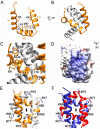
Structure of the DXV VP3 dsRNA-binding domain. (A) Ribbon diagram of the structure of the DXV VP3 N-terminal domain (residues 43 to 90) showing the secondary structure elements (α1 to α3), with the central hydrophobic core side chains depicted as sticks and explicitly labeled. (B) DXV VP3 N-terminal domain dimer, with one protomer shown in orange and the second one in gray. (C) Closeup view of residues involved in dimerization contacts. (D) Surface representation of the DXV VP3 residues involved in dsRNA binding. The surface is colored according to its electrostatic potential, with positive charges in blue and negative charges in red. Residues forming patch A are explicitly labeled. (E and F) Comparison of the VP3 N-terminal domain dimers in the P6322 (orange/gray monomers) (E) and P41212 (red/blue monomers) (F) crystals. In the P6322 crystals, dimers are formed through the crystallographic 2-fold axes. In the P41212 crystal form, a quasiequivalent dimer is formed in the asymmetric unit by means of a pseudo-2-fold molecular axis. In both panels, the positively charged residues in each monomer (H60, R75, R77, and K85) forming the flat surface in panel D are shown as sticks and explicitly labeled.
FIG 4
FIG 4
Stability studies of the DXV VP3 wild type and charge inversion mutants. (A) SDS-PAGE of the VP3 wild-type protein and PA to PD mutants. (C) Temperature-versus-fluorescence unfolding curves for the DXV VP3 wild type and PA to PD mutants. The color codes are the same as those in Fig. 1A.
FIG 5
FIG 5
Binding studies of DXV VP3-dsRNA complexes by electrophoretic mobility shift assays. EMSAs were performed with wild-type VP3 (A) and patch substitutions (B to E). Wild-type and substituted VP3 proteins (1 and 3 μg, indicated as + and ++++, respectively) were incubated with constant amounts of IBDV genomic dsRNAs (7.5 nM) before resolving the protein-RNA complexes in 1% agarose gels. Gels were stained with EtBr and photographed under UV light. In panels A and B, the VP3 protein and dsRNA were individually loaded as negative controls.
FIG 6
FIG 6
Kinetics of DXV VP3 binding to dsRNA measured by SPR. (A) Schematic representation of the SPR setup to produce dsRNAs of different lengths on the chip surface. The sequences of the oligonucleotides used in these assays (Table 2) were designed to minimize the formation of heterogeneous RNA duplexes arising from partial oligonucleotide annealing. Indeed, the longest fully complementary stretch allowing the formation of spurious RNA duplexes with the described oligonucleotides is 4 nucleotides. Even then, annealing would generate duplexes with melting temperatures (Tm) nearing 10°C at 150 mM NaCl, a temperature exceedingly lower than that used in our experiments (25°C). In contrast, correct annealing of the 24-, 21-, 15-, 11-, and 9-mer oligonucleotides to their immobilized 24-mer counterpart would generate duplexes with Tms of 59.9°C, 53°C, 45.8°C, 37.4°C, and 36°C, respectively. Although we cannot completely rule out the presence of a minimal population of heterogenous duplexes, their contribution to the results of the assay would be negligible. (B) Sensorgram showing binding responses of DXV VP3 to dsRNA molecules of different lengths. (C) Sensorgrams for binding to a 24-bp dsRNA at different VP3 concentrations. Each injection was performed in duplicate. Fitting curves are shown with thin black lines. (D) Comparative binding of the VP3 wild type and PA, PB, PC, and PD mutants to the 24-bp dsRNA.
FIG 7
FIG 7
VP3 of DXV suppresses RNA silencing in insect cells. (A) eGFP silencing in Hi5 cells. eGFP was produced in Hi5 cells by transient transfection and silenced by coexpression of dsRNA (seGFP) containing the eGFP-coding sequence. Silencing inhibition was tested by coexpressing DXV VP3 or patch-substituted variants (PAmut to PDmut). Images were captured at different time points after the initiation of VP3 production (24, 48, 72, and 96 h). (B) eGFP fluorescence intensity quantification of images at 96 h. Values are normalized with respect to the eGFP control, and error bars represent the standard deviations (SD) from 3 replicates. (C) Western blot analysis of cotransfected cell lysates using anti-GFP and anti-His antibody was used to monitor the expression levels of eGFP and DXV VP3 variants. A Coomassie blue-stained 12% SDS-PAGE gel is shown below as a loading control.
FIG 8
FIG 8
Binding modes of VSSs from different viral families. Shown are cartoon representations (left) and electrostatic surfaces (right) for carnation Italian ringspot virus p19 (PDB accession number 1RPU) (40) (a), influenza A virus NS1 (PDB accession number 2ZKO) (57) (b), Flock House virus B2 (PDB accession number 2AZ0) (36) (c), rice hoja blanca virus NS3 (PDB accession number 3AJF) (77) (d), the DXV VP3 N-terminal domain (this work) (e), tomato aspermy virus 2b (PDB accession number 2ZI0) (58) (f), beet yellows virus p21 (PDB accession number 2CWO) (59) (g), Marburg virus VP35 (PDB accession number 4GHA) (78) (h), Ebola virus VP35 (PDB accession number 3KS8) (79) (i), and IBDV VP3 (PDB accession number 2R18) (50) (j). For those structures determined in complex with dsRNA, the nucleic acid is represented as rainbow-colored ribbons; for the other structures, the location of the predicted RNA-binding site is indicated with a black circle. A schematic representation of the binding mode is also shown for each VSS, with the dsRNA represented by two orange dented lines and the protein domains as colored circles.

Similar articles

Cited by

References

    1. Delmas B, Mundt E, Vakharia VN, Wu JL. 2011. Birnaviridae, p 499–507. In King AMQ, Adams MJ, Carstens EB, Lefkowitz EJ (ed), Virus taxonomy: classification and nomenclature of viruses. Ninth report of the International Committee on Taxonomy of Viruses. Elsevier Academic Press, San Diego, CA.
    1. Coulibaly F, Chevalier C, Gutsche I, Pous J, Navaza J, Bressanelli S, Delmas B, Rey FA. 2005. The birnavirus crystal structure reveals structural relationships among icosahedral viruses. Cell 120:761–772. doi:10.1016/j.cell.2005.01.009. - DOI - PubMed
    1. Birghan C, Mundt E, Gorbalenya AE. 2000. A non-canonical lon proteinase lacking the ATPase domain employs the Ser-Lys catalytic dyad to exercise broad control over the life cycle of a double-stranded RNA virus. EMBO J 19:114–123. doi:10.1093/emboj/19.1.114. - DOI - PMC - PubMed
    1. Sánchez AB, Rodriguez JF. 1999. Proteolytic processing in infectious bursal disease virus: identification of the polyprotein cleavage sites by site-directed mutagenesis. Virology 262:190–199. doi:10.1006/viro.1999.9910. - DOI - PubMed
    1. Chung HK, Kordyban S, Cameron L, Dobos P. 1996. Sequence analysis of the bicistronic Drosophila-X virus genome segment A and its encoded polypeptides. Virology 225:359–368. doi:10.1006/viro.1996.0610. - DOI - PubMed

LinkOut - more resources