. 2021 Feb 15;95(4):e01869-20.
doi: 10.1128/JVI.01869-20. Epub 2020 Nov 25.

The Human Adenovirus Type 2 Transcriptome: An Amazing Complexity of Alternatively Spliced mRNAs

Affiliations

The Human Adenovirus Type 2 Transcriptome: An Amazing Complexity of Alternatively Spliced mRNAs

Amanda Westergren Jakobsson et al. J Virol. .

Abstract

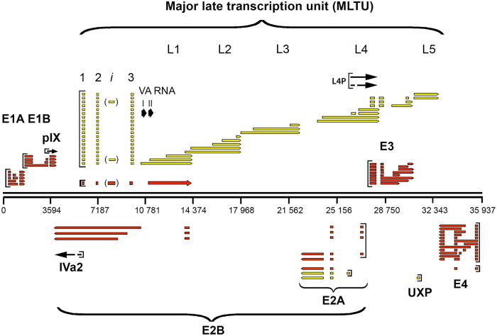
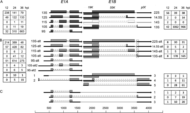
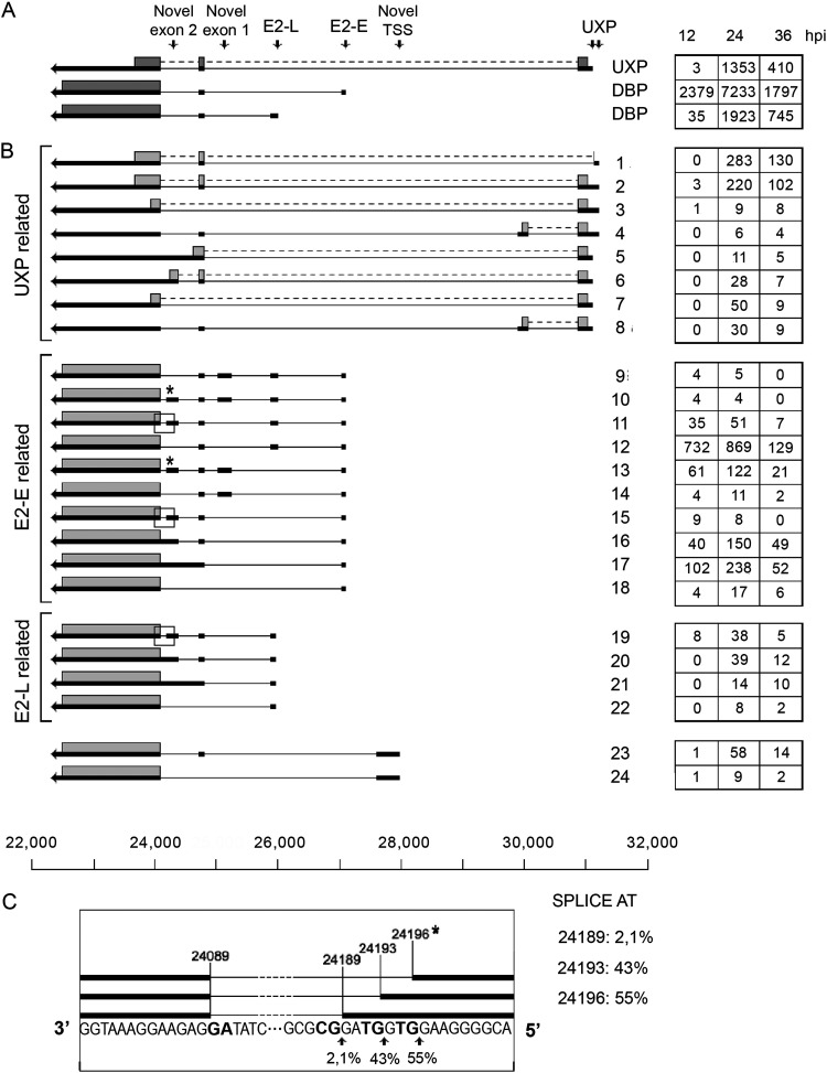
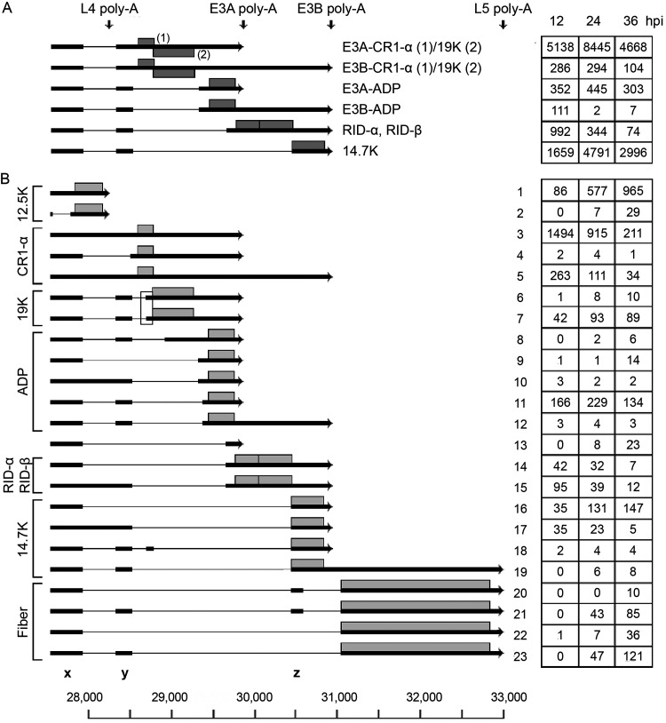
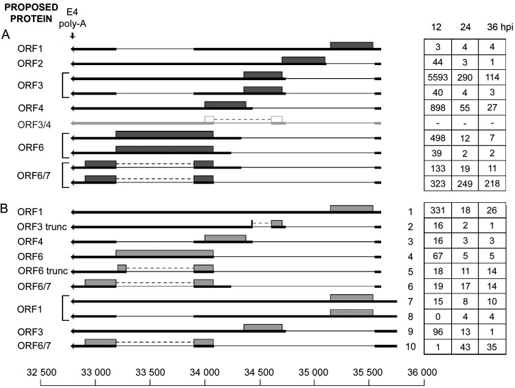
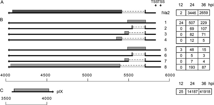
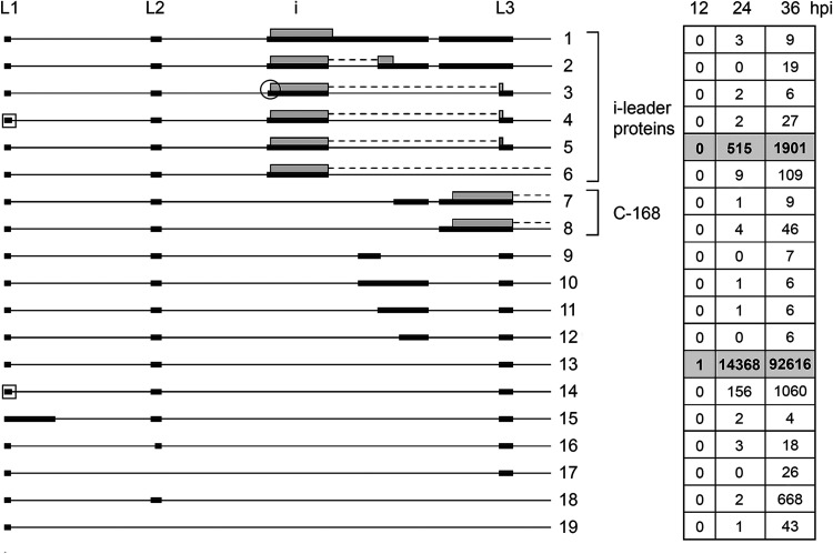
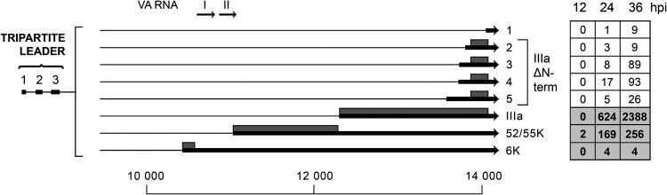
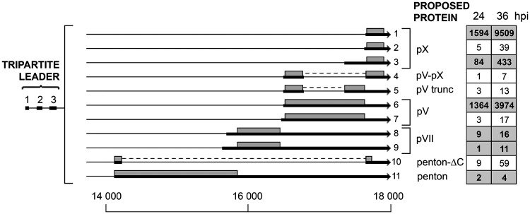
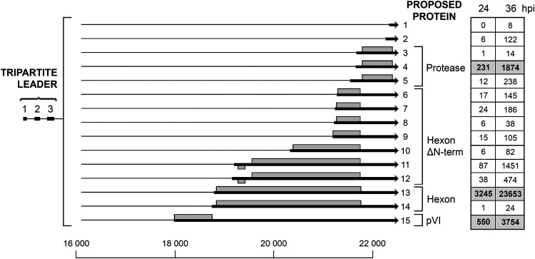
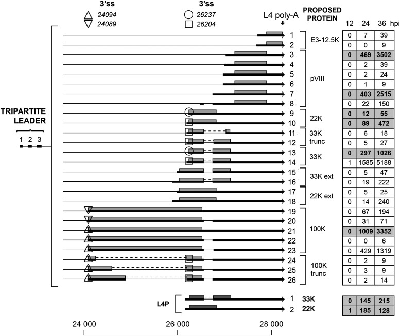
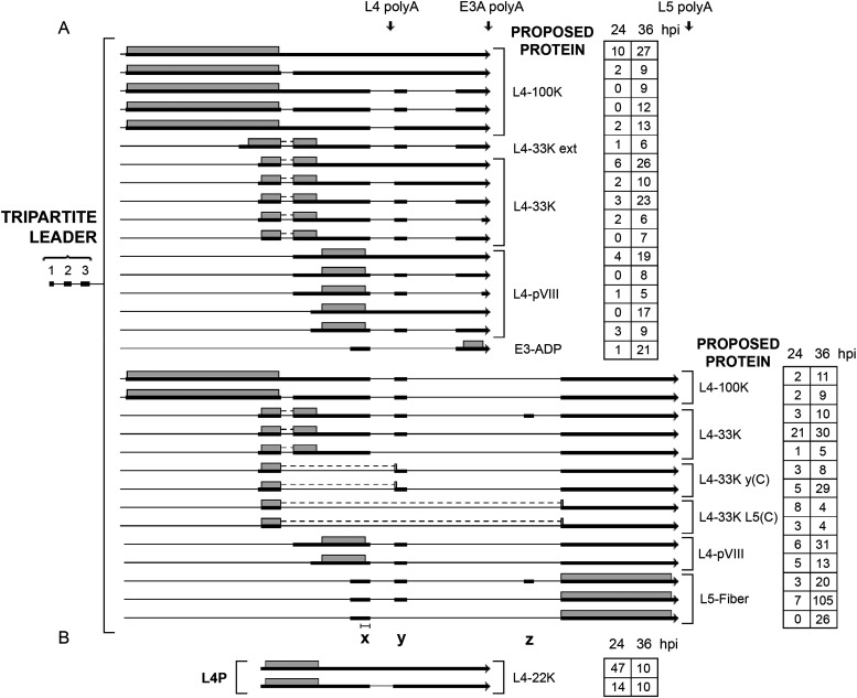
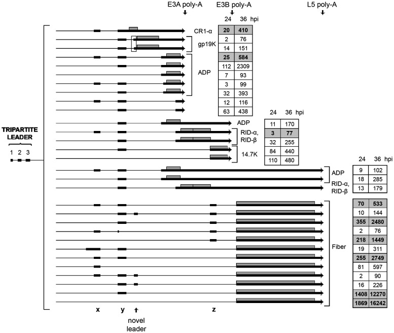
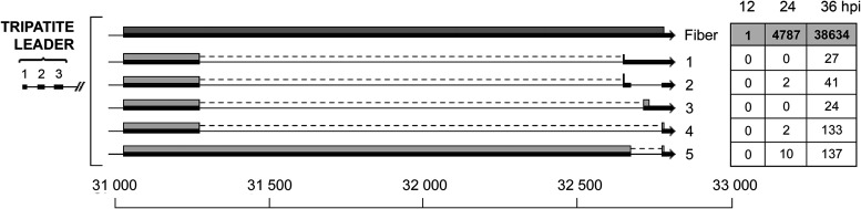
We have used the Nanopore long-read sequencing platform to demonstrate how amazingly complex the human adenovirus type 2 (Ad2) transcriptome is with a flexible splicing machinery producing a range of novel mRNAs both from the early and late transcription units. In total we report more than 900 alternatively spliced mRNAs produced from the Ad2 transcriptome whereof more than 850 are novel mRNAs. A surprising finding was that more than 50% of all E1A transcripts extended upstream of the previously defined transcriptional start site. The novel start sites mapped close to the inverted terminal repeat (ITR) and within the E1A enhancer region. We speculate that novel promoters or enhancer driven transcription, so-called eRNA transcription, is responsible for producing these novel mRNAs. Their existence was verified by a peptide in the Ad2 proteome that was unique for the E1A ITR mRNA. Although we show a high complexity of alternative splicing from most early and late regions, the E3 region was by far the most complex when expressed at late times of infection. More than 400 alternatively spliced mRNAs were observed in this region alone. These mRNAs included extended L4 mRNAs containing E3 and L5 sequences and readthrough mRNAs combining E3 and L5 sequences. Our findings demonstrate that the virus has a remarkable capacity to produce novel exon combinations, which will offer the virus an evolutionary advantage to change the gene expression repertoire and protein production in an evolving environment.IMPORTANCE Work in the adenovirus system led to the groundbreaking discovery of RNA splicing and alternative RNA splicing in 1977. These mechanisms are essential in mammalian evolution by increasing the coding capacity of a genome. Here, we have used a long-read sequencing technology to characterize the complexity of human adenovirus pre-mRNA splicing in detail. It is mindboggling that the viral genome, which only houses around 36,000 bp, not being much larger than a single cellular gene, generates more than 900 alternatively spliced mRNAs. Recently, adenoviruses have been used as the backbone in several promising SARS-CoV-2 vaccines. Further improvement of adenovirus-based vaccines demands that the virus can be tamed into an innocent carrier of foreign genes. This requires a full understanding of the components that govern adenovirus replication and gene expression.


Figures

FIG 1
FIG 1
The Ad2 transcriptome before Nanopore sequencing. Schematic view of adenovirus mRNAs expressed from the rightward- and leftward-transcribed strands. The mRNAs are color coded based on time of expression: early transcripts are shown in red, late mRNAs are in yellow, and mRNAs expressed at intermediate times of infection are represented with black arrows.
FIG 2
FIG 2
The E1A and E1B family of mRNAs. (A) Previously characterized mRNAs from the E1 region. The characterized E1B 14S mRNA (16) is shown in light gray, since the number of reads was below our cutoff. (B) Novel E1 mRNAs identified. (C) Structure of the most abundant E1A-E1B fusion mRNAs. The numbers of reads identified at 12, 24, and 36 hpi are shown within the boxes on the left (E1A) or on the right (E1B). The squares mark the E1B mRNAs using the alternative minor 3,591 3′ splice site. Thick lines denotes exons, thin lines introns. Gray boxes indicate the proposed protein(s) expressed from respective mRNA. For a complete summary of the triple-verified E1 mRNAs, see Table S1.
FIG 3
FIG 3
The E2A and UXP family of mRNAs. (A) Previously characterized E2A and UXP mRNAs. (B) Novel E2A and UXP mRNAs grouped into mRNAs with the same transcriptional start site. The numbers of reads identified at 12, 24, and 36 hpi are shown in boxes on the right. Thick lines denote exons, thin lines introns. Gray boxes indicate the proposed proteins expressed from respective mRNA. The asterisk and the boxed areas in panel B indicate a microheterogeneity in 5′ splice site usage at novel exon 2, which is shown expanded in panel C. TSS, transcriptional start site. For a complete summary of the triple-verified E2 and UXP mRNAs, see Table S1.
FIG 4
FIG 4
The E3 family of mRNAs. (A) Previously characterized E3 mRNAs with the likely proteins translated shown as gray boxes. (B) Novel E3 mRNAs grouped into mRNAs based on the likely protein encoded (gray boxes). The boxed area shows two gp19K mRNAs that use two alternative 3′ splice sites separated by 16 nucleotides. The number of reads identified at 12, 24 and 36 hpi are shown within boxes on the right. Thick lines denotes exons, thin lines introns. Poly-A, polyadenylation site. For a complete summary of the triple-verified E3 mRNAs, see Table S1.
FIG 5
FIG 5
The E4 family of mRNAs. (A) Previously characterized E4 mRNAs, with the likely proteins translated shown as gray boxes. The ORF3/4 mRNA is shown in light gray, since we could not detect reads corresponding to this mRNA in our Nanopore data. (B) Novel E4 mRNAs grouped based on the promoter position. The numbers of reads identified at 12, 24, and 36 hpi are shown in boxes to the right. Thick lines denotes exons, thin lines introns. Gray boxes indicate the proposed proteins expressed from respective mRNA. Poly-A, polyadenylation site. For a complete summary of the triple-verified E4 mRNAs, see Table S1.
FIG 6
FIG 6
The IVa2 and pIX family of mRNAs. (A) Spliced structure of the previously characterized IVa2 mRNA. (B) Novel IVa2 mRNAs grouped based on three apparent promoter positions. (C) Unspliced pIX mRNA. The numbers of reads identified at 12, 24, and 36 hpi are shown in boxes on the right. Thick lines denotes exons, thin lines introns. Gray boxes indicate the proposed proteins expressed from respective mRNA. TSS, transcriptional start site. For a complete summary of the triple-verified mRNAs from these regions, see Table S1.
FIG 7
FIG 7
Tripartite leader splicing. Alternative splicing events detected in the tripartite leader region are shown. Transcripts have been organized into groups to highlight the proposed proteins being translated and structural similarities between noncoding RNAs. The numbers of reads identified at 12, 24, and 36 hpi are shown in boxes to the right, with previously described transcripts in bold in shaded boxes. Thick lines denotes exons, thin lines introns. The square box indicates an alternative L1 5′ splice site located 4 nucleotides downstream of the major 5′ splice site. The encircled i-leader 3′ splice site is located 9 nucleotides downstream of the major i-leader 3′ splice site. Gray boxes indicate the proposed i-leader and C-168 proteins expressed from respective mRNA. For a complete summary of the triple-verified transcripts, see Table S1.
FIG 8
FIG 8
The L1 family of mRNAs. Alternative spliced mRNAs detected from late region 1 are shown. The numbers of reads identified at 12, 24, and 36 hpi are shown in boxes on the right, with previously described transcripts in bold in shaded boxes. Thick lines denotes exons, thin lines introns. Gray boxes indicate the proposed proteins expressed from respective mRNA. For a complete summary of the triple-verified L1 mRNAs, see Table S1.
FIG 9
FIG 9
The L2 family of mRNAs. Alternative spliced mRNAs detected from late region 2 are shown. The numbers of reads identified at 24 and 36 hpi are shown in boxes on the right, with previously described transcripts highlighted in bold in shaded boxes. Thick lines denotes exons, thin lines introns. Gray boxes indicate the proposed proteins expressed from respective mRNA. For a complete summary of the triple-verified L2 mRNAs, see Table S1.
FIG 10
FIG 10
The L3 family of mRNAs. The figure shows the alternative spliced mRNAs detected from late region 3. The number of reads identified at 24 and 36 hpi are shown in boxes on the right, with previously described transcripts in bold in shaded boxes. Thick lines denotes exons, thin lines introns. Gray boxes indicate the proposed proteins expressed from respective mRNA. For a complete summary of the triple-verified L3 mRNAs, see Table S1.
FIG 11
FIG 11
The L4 family of mRNAs. Alternative spliced mRNAs detected from late region 4 are shown. The numbers of reads identified at 12, 24, and 36 hpi are shown in boxes on the right, with previously described transcripts in bold in shaded boxes. Thick lines denotes exons, thin lines introns. Triangles (up or down) show microheterogeneities in 100K 3′ splice site usage. Circles and squares similarly show heterogeneities in 22K and 33K 3′ splice site usage. Gray boxes indicate the proposed proteins expressed from the respective mRNA. Poly-A, polyadenylation site. For a complete summary of the triple-verified L4 mRNAs, see Table S1.
FIG 12
FIG 12
L4-E3-L5 readthrough mRNAs. (A) A novel class of mRNAs that we detected are L4 spliced mRNAs that bypass the normal L4 polyadenylation site and use the downstream E3A or L5 poly(A) sites. (B) Novel mRNAs initiated at the L4 promoter. The number of reads identified at 24 and 36 hpi are shown in boxes on the right. Thick lines denotes exons, thin lines introns. Gray boxes indicate the proposed proteins expressed from respective mRNA. The locations of the x, y, and z leaders are depicted at the bottom. The bar above the x leader shows the normal size of the x leader. Poly-A, polyadenylation site. For a complete summary of the triple-verified L4-E3-L5 mRNAs, see Table S1.
FIG 13
FIG 13
The complex splicing pattern of the E3 and L5 family of mRNAs expressed from the major late promoter at late times of infection. The number of reads identified at 24 and 36 hpi are shown in boxes on the right, with previously described transcripts in bold in shaded boxes. The boxed area shows two gp19K mRNAs that use two alternative 3′ splice sites separated by 16 nucleotides. Thick lines denotes exons, thin lines introns. Gray boxes indicate the proposed proteins expressed from respective mRNA. Poly-A, polyadenylation site. For a complete summary of the triple-verified L4-E3-L5 mRNAs, see Table S1.
FIG 14
FIG 14
Alternative splicing events interrupting the fiber coding sequence. The numbers of reads identified at 12, 24, and 36 hpi are shown in boxes on the right, with the previously described fiber mRNA in bold in a shaded box. Thick lines denotes exons, thin lines introns. Gray boxes indicate the proposed proteins expressed from respective mRNA. For a complete summary of the triple-verified fiber alternative splicing events, see Table S1.

Similar articles

Cited by

References

    1. Berget SM, Moore C, Sharp PA. 1977. Spliced segments at the 5' terminus of adenovirus 2 late mRNA. Proc Natl Acad Sci U S A 74:3171–3175. doi:10.1073/pnas.74.8.3171. - DOI - PMC - PubMed
    1. Chow LT, Gelinas RE, Broker TR, Roberts RJ. 1977. An amazing sequence arrangement at the 5' ends of adenovirus 2 messenger RNA. Cell 12:1–8. doi:10.1016/0092-8674(77)90180-5. - DOI - PubMed
    1. Chow LT, Broker TR, Lewis JB. 1979. Complex splicing patterns of RNAs from the early regions of adenovirus-2. J Mol Biol 134:265–303. doi:10.1016/0022-2836(79)90036-6. - DOI - PubMed
    1. Chow LT, Roberts JM, Lewis JB, Broker TR. 1977. A map of cytoplasmic RNA transcripts from lytic adenovirus type 2, determined by electron microscopy of RNA:DNA hybrids. Cell 11:819–836. doi:10.1016/0092-8674(77)90294-x. - DOI - PubMed
    1. Akusjärvi G, Pettersson U, Roberts RJ. 1986. Structure and function of the adenovirus-2 genome, p 53–95. In Doerfler W (ed), Adenovirus DNA: the viral genome and its expression. Martin Nijhoff Publishing, Boston, MA.