Review
. 2021 Jan;124(1):27-36.
doi: 10.1038/s41416-020-01172-1. Epub 2020 Nov 26.

The fibrotic and immune microenvironments as targetable drivers of metastasis

Affiliations
Review

The fibrotic and immune microenvironments as targetable drivers of metastasis

Luke Boulter et al. Br J Cancer. 2021 Jan.

Abstract

Although substantial progress has been made over the past 40 years in treating patients with cancer, effective therapies for those who are diagnosed with advanced metastatic disease are still few and far between. Cancer cells do not exist in isolation: rather, they exist within a complex microenvironment composed of stromal cells and extracellular matrix. Within this tumour microenvironment exists an interplay between the two main stromal cell subtypes, cancer-associated fibroblasts (CAFs) and immune cells, that are important in controlling metastasis. A complex network of paracrine signalling pathways between CAFs, immune cells and tumour cells are involved at multiple stages of the metastatic process, from invasion and intravasation at the primary tumour site to extravasation and colonisation in the metastatic site. Heterogeneity and plasticity within stromal cell populations also contribute to the complexity. Although many of these processes are likely to be common to a number of metastatic sites, we will describe in detail the interplay within the liver, a preferred site of metastasis for many tumours. A greater understanding of these networks provides opportunities for the design of new therapeutic approaches for targeting the metastatic disease.


Conflict of interest statement

The authors declare no competing interests.

Figures

Fig. 1
Fig. 1. The metastatic cascade.
Metastasis occurs in a number of steps starting with the invasion of tumour cells into the local stroma (1) and their intravasation (2) into the vascular or lymphatic system. In the case of haematogenous metastasis, they must survive the mechanical forces of the circulatory system and suppressive immune cells (3). Few circulating tumour cells (CTCs) survive the circulation but those that do then extravasate into the metastatic site (4). The systemic release of exosomes, inflammatory cytokines and growth factors by cancer and stromal cells in the primary tumour can ‘prime’ and remodel the microenvironment of distant organs to form a pre-metastatic niche that supports the outgrowth of DTCs before their arrival. Once in the metastatic site, disseminated tumour cells (DTCs) must overcome immune surveillance by resident immune cells, and can direct immune suppression by recruiting myeloid-derived suppressor cells. DTCs can also become dormant, induced by factors derived from metastatic niche stromal cells, or supported by signalling from endothelial cells, and then subsequently ‘awakened’ by signals from surrounding stromal cells and the extracellular matrix (ECM)., Reawakening of dormant DTCs can also be induced by exosomes, either released by metastatic niche stromal cells or systemically released by cancer-associated fibroblasts (CAFs) in the primary site., Proliferating DTCs form micrometastases and, finally, colonisation of the metastatic niche occurs and tumour cells form clinically relevant macrometastases, co-opting and recruiting local stromal cells to support metastatic cell growth (5). Created with BioRender.com.
Fig. 2
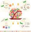
Fig. 2. Metastasis-promoting functions of cancer-associated fibroblasts.
Heterogeneous cancer-associated fibroblasts (CAFs) drive pro-tumorigenic and metastatic phenotypes via diverse mechanisms: (1) secretion of soluble factors such as growth factors, cytokines and lipids to promote tumour survival and metastasis; (2) secretion and remodelling of components of the extracellular matrix (ECM); (3) secretion of immunomodulatory factors, which contribute to changes in the tumour immune microenvironment; and (4) establishment of a pre-metastatic niche through the secretion of soluble factors and extracellular vesicles. Created with BioRender.com.
Fig. 3
Fig. 3. The metastatic microenvironment in the liver.
The liver provides a permissive environment for metastatic colonisation. (1) Circulating tumour cells (CTCs) within the liver microvasculature must survive immune attack from circulating immune cell populations. (2) Cells then enter the lumen of the hepatic sinusoid via specialised sieve-like fenestrated endothelial cells (liver sinusoidal endothelial cells; LSECs). (3) Cells are subjected to further immune surveillance mediated via cytokine and chemokine crosstalk between sinusoidal endothelial cells, Kupffer cells and lymphocytes. (4) The tumour cells must then establish themselves within the space of Dissé, where they recruit hepatic stellate cells (HSCs), myeloid-derived suppressor cells and lymphocytes to establish a metastatic-promoting environment required for the initiation of micrometastases. (5) The further outgrowth of metastases is accompanied by matrix deposition and stiffening mediated by activated HSCs and the formation of a local vasculature and recruitment of pro-inflammatory immune cells. Created with BioRender.com.

References

    1. Dillekås H, Rogers MS, Straume O. Are 90% of deaths from cancer caused by metastases? Cancer Med. 2019;8:5574–5576. - PMC - PubMed
    1. Lambert AW, Pattabiraman DR, Weinberg RA. Emerging biological principles of metastasis. Cell. 2017;168:670–691. - PMC - PubMed
    1. Welch DR, Hurst DR. Defining the hallmarks of metastasis. Cancer Res. 2019;79:3011–3027. - PMC - PubMed
    1. Hanahan D, Weinberg RA. Hallmarks of cancer: the next generation. Cell. 2011;144:646–674. - PubMed
    1. Acharyya S, Oskarsson T, Vanharanta S, Malladi S, Kim J, Morris PG, et al. A CXCL1 paracrine network links cancer chemoresistance and metastasis. Cell. 2012;150:165–178. - PMC - PubMed

Publication types