. 2020 Nov 25;10(1):20505.
doi: 10.1038/s41598-020-77554-5.

Label-free X-ray estimation of brain amyloid burden

Affiliations

Label-free X-ray estimation of brain amyloid burden

Eshan Dahal et al. Sci Rep. .

Abstract

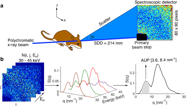
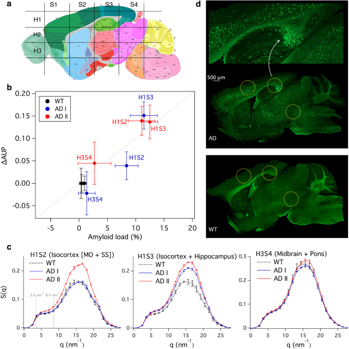
Amyloid plaque deposits in the brain are indicative of Alzheimer's and other diseases. Measurements of brain amyloid burden in small animals require laborious post-mortem histological analysis or resource-intensive, contrast-enhanced imaging techniques. We describe a label-free method based on spectral small-angle X-ray scattering with a polychromatic beam for in vivo estimation of brain amyloid burden. Our findings comparing 5XFAD versus wild-type mice correlate well with histology, showing promise for a fast and practical in vivo technique.


Conflict of interest statement

Patents have been filed pertaining to the described sSAXS based method for amyloid burden estimation, listing ED and AB as inventors. The other authors declare no competing interests.

Figures

Figure 1
Figure 1
Schematic of the experimental sSAXS setup and data processing steps. (a) The mouse head is irradiated at select locations with a collimated beam of polychromatic X rays of 1 mm diameter. A 2D spectroscopic detector (CdTe) is used to collect SAXS data simultaneously in angle- and energy-dispersive modes for each location. The sample to detector distance (SDD) was 214 mm (b) sSAXS data processing steps in 30–45 keV energy range are illustrated using an amyloid model from BSA with two Bragg peaks at 6.04 and 13.24nm-1. The 2D detector data (left) showing counts in each pixel per energy bin is converted to scattering cross section, S(q), per energy bin (middle) after applying a transmission correction. The S(q) is then summed (Δq=1.2nm-1) from 30 to 45 keV to combine scattering information in all energy bins and to calculate the area under the peak (AUP) from 3.6 to 8.4nm-1 (right).
Figure 2
Figure 2
Amyloid burden estimations in intact heads corresponding to 5XFAD mice and WT controls. (a) Approximate studied locations in the mouse brain in comparison to an anatomic reference image from the Allen Mouse Brain Atlas containing major brain regions in a sagittal plane. (b) Correlation between the ΔAUP calculated from the sSAXS method and the amyloid load estimates derived from histology for three locations (Pearson r=0.94,P<0.001). (c) Corresponding S(q) of AD and WT mice used for ΔAUP calculation after studying their heads with an intact skull in each location for 300 s. Error bars represent ± standard deviation from three measurements for each location. (d) Representative histology images of AD and WT brain slices using Thioflavin S. The zoomed image (top) is focused on the hippocampus of AD brain slice.

References

    1. Aguzzi A, O’connor T. Protein aggregation diseases: Pathogenicity and therapeutic perspectives. Nat. Rev. Drug Discov. 2010;9:237–248. doi: 10.1038/nrd3050. - DOI - PubMed
    1. Araki K, et al. Parkinson’s disease is a type of amyloidosis featuring accumulation of amyloid fibrils of α-synuclein. Proc. Natl. Acad. Sci. 2019;116:17963–17969. doi: 10.1073/pnas.1906124116. - DOI - PMC - PubMed
    1. Landau SM, et al. Amyloid-β imaging with Pittsburgh compound B and florbetapir: Comparing radiotracers and quantification methods. J. Nucl. Med. 2013;54:70–77. doi: 10.2967/jnumed.112.109009. - DOI - PMC - PubMed
    1. Klunk WE, et al. Binding of the positron emission tomography tracer Pittsburgh compound-B reflects the amount of amyloid-β in Alzheimer’s disease brain but not in transgenic mouse brain. J. Neurosci. 2005;25:10598–10606. doi: 10.1523/JNEUROSCI.2990-05.2005. - DOI - PMC - PubMed
    1. Kuntner C, et al. Limitations of small animal PET imaging with [18F]FDDNP and FDG for quantitative studies in a transgenic mouse model of Alzheimer’s disease. Mol. Imaging Biol. 2009;11:236–240. doi: 10.1007/s11307-009-0198-z. - DOI - PubMed

Publication types

Substances

LinkOut - more resources