

Acral lentiginous melanoma in situ: dermoscopic features and management strategy
- PMID: 33239715
- PMCID: PMC7688656
- DOI: 10.1038/s41598-020-77425-z
Acral lentiginous melanoma in situ: dermoscopic features and management strategy
Abstract
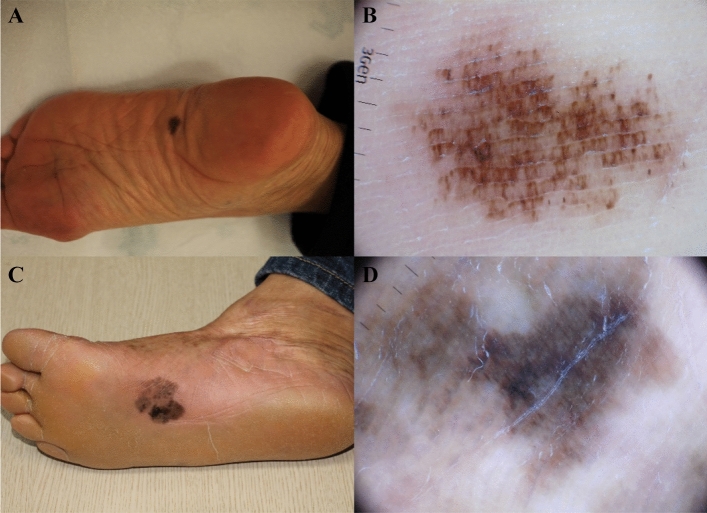
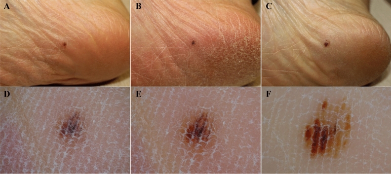
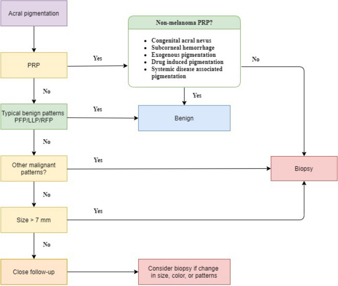
Diagnosis of acral lentiginous melanoma in situ (ALMIS) is challenging. However, data regarding ALMIS are limited in the literature. The aim of this study was to investigate the clinical and dermoscopic features of ALMIS on palmoplantar surfaces. Patients with ALMIS and available dermoscopic images were retrospectively reviewed at our institution between January 2013 and February 2020. Clinical and dermoscopic features were analysed and compared between small (< 15 mm) and large (≥ 15 mm) ALMIS. Twenty-one patients with ALMIS were included in this study. Mean patient age was 58.5 (range 39-76) years; most lesions were located on the sole (90.5%). The mean maximal diameter was 19.9 ± 13.7 mm (mean ± standard deviation). Statistical analysis of dermoscopic features revealed that parallel ridge patterns (54.5% vs. 100%, P = 0.035), irregular diffuse pigmentation (27.3% vs. 100%, P = 0.001) and grey colour (18.2% vs. 90%, P = 0.002) were significantly less frequent in small lesions than in large lesions. We have also illustrated two unique cases of small ALMIS; their evolution and follow-up dermoscopic examination are provided. In conclusion, this study described detailed dermoscopic findings of ALMIS. Based on the present study and a review of the literature, we proposed a dermoscopic algorithm for the diagnosis of ALMIS.
Conflict of interest statement
The authors declare no competing interests.
Figures
References
-
- Reed, R. J. New Concepts in Surgical Pathology of the Skin. (John Wiley & Sons, 1976).
MeSH terms
LinkOut - more resources
Full Text Sources
Medical
