

Comment
doi: 10.1038/s41433-020-01283-2.
Epub 2020 Nov 25.
Reply to Editorial: Interpretation of OCT and fundus findings in COVID-19 patients in recent Lancet publication
Affiliations
- PMID: 33239764
- PMCID: PMC7687980
- DOI: 10.1038/s41433-020-01283-2
Item in Clipboard
Comment
Reply to Editorial: Interpretation of OCT and fundus findings in COVID-19 patients in recent Lancet publication
Eye (Lond).
2021 Dec.
No abstract available
Conflict of interest statement
The authors declare that they have no conflict of interest.
Figures
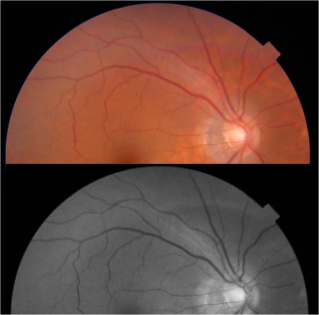
The pictures were taken 3 months after first evaluation and shows the absence of the previously reported cotton wool spot lesion.
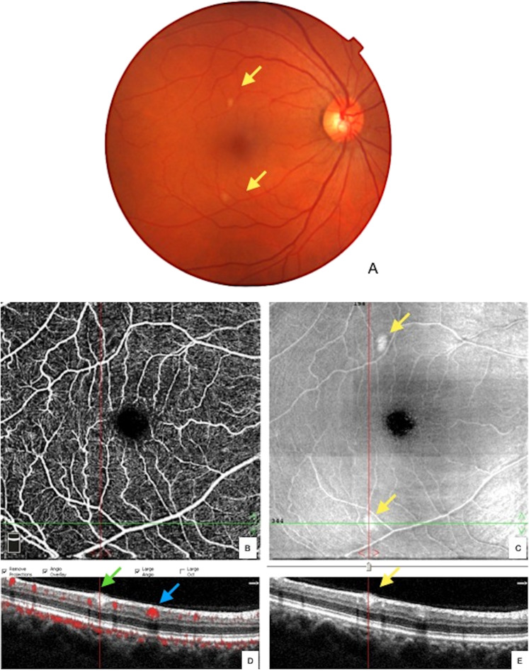
The images depict a hyperreflective lesion at the level of the inner plexiform and ganglion cell layer (yellow arrows), which aligns with inferior lesion at the enface OCT image as highlighted by in red line (c, d, e). The OCT-angiography indicates no flow void at that same site (b). The red dot flow overlay on the B scan OCT image, based upon OCT angiogram data, differentiates this lesion (green arrow) and from an adjacent blood vessel cross-section by the presence of the red overlay within the lumen marking active blood flow (blue arrow). The density within the vessel results in a shadow projected deep into the underlying retinal layers (d), which the lesion produces no shadow artifact.
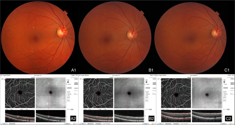
a1 and a2 were taken on 1st evaluation, b1 and b2 were taken after 37 days. c1 and c2 were taken after another 28 days.
Comment on
-
Retinal findings in patients with COVID-19.Lancet. 2020 May 23;395(10237):1610. doi: 10.1016/S0140-6736(20)31014-X. Epub 2020 May 12. Lancet. 2020. PMID: 32405105 Free PMC article. No abstract available.
References
-
- Landecho MF, Yuste JR, Gándara E, Sunsundegui P, Alcaide AB, García-Layana A. COVID-19 retinal microangiopathy as an in vivo biomarker of systemic vascular disease? J Intern Med. 2020. 10.1111/joim.13156. - PubMed
Publication types
MeSH terms
LinkOut - more resources
Full Text Sources
Medical
