Review
. 2020 Nov;587(7835):567-576.
doi: 10.1038/s41586-020-2933-1. Epub 2020 Nov 25.

Discoveries in structure and physiology of mechanically activated ion channels

Affiliations
Review

Discoveries in structure and physiology of mechanically activated ion channels

J M Kefauver et al. Nature. 2020 Nov.

Abstract

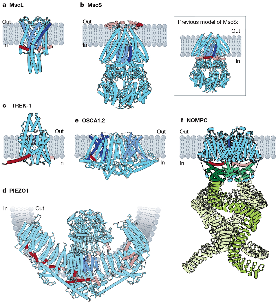
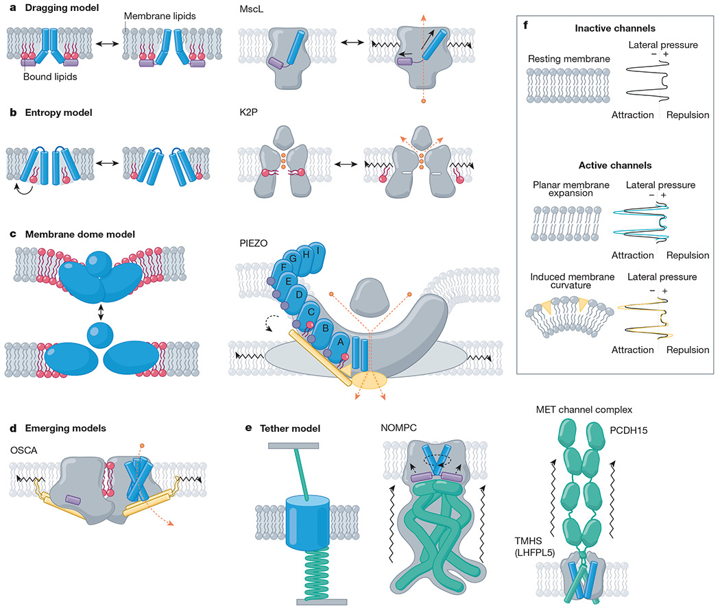
The ability to sense physical forces is conserved across all organisms. Cells convert mechanical stimuli into electrical or chemical signals via mechanically activated ion channels. In recent years, the identification of new families of mechanosensitive ion channels-such as PIEZO and OSCA/TMEM63 channels-along with surprising insights into well-studied mechanosensitive channels have driven further developments in the mechanotransduction field. Several well-characterized mechanosensory roles such as touch, blood-pressure sensing and hearing are now linked with primary mechanotransducers. Unanticipated roles of mechanical force sensing continue to be uncovered. Furthermore, high-resolution structures representative of nearly every family of mechanically activated channel described so far have underscored their diversity while advancing our understanding of the biophysical mechanisms of pressure sensing. Here we summarize recent discoveries in the physiology and structures of known mechanically activated ion channel families and discuss their implications for understanding the mechanisms of mechanical force sensing.


Conflict of interest statement

Competing interests The authors declare no competing interests.

Figures

Fig. 1 |
Fig. 1 |. Structures of mechanically activated ion channels.
Many mechanically activated channels seem to share a common feature: amphipathic helices (dark red on subunit A, rose on all other subunits) connected directly or indirectly to pore-lining regions (dark blue on subunit A, cornflower on all other subunits). a, Cartoon model of MscL (Protein Data Bank (PDB): 2OAR). Pore-lining TM1 (blue) is connected to the amphipathic S1 helix (red). b, Cartoon models of MscS. Main, a recent structure of MscS in nanodiscs (PDB: 6PWP), with the amphipathic anchor domain (red) sitting on the external membrane leaflet. Inset, the previous model of MscS (PDB: 2OAU), with the pore-lining TM3a helix (blue) completely embedded within the membrane and TM3b (red) predicted to be an amphipathic segment at the cytoplasmic leaflet. c, Cartoon model of TREK-1 (PDB: 6CQ6). The pore domains (blue) are gated by a C-type mechanism. The amphipathic C-tail (red) extends below the M4 helix. d, Cartoon model of PIEZO1 (PDB: 5Z10). Beneath the extracellular cap, two TM helices from each subunit line the pore (blue). In the domain-swapped blades, several amphipathic helices (red) line the cytoplasmic leaflet. e, Cartoon model of OSCA1.2 (PDB: 6MGV). Five helices (blue) line each of the two putative pores of OSCA1.2 and an amphipathic helix (red) sits on the opposite face of each subunit. f, Cartoon model of NOMPC (PDB: 5VK4). Each NOMPC subunit has an amphipathic TRP domain (red), a pore helix (blue) and a large spring-like ankyrin repeat domain (green).
Fig. 2 |
Fig. 2 |. Mechanistic models of mechanically activated ion channel gating.
Proposed mechanistic models with channel family examples. Amphipathic helices (violet), TM helices (blue), bound lipids (red), beam-like features (gold), tethers (emerald), ions (orange) and membrane lipids (grey) are indicated. a, Left, the dragging model. Lipids interact with an amphipathic helix and drag it outwards upon membrane expansion. Right, MscL, for example, has an amphipathic helix on the internal leaflet (helix S1, violet) that drives a tilt to the pore-lining helix (TM1, blue) as it is ‘dragged’ outward under tension. b, Left, entropy model. Lipids reside in hydrophobic pockets in the closed state and exit these pockets under membrane tension, inducing a conformational change. Right, K2P, for example, has a fenestration occupied by lipid acyl tails (red) when inactive, whereas in active channels, this fenestration is closed and lipids are absent. c, Left, membrane dome model. Channel curvature within the membrane stores energy. Right, PIEZOs, for example, expand and flatten, gating the pore via interactions between the beam domain, the anchor domain and the CTD (gold). d, Emerging models. OSCA channels have lipid-occupied pores and an intersubunit cleft (red), an amphipathic helix (violet) on the inner leaflet, and a beam-like domain (gold) connected to pore-lining helices, which terminates a membrane-entrant hook domain (gold); all of these domains could have a role in gating. e, Left, the tether model. Force is transmitted to the channel via a tether to the extracellular matrix, the cytoskeleton or both. For example, NOMPC (middle) is tethered to microtubules via its ankyrin repeat domain (emerald). Right, the MET channel complex is tethered to the neighbouring stereocilium via the tip link (PCDH15; emerald). f, Top, resting membrane tension. The transbilayer pressure profile reflects the lateral pressure experienced through the bilayer as a consequence of repulsion (positive pressure) of the lipid head groups, attraction (negative pressure) due to surface tension at the glycerol backbone, and steric hindrance (positive pressure) between the lipid tails. Bottom, model membranes under tension. Planar membrane expansion thins the bilayer and increases the area occupied by each lipid. Membrane curvature is induced when suction is applied to the membrane or conical-shaped amphipathic compounds insert into the bilayer,.
Fig. 3 |
Fig. 3 |. Lipids observed in structures of mechanosensitive ion channels.
a, Lipids are observed in three locations in MscS structures. (1) one lipid per subunit is ‘hooked’ at the periplasmic leaflet,; (2) densities ascribed to lipid acyl chains reside inside the pore, (PDB: 6PWN). (3) Two additional lipids per protomer are observed parallel to TM3b, below the membrane leaflet (PDB: 6RLD). b, In inactive structures of TRAAK (PDB: 4WWF), an acyl tail (green) occupies a fenestration below the selectivity filter. c, Two lipid-like densities are observed in the PIEZO1 structure (PDB: 6BPZ): (1) in the region between the anchor domain and piezo repeat A, and (2) between piezo repeats B and C (second and third from the pore, respectively).
Fig. 4 |
Fig. 4 |. The MET channel complex.
a, Model of a hair cell. b, MET channel complex components: TMC1 or TMC2, TMIE and LHFPL5 are localized to the stereocilia tips. Tip links are formed by dimers of PCDH15 and cadherin 23 (CDH23). c, Cartoon model of components of the MET channel complex: PCDH15 forms the lower half of the tip link (green) and LHFPL5 is a dimeric TM protein (blue) (PDB: 6C13 and 6C14).

References

    1. Anishkin A, Loukin SH, Teng J & Kung C Feeling the hidden mechanical forces in lipid bilayer is an original sense. Proc. Natl Acad. Sci. USA 111, 7898–7905 (2014). - PMC - PubMed
    1. Arnadóttir J & Chalfie M Eukaryotic mechanosensitive channels. Annu. Rev. Biophys 39, 111–137 (2010). - PubMed
    1. Ranade SS, Syeda R & Patapoutian A Mechanically activated ion channels. Neuron 87, 1162–1179 (2015). - PMC - PubMed
    1. Cox CD, Bavi N & Martinac B Bacterial mechanosensors. Annu. Rev. Physiol 80, 71–93 (2018). - PubMed
    1. Chalfie M Neurosensory mechanotransduction. Nat. Rev. Mol. Cell Biol 10, 44–52 (2009). - PubMed

Publication types

LinkOut - more resources