. 2020 Nov 9:11:588685.
doi: 10.3389/fendo.2020.588685. eCollection 2020.

The Pervasive Effects of ER Stress on a Typical Endocrine Cell: Dedifferentiation, Mesenchymal Shift and Antioxidant Response in the Thyrocyte

Affiliations

The Pervasive Effects of ER Stress on a Typical Endocrine Cell: Dedifferentiation, Mesenchymal Shift and Antioxidant Response in the Thyrocyte

Luca Ulianich et al. Front Endocrinol (Lausanne). .

Abstract

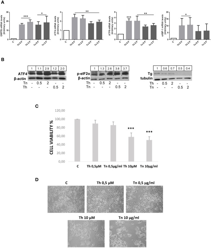
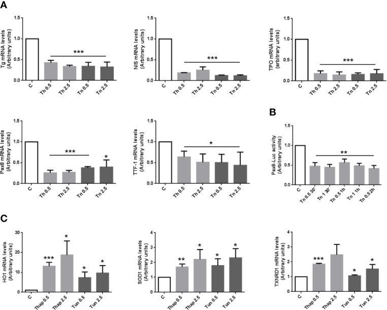
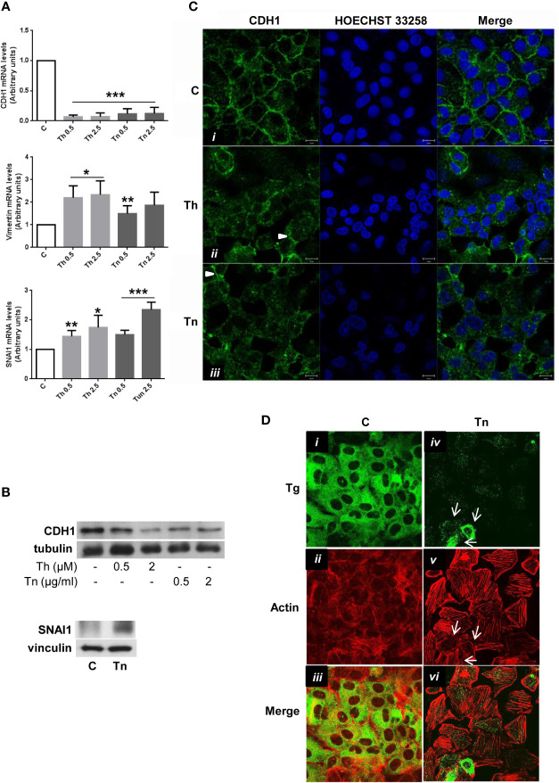
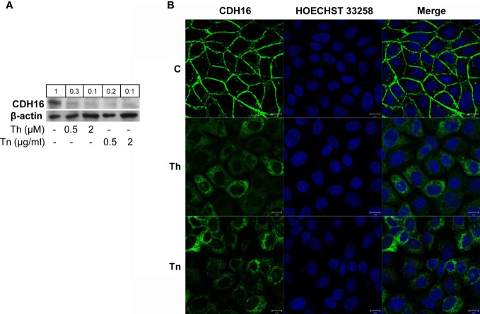
The endoplasmic reticulum stress and the unfolded protein response are triggered following an imbalance between protein load and protein folding. Until recently, two possible outcomes of the unfolded protein response have been considered: life or death. We sought to substantiate a third alternative, dedifferentiation, mesenchymal shift, and activation of the antioxidant response by using typical endocrine cells, i.e. thyroid cells. The thyroid is a unique system both of endoplasmic reticulum stress (a single protein, thyroglobulin represents the majority of proteins synthesized in the endoplasmic reticulum by the thyrocyte) and of polarized epithelium (the single layer of thyrocytes delimiting the follicle). Following endoplasmic reticulum stress, in thyroid cells the folding of thyroglobulin was disrupted. The mRNAs of unfolded protein response were induced or spliced (X-box binding protein-1). Differentiation was inhibited: mRNA levels of thyroid specific genes, and of thyroid transcription factors were dramatically downregulated, at least in part, transcriptionally. The dedifferentiating response was accompanied by an upregulation of mRNAs of antioxidant genes. Moreover, cadherin-1, and the thyroid (and kidney)-specific cadherin-16 mRNAs were downregulated, vimentin, and SNAI1 mRNAs were upregulated. In addition, loss of cortical actin and stress fibers formation were observed. Together, these data indicate that ER stress in thyroid cells induces dedifferentiation, loss of epithelial organization, shift towards a mesenchymal phenotype, and activation of the antioxidant response, highlighting, at the same time, a new and wide strategy to achieve survival following ER stress, and, as a sort of the other side of the coin, a possible new molecular mechanism of decline/loss of function leading to a deficit of thyroid hormones formation.

Keywords: ER stress; antioxidant response; dedifferentiation; mesenchymal phenotype; thyroid.


Figures

Figure 1
Figure 1
Th/Tn induce ER stress and UPR activation in PCCl3 cells without appreciably affecting viability. (A) Cells were plated in 100 mm diameter plates to about 80% confluence 24 h before treatments. Cells were treated or mock treated for 30 min with various concentrations of Th/Tn, followed by 24 h in complete medium without Th/Tn. Total RNA was extracted with the TRIzol reagent, according to the manufacturer’s protocol. Quantitative real-time RT-PCR analysis was performed as described in Materials and Methods. PCCl3 cells vehicle-treated (C) or treated with increasing concentrations of Th/Tn. *p < 0.05, **p < 0.01, ***p < 0.001, of each group respect to control. (B) Cells were plated in 60 mm diameter plates to about 80% confluence 24 h before treatments. Cells were treated or mock treated for 30 min with various concentrations of Th/Tn, followed by 24 h in complete medium without Th/Tn. Western blots of total protein extracts from PCCl3 cells vehicle-treated or treated with increasing concentrations of Th/Tn. The ratio of the densitometric values ATF4/β-actin, p-eIF2-α/β-actin, and Tg/tubulin is reported. (C, D) Cells were plated in 35 mm diameter plates to about 50% confluence 48 h before treatments. Cells were treated or mock treated for 30 min with various concentrations of Th/Tn, followed by 24 h in complete medium without Th/Tn. Cells were photographed by a Nikon Eclipse TS100 inverted microscope. Successively, MTT (0.5 µg/ml) was added to the cells for a 4-h incubation and cells were lysed in acidified isopropanol/HCl 0.04N. The lysates were subsequently read on a spectrophotometer at 550 nm (Bio-rad, Richmond, CA, USA) after a 1:2 dilution with water. The results were expressed as percent viability compared to controls. ***p < 0.001, of each group respect to control.
Figure 2
Figure 2
ER stress induces an inhibition of differentiation with a mechanism, at least in part, transcriptional, and an antioxidant response in PCCl3 cells. (A) Cells were plated in 100 mm diameter plates to about 80% confluence 24 h before treatments. Cells were treated or mock treated for 30 min with various concentrations of Th/Tn, followed by 24 h in complete medium without Th/Tn. Total RNA was extracted with the TRIzol reagent, according to the manufacturer’s protocol. Quantitative real-time RT-PCR analysis was performed as described in Materials and Methods. PCCl3 cells vehicle-treated (C) or treated with increasing concentrations of Th/Tn. *p < 0.05, ***p < 0.001, of each group respect to control. (B) Cells were plated in six-well plates to about 80% confluence 24 h before transfection. PCCl3 cells transfected with 2.5 μg of luciferase reporter plasmid and 0.5 μg of pRL-TK vector with 5 µl Lipofectamine 2000, as reported in Materials and Methods. Twenty-four hours after transfection cells were vehicle-treated or treated with 0.5 or 1.0 μg/ml Tn for 30, 60, and 120 min and harvested after 24 h in medium without Tn. Firefly and renilla activities were determined in cell lysates using the Dual-Luciferase Reporter Assay System and a luminometer. Results were expressed as the ratio of firefly to renilla activity. **p < 0.01, of each group respect to control. (C) Cells were plated in 100 mm diameter plates to about 80% confluence 24 h before treatments. Cells were treated or mock treated for 30 min with various concentrations of Th/Tn, followed by 24 h in complete medium without Th/Tn. Total RNA was extracted with the TRIzol reagent, according to the manufacturer’s protocol. Quantitative real-time RT-PCR analysis was performed as described in Materials and Methods. PCCl3 cells vehicle-treated (C) or treated with increasing concentrations of Th/Tn. *p < 0.05, **p < 0.01, ***p < 0.001, of each group respect to control.
Figure 3
Figure 3
ER stress induces a shift towards a mesenchymal phenotype in PCCl3 cells. (A) Cells were plated in 100 mm diameter plates to about 80% confluence 24 h before treatments. Cells were treated or mock treated for 30 min with various concentrations of Th/Tn, followed by 24 h in complete medium without Th/Tn. Total RNA was extracted with the TRIzol reagent, according to the manufacturer’s protocol. Quantitative real-time RT-PCR analysis was performed as described in Materials and Methods. PCCl3 cells vehicle-treated (C) or treated with increasing concentrations of Th/Tn. *p < 0.05, **p < 0.01, ***p < 0.001, of each group respect to control. (B) Cells were plated in 60 mm diameter plates to about 80% confluence 24 h before treatments. Cells were treated or mock treated for 30 min with various concentrations of Th/Tn, followed by 24 h in complete medium without Th/Tn. Western blots of total protein extracts from PCCl3 cells vehicle-treated or treated with increasing concentrations of Th/Tn (CDH1) or with 0.5 μg/ml Tn (SNAI1). (C) PCCl3 cells were grown on glass coverslips for 48 h, then were vehicle-treated (i) or treated for 30 min with 0.5 μM Th or 0.5 μg/ml Tn (ii, iii, respectively). The medium was then replaced with medium without Th/Tn and cells incubated for 24 h. Cells were fixed in 4% paraformaldehyde in PBS for 20 min, washed twice in 50 mm NH4Cl in PBS, and permeabilized for 5 min in 0.1% Triton X-100 in PBS. Cells were double stained with anti-CDH1 antibodies and HOECHST 33258. Following Th/Tn treatments, the signal for CDH1 decreased. Arrowheads in (ii, iii) indicate residual CDH1 localized at the remaining cell-cell contacts. Bars, 10 μm. (D) PCCl3 cells were grown on glass coverslips for 48 h, then were vehicle-treated (i, ii, iii) or treated for 30 min with 0.5 μg/ml Tn (iv, v, vi). The medium was then replaced with medium without Tn and cells incubated for 24 h. Cells were fixed in 4% paraformaldehyde in PBS for 20 min, washed twice in 50 mm NH4Cl in PBS, and permeabilized for 5 min in 0.1% Triton X-100 in PBS. Cells were double-stained with anti-Tg antibodies and rhodamine-conjugated phalloidin. In control cells, rhodamine-conjugated phalloidin staining is mainly at the level of cortical actin. Following Tn treatment, the signal for Tg decreased and stress fibers were formed. Arrows indicate: few cells expressing various amounts of residual Tg (iv), the correlation between residual Tg expression and partially, not fully, formed stress fibers (v), and, consequently, the lack of overlap between Tg and actin signals (vi).
Figure 4
Figure 4
ER stress induces CDH16 downregulation in FRT cells. (A) Cells were plated in 60 mm diameter plates to about 80% confluence 24 h before treatments. Cells were treated or mock treated for 30 min with various concentrations of Th/Tn, followed by 24 h in complete medium without Th/Tn. Western blots of total protein extracts from FRT cells vehicle-treated or treated with increasing concentrations of Th/Tn. (B) FRT cells were grown on glass coverslips for 48 h, then were vehicle-treated (i) or treated for 30 min with 0.5 μM Th or 0.5 μg/ml Tn (ii, iii, respectively). The medium was then replaced with medium without Th/Tn and cells incubated for 24 h. Then, cells were fixed in 4% paraformaldehyde in PBS for 20 min, washed twice in 50 mm NH4Cl in PBS, and permeabilized for 5 min in 0.1% Triton X-100 in PBS. Cells were double stained with anti-CDH16 antibodies and HOECHST 33258. Following Th/Tn treatments, the signal for CDH16 dramatically decreased, and cell-cell contacts were lost. Bars, 10 μm.

References

    1. Walter P, Ron D. The unfolded protein response: from stress pathway to homeostatic regulation. Science (2011) 334:1081–6. 10.1126/science.1209038 - DOI - PubMed
    1. Rutkowski DT, Arnold SM, Miller CN, Wu J, Li J, Gunnison KM, et al. Adaptation to ER stress is mediated by differential stabilities of prosurvival and pro-apoptotic mRNAs and proteins. PloS Biol (2006) 4:11. 10.1371/journal.pbio.0040374 - DOI - PMC - PubMed
    1. Lin JH, Li H, Yasumura D, Cohen HR, Zhang C, Panning B, et al. IRE1 signaling affects cell fate during the unfolded protein response. Science (2007) 318:944–9. 10.1126/science.1146361 - DOI - PMC - PubMed
    1. Han D, Lerner AG, Vande Walle L, Upton JP, Xu W, Hagen A, et al. IRE1alpha kinase activation modes control alternate endoribonuclease outputs to determine divergent cell fates. Cell (2009) 138:562–75. 10.1016/j.cell.2009.07.017 - DOI - PMC - PubMed
    1. Yang L, Carlson SG, McBurney D, Horton WE., Jr Multiple signals induce endoplasmic reticulum stress in both primary and immortalized chondrocytes resulting in loss of differentiation, impaired cell growth and apoptosis. J Biol Chem (2005) 280:31156–65. 10.1074/jbc.M501069200 - DOI - PubMed

Publication types