Review
. 2020 Nov 9:11:581119.
doi: 10.3389/fimmu.2020.581119. eCollection 2020.

Higher Incidence of B Cell Malignancies in Primary Immunodeficiencies: A Combination of Intrinsic Genomic Instability and Exocytosis Defects at the Immunological Synapse

Affiliations
Review

Higher Incidence of B Cell Malignancies in Primary Immunodeficiencies: A Combination of Intrinsic Genomic Instability and Exocytosis Defects at the Immunological Synapse

Jérôme Mastio et al. Front Immunol. .

Abstract

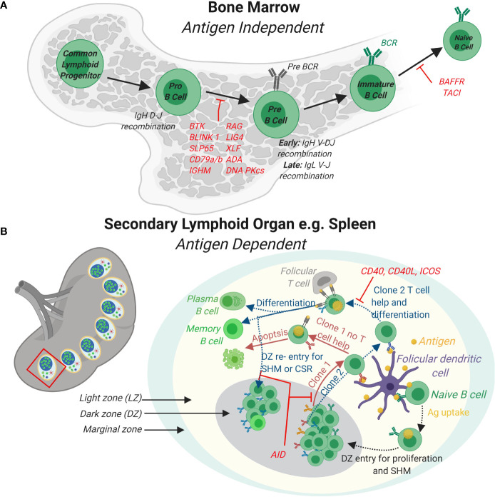
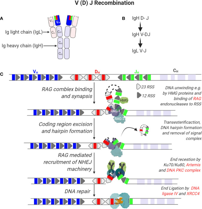
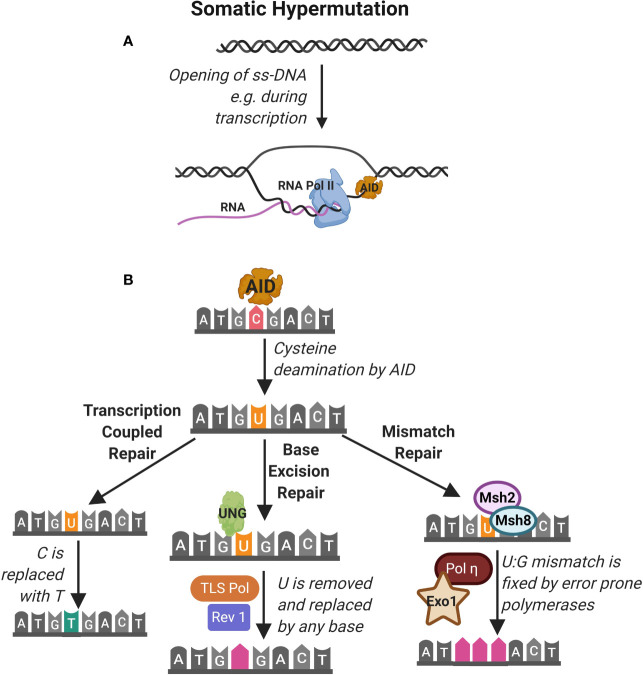
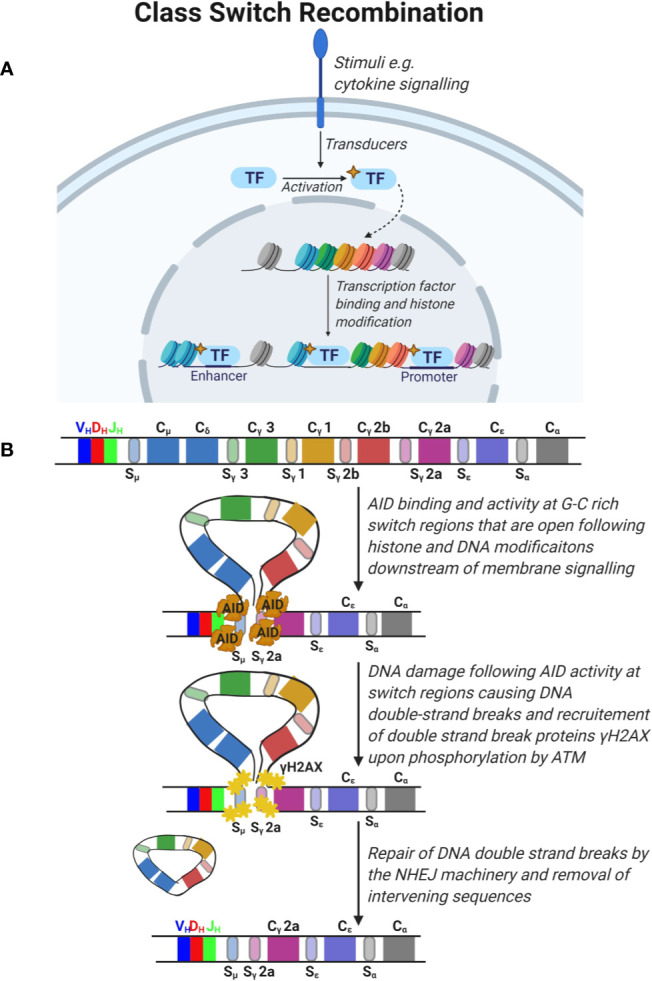
Congenital defects of the immune system called primary immunodeficiency disorders (PID) describe a group of diseases characterized by a decrease, an absence, or a malfunction of at least one part of the immune system. As a result, PID patients are more prone to develop life-threatening complications, including cancer. PID currently include over 400 different disorders, however, the variety of PID-related cancers is narrow. We discuss here reasons for this clinical phenotype. Namely, PID can lead to cell intrinsic failure to control cell transformation, failure to activate tumor surveillance by cytotoxic cells or both. As the most frequent tumors seen among PID patients stem from faulty lymphocyte development leading to leukemia and lymphoma, we focus on the extensive genomic alterations needed to create the vast diversity of B and T lymphocytes with potential to recognize any pathogen and why defects in these processes lead to malignancies in the immunodeficient environment of PID patients. In the second part of the review, we discuss PID affecting tumor surveillance and especially membrane trafficking defects caused by altered exocytosis and regulation of the actin cytoskeleton. As an impairment of these membrane trafficking pathways often results in dysfunctional effector immune cells, tumor cell immune evasion is elevated in PID. By considering new anti-cancer treatment concepts, such as transfer of genetically engineered immune cells, restoration of anti-tumor immunity in PID patients could be an approach to complement standard therapies.

Keywords: B cells; actin cytoskeleton; cancer; cytotoxic cells; exocytosis; immunological synapse; membrane trafficking; primary immunodeficiencies.


Figures

Figure 1
Figure 1
B cell development in the bone marrow and periphery. (A) In the bone marrow, B cells develop from Common Lymphoid Progenitor cells to naïve B cells expressing a functional B cell receptor (BCR) in a series of developmental steps. Firstly, expression of lymphocyte specific RAG complexes triggers Pro-B cell development by initiating the recombination of the D and J segments of the Immunoglobulin heavy chain (IgH) and expression of surrogate light chains. Further differentiation of Pre-B cells leads to the rearrangement of the complete IgH and also Ig light (IgL) gene. Immature B cells with functional BCRs receive stimulatory signals completing naïve B cell development in the bone marrow. (B) In response to antigens, naïve B cells develop further into effector B cells in peripheral lymphoid organs such as the spleen (shown) or lymph nodes (not shown). In the spleen, germinal centers (GCs) that are specialized structures that contain follicular dendritic cells and T follicular helper cells are formed. A GC contains a light zone (LZ) and a dark zone (DZ). The marginal zone surrounds the GC. Naïve B cells take up antigen presented by follicular dendritic cells in the LZ. BCR signaling induces B cell proliferation in the DZ and triggers AID somatic hyper mutation (SHM) of the BCR. B cells expressing newly formed clones (e.g. clones 1 and 2) re-enter the LZ to take up antigen from follicular DCs and present these via major histocompatibility complex (MHC) II to T follicular helper cells. Clones (e.g. clone 1) that do not receive T cell help via CD40-CD40 L signaling axis fail to differentiate and survive. Clones that receive T cell help (e.g. clone 2) differentiate to antibody secreting plasma cells or long-lived memory cell or re-enter the DZ for further SHM and class switch recombination (CSR). Loss of function mutations for genes involved and that cause PID are indicated in red.
Figure 2
Figure 2
Recombination of immunoglobulin V (variable), D (diversity), and J (joining) segments of immunoglobulin heavy and light chains. (A) Tertiary structure of an antibody indicating two molecules of immunoglobulin heavy (IgH) and light chains (IgL). Each IgH is composed of a variable region (VH) derived from the V(D)J segments of the IgH gene and constant (CH) regions. On the other hand, IgL is composed of a variable region (VL) derived from recombining V and J segments of the IgL gene and smaller constant regions (CL). (B) Sequence of immunoglobin gene segment recombination. (C) The IgH gene is composed of V, D, and J gene segments that are flanked by recombination signal sequences (RSS) that are 23 or 12 nucleotides long. In the first step of IgH recombination, D and J segments access is enabled by unwinding of DNA in these regions by e.g. HMG proteins enabling the binding of a complex of recombination-activating genes 1 and 2 (RAG1/2) randomly to any of these segments. Endonuclease activity by RAG1/2 enables removal of intervening sequences, formation of DNA hairpins and recruitment of the non-homologous end joining DNA repair machinery. Here, hairpins are opened by the activity of Ku70/80, Artemis and DNA PKC complexes while ends are ligated by the activity of XRCC4 and DNA ligase IV.
Figure 3
Figure 3
Somatic hypermutation. (A) Somatic hypermutation of the immunoglobulin heavy chain is mediated by the activity of the enzyme activation-induced cytidine deaminase (AID). AID accesses nucleotides on single strand DNA (ss-DNA) during transcription of RNA e.g. by RNA polymerase II (RNA pol II). (B) AID deaminates cytidine (C) nucleotides converting them to uracil (U). This is identified as DNA damage that can be repaired by (1) transcription coupled repair where the uracil is transcribed as thymidine (T), (2) base excision repair (BER) pathway where Uracil-DNA glycosylase (UNG) detects and excises U leading to replacement by any base by the TLS polymerase and Rev1 and (3) mismatch repair (MMR) pathway where Msh2 and Msh8 identify the U and repair mismatch using error prone Pol and Exo1.
Figure 4
Figure 4
Class switch recombination. (A) Signals from the microenvironment via cytokine signaling are transduced in B cells to activate specific transcription factors which translocate to the nucleus and promote the opening of condensed DNA regions at specific areas and expose the switch regions 5’ of the immunoglobulin constant (C) segments. (B) The constant region of the immunoglobulin gene is composed of constant segments that encode different antibody subclasses. Constant regions are flanked by G-C rich switch regions. Deaminase activity of activation-induced cytidine deaminase (AID) leads to DNA damage at these switch regions triggering the double strand break repair response by ATM which phosphorylates H2AX and recruitment of the non-homologous end joining (NHEJ) machinery. This leads to DNA double strand break repair and removal of intervening sequences.
Figure 5
Figure 5
Regulation of actin filament turnover. (A) Guanine exchange factors such as Vav1 are activated downstream of receptor tyrosine kinase (RTK) signaling. Upon phosphorylation of its SH2 domain, Vav1 undergoes intramolecular reorganization that releases its nucleotide exchanging Dbl homology (DH)-pleckstrin homology (PH). Other important GEFs in immune cells are the DOCK family members (not shown). DOCKs are structurally different from the Vav family members in important ways. Namely, they contain lipid-binding domains (and thus are directly recruited to membranes) and lack DH-PH domains requiring association with the Elmo family of proteins to exchange nucleotides. (B) The activity of typical Rho GTPases is regulated by 1) GTP cycling and 2) intracellular retention by Rho GDI proteins. Rho GEFs such as Vav1, DOCK2 or DOCK8 enhance the exchange of GDP to GTP while Rho GAPs switch off Rho GTPases by enhancing the exchange of GTP with GDP. An important mechanism of signaling specificity is the preference of certain GEFs for certain Rho GTPases e.g. DOCK-2 to Rac, Gef-1 to CDC42 (not shown). (C) Nucleation promoting factors such as WASp are activated by Rho GTPases by relieving intramolecular folding (shown) or as in the case of the NPF WAVE regulatory complex by stabilizing a specific orientation (not shown). Inactive WASp in the cytoplasm is autoinhibited through interactions of its Basic Region (BR) with the Acidic (A) domain as well as its GTPase binding domain (GBD) with the central (C) domain. Interaction with activated CDC42 (through the GBD) and recruitment to the plasma membrane (through PIP2) relieves this inhibited conformation. Through its WH1 domain WASp also interacts with WIP which regulates its activity. (D) Activated membrane-proximal WASp interacts with the Arp2/3 complex to mediate the nucleation of new filaments at a 70° angle by direct binding of its CA domains to Arp2/3 and to profilin and actin through its verprolin (V) domain. Other important actin nucleators are the formins that are also similarly activated by relief of autoinhibitory intramolecular folding following interaction with Rho GTPases downstream of tyrosine kinase signaling (not shown). (E) Receptor tyrosine kinases also activate filament breakdown which is important in maintaining a pool of profilin actin monomers that can be used for de novo actin polymerization as well as feedback regulation of activation. A key protein in actin severing is cofilin which enhances filament severing and breakdown by decorating actin filaments. Cofilin is activated by LIMK mediated phosphorylation.
Figure 6
Figure 6
“Push and pull” model for MTOC polarization at the IS and DAG gradient at the cSMAC in CD8+ T cells. On the left part of the figure, the MTOC is relocated at the IS thanks the joint effort of both dynein and myosin IIA. Upon actin depletion at the cSMAC, dynein is anchored at the synaptic membrane from where it “pulls” on the microtubules to reposition the MTOC closer to the IS. At the same time, myosin IIA “pushes” microtubules from the opposite cell side. The right part of the figure depicts the diacylglycerol (DAG) gradient present at the IS. This gradient, which increases from the outside to the inside of the IS, guides the microtubule-driven MTOC reorientation at the IS. Several key enzymes needed to generate this DAG gradient are described more in detail in the text. Note that a simplified version of the LAT signalosome is depicted. The molecules highlighted in red have been found mutated in some PID.
Figure 7
Figure 7
Terminal transport of lytic granules from the polarized MTOC to the synaptic membrane. The terminal transport of lytic granules from the polarized MTOC to the synaptic membrane differs between CD8+ T cells (on the left) and NK cells (on the right). In CD8+ T cells, lytic granules first converge to the MTOC in a retrograde dynein-dependent process. Then an anterograde transport mediated by Rab27a, kinesin-1 and Slp3 allows lytic granules to cover the small distance separating the polarized MTOC from the membrane. In NK cells, such an anterograde final transport has not been described so far. Instead, lytic granules are first associated with myosin IIA. Myosin IIA later helps lytic granules to navigate through the synaptic actin network by interacting with F-actin. Concomitantly, coronin 1A is required to reduce the density of the actin mesh, thereby facilitating lytic granules approach to the membrane. The molecules highlighted in red have been found mutated in some PID.
Figure 8
Figure 8
Docking, tethering, priming and fusion of lytic granules at the IS. Upon immune cell activation, synthesis of mature lytic granules occurs (step # 1). Several molecules, including LYST and the AP-3 complex, are involved in this process (not shown here). These two proteins are causative in the Chediak-Higashi syndrome and the type 2 Hermansky-Pudlak syndrome, respectively. Once mature lytic granules have reached the IS, sequential steps must take place to guarantee their proper docking, tethering, priming and fusion with the synaptic membrane. Before the arrival of lytic granules, Rab11a+ recycling endosomes fuse with the plasma membrane through a VAMP8-syntaxin 4 interaction (step # 2). Upon this fusion, syntaxin 11/STXBP2 complexes are deposited at the plasma membrane. After newly arrived lytic granules are docked at the IS (step # 3), tethering factors ensure that these granules remain firmly in place. This is mediated by several tethering proteins, such as Munc13-4 and Slp2a (step # 4). Simultaneously, Munc13-4 primes the lytic granules for the final fusion stage. Syntaxin 11/STXBP2 complexes, formerly brought by recycling endosomes, mediate the fusion of lytic granules with the synaptic membrane by interacting with VAMP7. After this fusion step, cytotoxic molecules present within the lytic granules are released within the synaptic cleft (step # 5). Note that the interfacial actin protrusions described in CD8+ T cells by Tamzalit et al. (215) are not shown.

References

    1. Tangye SG, Al-Herz W, Bousfiha A, Chatila T, Cunningham-Rundles C, Etzioni A, et al. Human Inborn Errors of Immunity: 2019 Update on the Classification from the International Union of Immunological Societies Expert Committee. J Clin Immunol (2020) 40(1):24–64. 10.1007/s10875-019-00737-x - DOI - PMC - PubMed
    1. Bousfiha A, Jeddane L, Picard C, Al-Herz W, Ailal F, Chatila T, et al. Human Inborn Errors of Immunity: 2019 Update of the IUIS Phenotypical Classification. J Clin Immunol (2020) 40(1):66–81. 10.1007/s10875-020-00758-x - DOI - PMC - PubMed
    1. de Wit J, Brada RJK, Van Veldhuizen J, Dalm V, Pasmans S. Skin disorders are prominent features in primary immunodeficiency diseases: A systematic overview of current data. Allergy (2019) 74(3):464–82. 10.1111/all.13681 - DOI - PubMed
    1. Haas OA. Primary Immunodeficiency and Cancer Predisposition Revisited: Embedding Two Closely Related Concepts Into an Integrative Conceptual Framework. Front Immunol (2018) 9:3136. 10.3389/fimmu.2018.03136 - DOI - PMC - PubMed
    1. Gallo V, Dotta L, Giardino G, Cirillo E, Lougaris V, D’assante R, et al. Diagnostics of Primary Immunodeficiencies through Next-Generation Sequencing. Front Immunol (2016) 7:466. 10.3389/fimmu.2016.00466 - DOI - PMC - PubMed

Publication types

MeSH terms