Review
. 2020 Oct 12;7(22):2002030.
doi: 10.1002/advs.202002030. eCollection 2020 Nov.

Is it Time for Reviewer 3 to Request Human Organ Chip Experiments Instead of Animal Validation Studies?

Affiliations
Review

Is it Time for Reviewer 3 to Request Human Organ Chip Experiments Instead of Animal Validation Studies?

Donald E Ingber. Adv Sci (Weinh). .

Abstract

For the past century, experimental data obtained from animal studies have been required by reviewers of scientific articles and grant applications to validate the physiological relevance of in vitro results. At the same time, pharmaceutical researchers and regulatory agencies recognize that results from preclinical animal models frequently fail to predict drug responses in humans. This Progress Report reviews recent advances in human organ-on-a-chip (Organ Chip) microfluidic culture technology, both with single Organ Chips and fluidically coupled human "Body-on-Chips" platforms, which demonstrate their ability to recapitulate human physiology and disease states, as well as human patient responses to clinically relevant drug pharmacokinetic exposures, with higher fidelity than other in vitro models or animal studies. These findings raise the question of whether continuing to require results of animal testing for publication or grant funding still makes scientific or ethical sense, and if more physiologically relevant human Organ Chip models might better serve this purpose. This issue is addressed in this article in context of the history of the field, and advantages and disadvantages of Organ Chip approaches versus animal models are discussed that should be considered by the wider research community.

Keywords: microfluidics; microphysiological systems; organoids; organ‐on‐a‐chip; preclinical studies.


Conflict of interest statement

The author has the following potential conflicts: Emulate Inc., equity, consulting, chair of SAB; BOA Biomedical Inc., equity, consulting, chair of SAB, board member; Free Flow Medical Device, equity; SynDevRx, equity; Consortia Rx, equity, board member; Roche, consulting; Astrazeneca, sponsored research; Fulcrum Therapeutics, sponsored research; Kraft Heinz, sponsored research; and inventor of multiple patent applications.

Figures

Figure 1
Figure 1
Potential in vitro replacements for animal testing. Schematics of mice, organoids that grow as small closed spheres which undergo organ‐specific cyto‐ and histo‐differentiation within 3D ECM gels, and microfluidic Organ Chips that may be lined with cells from organoids (dashed line), iPS cell‐derived cells, or primary cells. Organ Chips reconstitute tissue–tissue interfaces between organ‐specific epithelium and endothelium, vascular perfusion, interstitial flow or air–liquid interfaces, and mechanical cues (e.g., by applying cyclic strain via application of suction to hollow side chambers) to mimic breathing or peristalsis motions. Circulating or resident immune cells, connective tissue cells, nerve cells, and other cell types can be integrated into the Organ Chips as needed to recapitulate increasing levels of complexity.
Figure 2
Figure 2
Human Lung Airway Chip recreates airway epithelial architecture and replicates patient responses to cigarette smoke in vitro. A) Still image captured from a video recording from the side of cilia beating on the apical surface of the differentiated airway epithelium on‐chip. B) Scanning electron micrograph of the apical surface of the airway epithelium formed on‐chip showing ciliated cells (blue) and non‐ciliated cells (brown). C) Graph showing changes in interleukin 8 (IL‐8) secretion in Lung Airway Chips lined with epithelial cells isolated from normal or COPD patients, with or without exposure to whole cigarette smoke for 75 min (smoking) (**p < 0.01). D) Heatmap comparing expression of 29 genes associated with cellular oxidation–reduction in lung airway epithelial cells obtained from small airways of different normal human smokers compared with samples obtained from three Lung Airway Chips that were exposed to whole cigarette smoke on‐chip. Note that the patterns of induced and suppressed genes in the Airway Chip mimic those seen in human patients. (B) Reproduced with permission.[ 18 ] Copyright 2016, Springer Nature. (C,D) Reproduced with permission.[ 19 ] Copyright 2016, Cell Press.
Figure 3
Figure 3
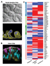
Human Intestine Chips lined by cells isolated from patient‐derived organoids exhibit differentiated structures and functions that closely resemble those displayed by living intestine in vivo. A) Differential interference microscopic image (top) and immunofluorescence microscopic image of F‐actin staining in green (bottom) of vertical sections through an Intestine Chip showing villus protrusions formed by primary intestinal (duodenal) epithelium cultured on‐chip for 12 days. B) Immunofluorescence microscopic image of a vertical section through a human Colon Chip showing a high polarized epithelium with basolateral adherens junctions labeled with E‐cadherin (green) and brush border stained for F‐actin (white) restricted to the apical regions, Hoechst stained nuclei (blue) localized at the cell base, and goblet cells stained for Muc2 (magenta). C) A curated heatmap showing gene expression profiles in the mechanically active Intestine Chip (with fluid flow and cyclic stretching to mimic peristalsis‐like motions) lined by cells from patient‐derived duodenal organoids, the duodenal organoids from which the cells were derived, and duodenum in vivo. Note that the expression of pattern of the Intestine Chip more closely resembles native intestine compared to the organoids. (A,C) Reproduced with permission.[ 26 ] Copyright 2018, Springer Nature. (B) Reproduced with permission.[ 27 ] Copyright 2020, Elsevier Inc.
Figure 4
Figure 4
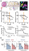
Prediction of clinically observed hematotoxicities at patient‐relevant drug exposures using a human Bone Marrow Chip. A) Left, schematic of bone and insert showing normal human marrow histology. Left middle, schematic of the human Bone Marrow Chip at the time of seeding, showing dispersed CD34+ progenitor cells and bone marrow stromal cells in an ECM gel filling the top channel, and a vascular endothelium beginning to line the adjacent channel. Right middle, within 2 weeks of culture, the endothelium covers all four sides of the lower channel, creating a vascular lumen, and the CD34+ cells undergo expansion and multilineage differentiation. Right, immunofluorescence view of a cross‐section through the ECM gel grown on‐chip for 14 days (yellow, megakaryocyte lineage; magenta, erythroid lineage; blue, neutrophil and other hematopoietic lineages; EC, endothelial cell). B) Effects of treating the Bone Marrow Chips, suspension cultures, and static ECM gel co‐cultures for 48 h with various doses of the cancer drug, 5‐fluorouracil (5‐FU) on total cell number (left) versus only neutrophil lineage cells (right)(***p < 0.001). The range of patient plasma 5‐FU concentrations for a 2 day infusion that are known to cause myelosuppression is indicated in orange. Note that the Bone Marrow Chip replicates this sensitivity to a clinically relevant dose exposure whereas the other in vitro models do not. C) Graphs showing that dynamic changes in concentrations of the cancer drug AZD2811 measured by mass spectrometry in chip outlet (circles), which were used to fit PK models of Bone Marrow Chip drug exposure (black line) for 2 and 48 h infusions. Note that these drug exposure profiles that were generated in the microfluidic Bone Marrow Chip closely resembled plasma levels of AZD2811 in vivo (grey), which were simulated for an average patient at a range of clinical doses based on the known PK characteristics of AZD2811. D) Graphs showing the effects of infusing the Bone Marrow Chips with varying doses of AZD2811 for 2 versus 48 h; total neutrophil (blue) and erythroid (red) cell numbers on day 12 are shown (***p < 0.001). Note that the clinical observation of unusual regimen‐specific neutropenia and anemia with 2 h dosing, but only neutropenia with 48 h dosing, was replicated on‐chip. Reproduced with permission.[ 36 ] Copyright 2020, Springer Nature.
Figure 5
Figure 5
A human Liver Chip recapitulates species‐specific drug toxicities in rat, dog, and human. A) Schematic of the Liver Chip that contains an upper parenchymal channel lined by primary rat, dog, or human hepatocytes grown in ECM sandwich, while species‐specific liver sinusoidal endothelial cells (LSECs), Kupffer cells, and stellate cells are cultured on the opposite side of the same membrane in the lower vascular channel. B) Previously observed species‐specific effects of the drug bosentan on albumin secretion were recreated in microfluidic human, dog, and rat Liver Chips (Chip, open circles) whereas this was not replicated in static hepatocyte sandwich monocultures (Plate, closed triangles). Reproduced with permission.[ 46 ] Copyright 2019, American Association for the Advancement of Science.
Figure 6
Figure 6
Multi‐Organ Chip model of the human neurovascular unit. A) Schematic of the influx Blood–Brain Barrier Chip containing brain microvascular endothelium (magenta) in its vascular channel separated by a porous membrane from brain astrocytes (blue) and pericytes (yellow) in the parenchymal channel through which medium mimicking cerebral spinal fluid (CSF) flows. The CSF fluid is transferred to a similar channel within a Brain Chip that is separated by a porous membrane from a channel containing cultured human brain neuronal cells (green) and astrocytes (blue), and from there to the parenchymal channel of a second efflux Blood–Brain Barrier Chip. Medium mimicking blood is flowed separately through the lower vascular channel. B) 3D confocal microscopic reconstruction of the Blood–Brain Barrier chip viewed from the side showing a continuous endothelium stained for VE‐cadherin (purple) forming a lumen in the lower vascular channel, as well as pericytes (F‐actin, yellow) and astrocytes (GFAP, blue) on the top surface of the porous membrane in the upper channel. C) Confocal fluorescence microscopic view of networks of human brain neurons (β‐III‐tubulin, green) and astrocytes (glial fibrillary astrocytic protein, GFAP, blue) in the lower compartment of the Brain Chip. Reproduced with permission.[ 54 ] Copyright 2018, Springer Nature.
Figure 7
Figure 7
A first‐pass human Body‐on‐Chips model that predicts human PK parameters. A) Top, photographs of the Gut, Liver, and Kidney Chips that were fluidically coupled to each other and to an arterio‐venous mixer reservoir to create the first‐pass PK model in vitro. Bottom, diagrams of the different 2‐channel microfluidic chips with arrows indicating the manner in which the chips and reservoir are fluidically linked to each other and to the AV reservoir (red arrows indicate flow direction). B) Predictions of how nicotine blood concentrations will change over time for three different oral doses (different colored dashed lines) made by a physiological PK model using experimental data obtained from the human Body‐on‐Chips platform compared with previously published blood nicotine levels measured in patients who received orally administered nicotine in the form of nicotine gum (blue), pouched chewing tobacco known as “snus” (black), or loose snus (green) at three different doses (4, 9, and 13–16 mg). Note that the experimental platform was able to quantitatively predict these human PK behaviors. C) Graph showing changes in cisplatin concentrations in blood over time predicted by the same PK model using data from a Body‐on‐Chips configuration containing linked Liver, Kidney, and Bone Marrow Chips along with an arterio‐venous reservoir for infusion periods (dotted lines) of either 1 h (black) or 3 h (blue). Note that these predictions closely match previously published blood cisplatin levels measured in patients who received cisplatin injected intravenously over these same time periods (solid lines). Reproduced with permission.[ 70 ] Copyright 2020, Springer Nature.

References

    1. Akthar A., Camb. Q. Healthc. Ethics 2015, 24, 407. - PMC - PubMed
    1. Meigs L., Smirnova L., Rovida C., Leist M., Hartung T., Altex 2018, 35, 275. - PubMed
    1. Pound P., Ritskes‐Hoitinga M., J.Transl. Med. 2018, 16, 304. - PMC - PubMed
    1. Seok J., Warren H. S., Cuenca A. G., Mindrinos M. N., Baker H. V., Xu W., Richards D. R., McDonald‐Smith G. P., Gao H., Hennessy L., Finnerty C. C., López C. M., Honari S., Moore E. E., Minei J. P., Cuschieri J., Bankey P. E., Johnson J. L., Sperry J., Nathens A. B., Billiar T. R., West M. A., Jeschke M. G., Klein M. B., Gamelli R. L., Gibran N. S., Brownstein B. H., Miller‐Graziano C., Calvano S. E., Mason P. H., et al., Proc. Natl. Acad. Sci. U. S. A. 2013, 110, 3507. - PMC - PubMed
    1. Franco R., Cedazo‐Minguez A., Front. Pharmacol 2014, 5, 146. - PMC - PubMed

LinkOut - more resources