. 2020 Nov 6:8:601704.
doi: 10.3389/fbioe.2020.601704. eCollection 2020.

Hydrogels for 3D Neural Tissue Models: Understanding Cell-Material Interactions at a Molecular Level

Affiliations

Hydrogels for 3D Neural Tissue Models: Understanding Cell-Material Interactions at a Molecular Level

Catalina Vallejo-Giraldo et al. Front Bioeng Biotechnol. .

Abstract

The development of 3D neural tissue analogs is of great interest to a range of biomedical engineering applications including tissue engineering of neural interfaces, treatment of neurodegenerative diseases and in vitro assessment of cell-material interactions. Despite continued efforts to develop synthetic or biosynthetic hydrogels which promote the development of complex neural networks in 3D, successful long-term 3D approaches have been restricted to the use of biologically derived constructs. In this study a poly (vinyl alcohol) biosynthetic hydrogel functionalized with gelatin and sericin (PVA-SG), was used to understand the interplay between cell-cell communication and cell-material interaction. This was used to probe critical short-term interactions that determine the success or failure of neural network growth and ultimately the development of a useful model. Complex primary ventral mesencephalic (VM) neural cells were encapsulated in PVA-SG hydrogels and critical molecular cues that demonstrate mechanosensory interaction were examined. Neuronal presence was constant over the 10 day culture, but the astrocyte population decreased in number. The lack of astrocytic support led to a reduction in neural process outgrowth from 24.0 ± 1.3 μm on Day 7 to 7.0 ± 0.1 μm on Day 10. Subsequently, purified astrocytes were studied in isolation to understand the reasons behind PVA-SG hydrogel inability to support neural network development. It was proposed that the spatially restrictive nature (or tight mesh size) of PVA-SG hydrogels limited the astrocytic actin polymerization together with a cytoplasmic-nuclear translocation of YAP over time, causing an alteration in their cell cycle. This was confirmed by the evaluation of p27/Kip1 gene that was found to be upregulated by a twofold increase in expression at both Days 7 and 10 compared to Day 3, indicating the quiescent stage of the astrocytes in PVA-SG hydrogel. Cell migration was further studied by the quantification of MMP-2 production that was negligible compared to 2D controls, ranging from 2.7 ± 2.3% on Day 3 to 5.3 ± 2.9% on Day 10. This study demonstrates the importance of understanding astrocyte-material interactions at the molecular level, with the need to address spatial constraints in the 3D hydrogel environment. These findings will inform the design of future hydrogel constructs with greater capacity for remodeling by the cell population to create space for cell migration and neural process extension.

Keywords: 3D models; cell interaction; hydrogels; mechanosensory; neural tissue engineering; primary neuroprogenitors.


Figures

FIGURE 1
FIGURE 1
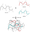
Schematic of the photopolymerization of PVA-SG hydrogels with visible light. Adapted with permission from Lim et al. (2013).
FIGURE 2
FIGURE 2
Physical characterization of 10 wt% PVA-SG hydrogels. (A) The mass swelling ratio of PVA-SG hydrogels increased at a steady rate, reaching a value of 18.9 ± 3.0 on day 28. This behavior was accompanied to a hydrolytic mass loss of the hydrogels of 11.8 ± 3.0% over 28 days after the initial sol fraction (B). An increase over time of the PVA-SG hydrogel mesh size was in line with the swelling and degradation behavior of the hydrogels during 28 days (C). N = 3.
FIGURE 3
FIGURE 3
Mechanical characterization of 10 wt% PVA-SG hydrogels. (A) AFM representative images of PVA-SG hydrogel surface right after photopolymerization (Day 0) and after 28 days of incubation in DPBS at 37°C (Day 28), respectively. The physical changes were quantified with roughness values of 40.11 ± 0.55 nm and 7.97 ± 0.11 nm on each of these days. (B) Young’s moduli of PVA-SG hydrogels obtained in wet condition at different time points showing the decrease of stiffness over time in line with the hydrogel physical behavior. *p < 0.05, N = 3.
FIGURE 4
FIGURE 4
Survival and growth of VM mixed cell population in 10 wt% PVA-SG hydrogels. (A) Fluorescent images of VM neural precursor cells encapsulated in PVA-SG hydrogels. Neurons are shown in red (β-tubulin III), astrocyte cells in green (anti-GFAP) and nuclei are visualized in blue (Hoechst). Scale bar = 50 μm, objective 63 × magnification. Cell density (%) analysis of astrocytes and neurons presence on each time point (days 3, 7, and 10) is presented in (B). An overall decrease in viability of astrocytes density was observed over time in the PVA-SG hydrogels, with a maintained neural presence. However, this neural presence was not accompanied by a healthy neurite outgrowth as depicted in (C). Neural length analysis showed a significant decrease in neurite outgrowth by day 10. *p < 0.05, N = 3.
FIGURE 5
FIGURE 5
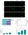
YAP and F-Actin expression of primary astrocytes encapsulated in 10 wt% PVA-SG hydrogels. (A) Fluorescent images of primary astrocytes in PVA-SG hydrogels. YAP expression is visualized in red, F-Actin in green, and nuclei are shown in blue (Hoechst). Scale bar = 50 μm, objective 63× magnification. (B) YAP localization was evaluated during 10 days and showed a cytoplasmic-nuclear translocation over this time, expression that was correlated with the cell area and circularity of the astrocytes shown in (C). The overtime cytoplasmic-nuclear translocation of YAP, correlated with the poor development of F-Actin and circular morphometric shapes of astrocytes encapsulated in the PVA-SG hydrogels. These unhealthy astrocytic shapes were confirmed with the low MMP-2 production of primary astrocytes encapsulated in the hydrogels shown in (D). Together, the functional expression of p27/Kip1 increased over time suggesting cell arrest and the poor migration and spreading of the astrocytes (E). *p < 0.05, N = 3.

References

    1. Aregueta-Robles U. A. (2017). Engineering a Living Electrode: Growth of Neural Networks Within a 3D Hydrogel.
    1. Aregueta-Robles U. A., Lim K. S., Martens P. J., Lovell N. H., Poole-Warren L. A., Green R. (2015). Producing 3D neuronal networks in hydrogels for living bionic device interfaces. Conf. Proc. IEEE Eng. Med. Biol. Soc. 2015 2600–2603. 10.1109/embc.2015.7318924 - DOI - PubMed
    1. Aregueta-Robles U. A., Martens P. J., Poole-Warren L. A., Green R. A. (2019). Tissue engineered hydrogels supporting 3D neural networks. Acta Biomater. 95 269–284. 10.1016/j.actbio.2018.11.044 - DOI - PubMed
    1. Aregueta-Robles U. A., Woolley A. J., Poole-Warren L. A., Lovell N. H., Green R. A. (2014). Organic electrode coatings for next-generation neural interfaces. Front. Neuroeng. 7:15. 10.3389/fneng.2014.00015 - DOI - PMC - PubMed
    1. Aregueta-Robles U. A., Martens P. J., Poole-Warren L. A., Green R. A. (2018). Tailoring 3D hydrogel systems for neuronal encapsulation in living electrodes. J. Poly. Sci. Part B 56 273–287. 10.1002/polb.24558 - DOI

LinkOut - more resources