. 2020 Nov 26;15(1):220.
doi: 10.1186/s11671-020-03451-5.

hGC33-Modified and Sorafenib-Loaded Nanoparticles have a Synergistic Anti-Hepatoma Effect by Inhibiting Wnt Signaling Pathway

Affiliations

hGC33-Modified and Sorafenib-Loaded Nanoparticles have a Synergistic Anti-Hepatoma Effect by Inhibiting Wnt Signaling Pathway

Jing Shen et al. Nanoscale Res Lett. .

Abstract

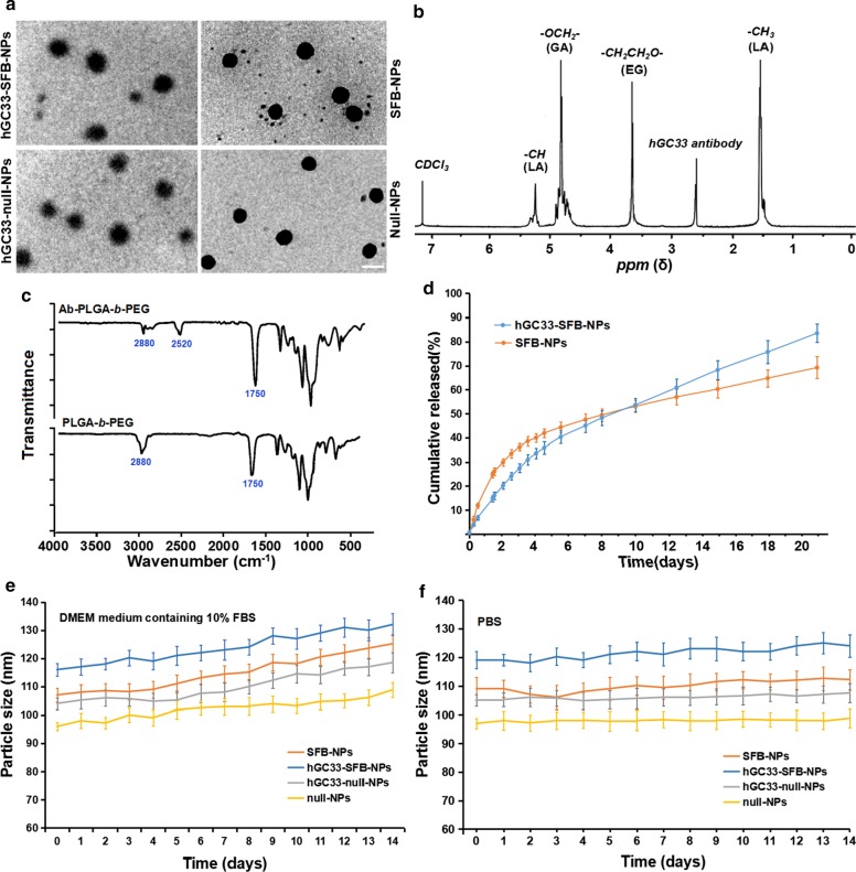
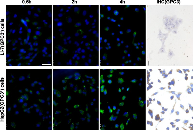
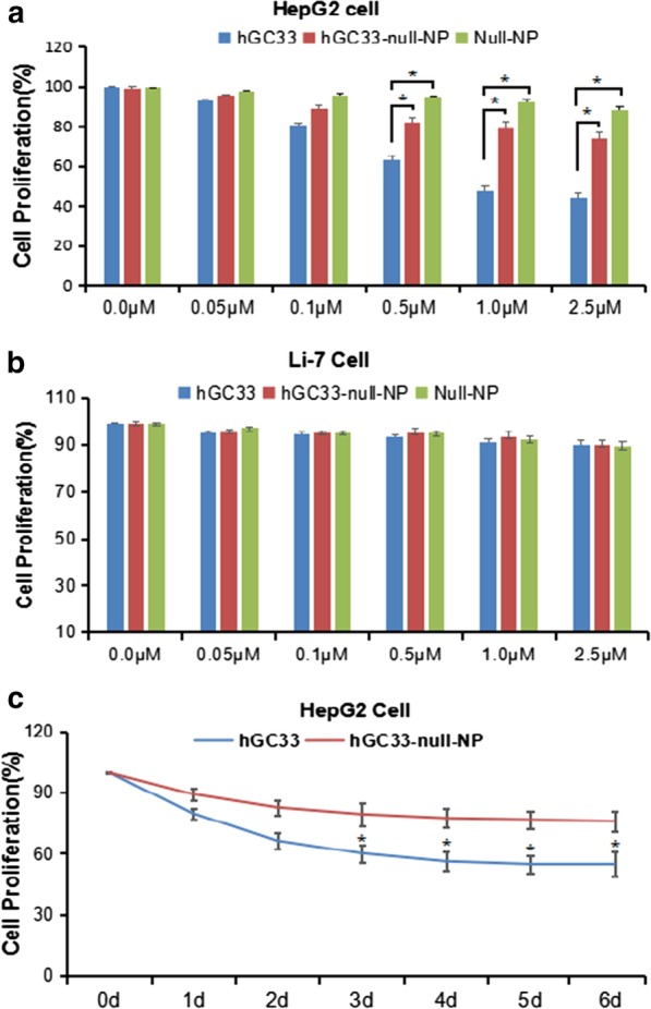
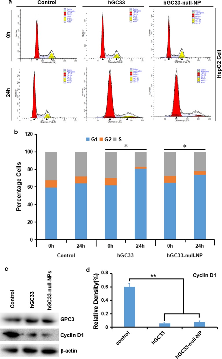
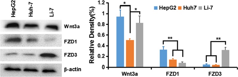
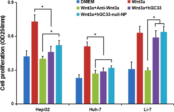
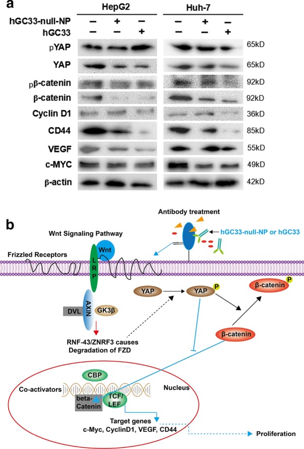
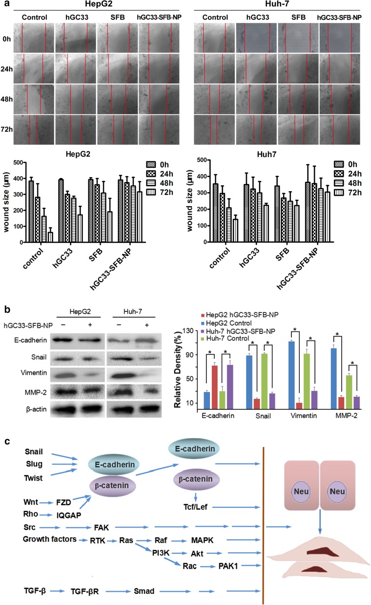
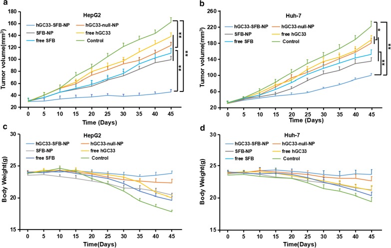
Delivery of tumor-specific inhibitors is a challenge in cancer treatment. Antibody-modified nanoparticles can deliver their loaded drugs to tumor cells that overexpress specific tumor-associated antigens. Here, we constructed sorafenib-loaded polyethylene glycol-b-PLGA polymer nanoparticles modified with antibody hGC33 to glypican-3 (GPC3 +), a membrane protein overexpressed in hepatocellular carcinoma. We found that hGC33-modified NPs (hGC33-SFB-NP) targeted GPC3+ hepatocellular carcinoma (HCC) cells by specifically binding to GPC3 on the surface of HCC cells, inhibited Wnt-induced signal transduction, and inhibited HCC cells in G0/1 by down-regulating cyclin D1 expression, thus attenuating HCC cell migration by inhibiting epithelial-mesenchymal transition. hGC33-SFB-NP inhibited the migration, cycle progression, and proliferation of HCC cells by inhibiting the Ras/Raf/MAPK pathway and the Wnt pathway in tandem with GPC3 molecules, respectively. hGC33-SFB-NP inhibited the growth of liver cancer in vivo and improved the survival rate of tumor-bearing mice. We conclude that hGC33 increases the targeting of SFB-NP to HCC cells. hGC33-SFB-NP synergistically inhibits the progression of HCC by blocking the Wnt pathway and the Ras/Raf/MAPK pathway.

Keywords: Glypican-3; Hepatocellular carcinoma; Targeted therapy; Wnt signal.


Conflict of interest statement

The authors declare that they have no competing interests.

Figures

Fig. 1
Fig. 1
Characterization of NPs. a TEM characterization of NPs, the scale bar indicates 100 nM; b the 1H NMR spectra of synthetic hGC33-PLGA-b-PEG in CDCl3; c FTIR spectra of hGC33-PEG-b-PLGA and PEG-b- PLGA; d cumulative release profiles of SFB-NP and hGC33-SFB-NP in PBS (pH = 7.4) at 37 °C; e size changes of NPs incubated in DMEM medium containing 10% FBS over 14 d; f size changes of NPs incubated in PBS over 14 d. SFB, sorafenib; TEM, transmission electron microscopy; NPs, nanoparticles; 1H NMR, 1H-nuclear magnetic resonance spectroscopy; FTIR, Fourier transform infrared spectroscopy
Fig. 2
Fig. 2
Expression of GPC3 and uptake of hGC33-coumarin6-NP in Li-7 and HepG2 cells. The cells, inoculated in the culture plate, were washed with PBS and incubated with 100 μg/ml hGC33-coumarin6-NP in DMEM for 2, 4, and 8 h. Nuclei were stained with DAPI, and the cells were fixed and detected by confocal laser scanning microscopy. GPC3 was not detected in Li-7 cells, but was highly expressed in HepG2 cells as detected by immunocytochemistry. The scale bar indicates 50 μM
Fig. 3
Fig. 3
The proliferation of HepG2 and Li-7 cells was inhibited by hGC33, null-NP, and hGC33-null-NP. a Cell proliferation test was performed on GPC3-positive HepG2 cells treated with hGC33, null-NP or hGC33-null-NP; b cell proliferation tests were conducted on GPC3 negative Li-7 cells treated with hGC33, null-NP, and hGC33-null-NP. The cells were incubated with 0–2.5 μM hGC33, null-NP, or hGC33-null-NP for 1 day. The proliferation of the cells was measured with MTT method and standardized as untreated cells. All values represent the mean ± SD. Compared with the control group (0 μM) without antibody treatment, *P < 0.01; c the representative results of response of HepG2 to hGC33, null-NP, and hGC33-null-NP in hGC33 treatment (1.0 μM)
Fig. 4
Fig. 4
hGC33-null-NP and hGC33-induced cell cycle arrest in G1 phase and inhibited cyclinD1 expression in HepG2 cells. a Representative cell–cycle diagram of various groups treated with hGC33-null-NP and hGC33. b Cell cycle analysis of various groups treated with hGC33-null-NP and hGC33. The HCC cells were incubated with 0.5 μm hGC33 or hGC33-null-NP (with the molar concentration of hGC33 as reference). *P < 0.05, the G1 phase of hGC33-null-NP or hGC33 cells was compared with that of 0 h cells. c, d After hGC33-null-NP or hGC33 treatment, cyclinD1 was significantly down regulated in HepG2 cells compared with that in the control group
Fig. 5
Fig. 5
Expression of Wnt3a, FZD1, and FZD3 in HCC cell lines
Fig. 6
Fig. 6
GPC3-activated Wnt signal transduction in HCC. Fifty percent Wnt3a DMEM medium was added with anti-wnt3a antibody, hGC33, or hGC33-null-NP. HepG2 (GPC3++), Huh-7 (GPC3+) and Li-7 (GPC3) cells were co-incubated for 48 h, and cell proliferation was measured by MTT assay. The data were expressed as mean ± SD (*P < 0.01)
Fig. 7
Fig. 7
Inhibition of Wnt3a-induced β-catenin signaling by hGC33-null-NP or hGC33. a Compared with the control group, Wnt/β-catenin signaling pathway in HepG2 and Huh-7 cells treated with hGC33-null-NP or hGC33 was inhibited, and the levels of β-catenin and YAP were decreased, while the increased phosphorylated β-catenin and phosphorylated YAP molecules were unstable, and degraded in cytoplasm. The decreased β-catenin was difficult to maintain in the nucleus and drive the expression of CyclinD1, CD44, VEGF, and c-MYC, which resulted in the decrease of cyclinD1, CD44, VEGF, and c-myc protein levels. b The mechanism pattern of Wnt/β-catenin signaling pathway inhibited by hGC33-null-NP or hGC33
Fig. 8
Fig. 8
Effect of hGC33-SFB-NP on EMT inhibition. a Compared with the control group, the hGC33-SFB-NP treatment group had less cell migration. Photographs were taken under an optical microscope (magnification, × 200). The error value represents the standard deviation of three independent experiments. *Compared with the control group, p < 0.01. b Compared with the control group, the EMT-related proteins snail, vimentin, and MMP-2 in HCCs treated with hGC33-SFB-NP decreased, whereas E-cadherin increased. c Molecular mechanism of EMT. EMT, epithelial–mesenchymal transition; MMP-2, matrix metalloproteinase-2; SFB, sorafenib; NP, nanoparticle
Fig. 9
Fig. 9
The effect of hGC33-SFB-NP on xenotransplantation of HCC in nude mice and the changes of mice weight. Liver cancer cells were inoculated subcutaneously on the back of each nude mouse (n = 10). After 10 days, the tumor bearing mice were treated with PBS (control), free hGC33, free SFB, hGC33-null-NP, SFB-NP, and hGC33-SFB-NP. Tumor size (a, b) and body weight (c, d) of mice were monitored at designated time points

Similar articles

Cited by

References

    1. Mingqian F, Wei G, Ruoqi W, Weizao C, Yan-Gao M, William DF, Xin WW, Dimiter SD, Mitchell H. Therapeutically targeting glypican-3 via a conformation-specific single-domain antibody in hepatocellular carcinoma. Proc Natl Acad Sci USA. 2013;110(12):E1083–E1091. doi: 10.1073/pnas.1217868110. - DOI - PMC - PubMed
    1. Gao W, Tang Z, Zhang Y, Feng M, Qian M, Dimitrov DS, Ho M. Immunotoxin targeting glypican-3 regresses liver cancer via dual inhibition of Wnt signaling and protein synthesis. Nat Commun. 2015;6:6536. doi: 10.1038/ncomms7536. - DOI - PMC - PubMed
    1. Kolluri A, Ho M. The role of glypican-3 in regulating Wnt, YAP, and hedgehog in liver cancer. Front Oncol. 2019;9:708. doi: 10.3389/fonc.2019.00708. - DOI - PMC - PubMed
    1. Sirisomboonlarp K, Udomsinprasert W, McConachie E, Woraruthai T, Poovorawan Y, Honsawek S. Increased serum glypican-3 is associated with liver stiffness and hepatic dysfunction in children with biliary atresia. Clin Exp Hepatol. 2019;5(1):48–54. doi: 10.5114/ceh.2019.83156. - DOI - PMC - PubMed
    1. Li N, Gao W, Zhang Y-F, Ho M. Glypicans as cancer therapeutic targets. Trends Cancer. 2018;4(11):741–754. doi: 10.1016/j.trecan.2018.09.004. - DOI - PMC - PubMed