Review
. 2021 Mar;78(6):2459-2483.
doi: 10.1007/s00018-020-03710-9. Epub 2020 Nov 26.

Modeling different types of diabetes using human pluripotent stem cells

Affiliations
Review

Modeling different types of diabetes using human pluripotent stem cells

Essam M Abdelalim. Cell Mol Life Sci. 2021 Mar.

Abstract

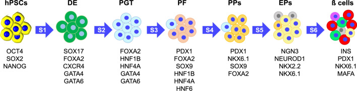
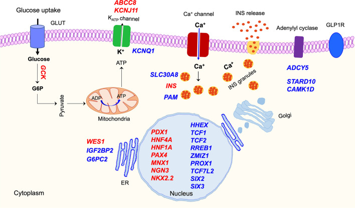
Diabetes mellitus (DM) is a metabolic disease characterized by chronic hyperglycemia as a result of progressive loss of pancreatic β cells, which could lead to several debilitating complications. Different paths, triggered by several genetic and environmental factors, lead to the loss of pancreatic β cells and/or function. Understanding these many paths to β cell damage or dysfunction could help in identifying therapeutic approaches specific for each path. Most of our knowledge about diabetes pathophysiology has been obtained from studies on animal models, which do not fully recapitulate human diabetes phenotypes. Currently, human pluripotent stem cell (hPSC) technology is a powerful tool for generating in vitro human models, which could provide key information about the disease pathogenesis and provide cells for personalized therapies. The recent progress in generating functional hPSC-derived β cells in combination with the rapid development in genomic and genome-editing technologies offer multiple options to understand the cellular and molecular mechanisms underlying the development of different types of diabetes. Recently, several in vitro hPSC-based strategies have been used for studying monogenic and polygenic forms of diabetes. This review summarizes the current knowledge about different hPSC-based diabetes models and how these models improved our current understanding of the pathophysiology of distinct forms of diabetes. Also, it highlights the progress in generating functional β cells in vitro, and discusses the current challenges and future perspectives related to the use of the in vitro hPSC-based strategies.

Keywords: Diabetes; Genome editing; Insulin; Pathogenesis; Precision medicine; hPSCs; β cells.


Conflict of interest statement

The author declares that this article content has no conflict of interest.

Figures

Fig. 1
Fig. 1
Schematic representation of the common hPSC-based approaches for modeling different types of diabetes. a Modeling of monogenic diabetes (MD) using hPSCs. iPSCs could be derived from a patient with MD (patient-iPSCs) with mutations in a single gene, followed by correcting the mutation using genome-editing tools. Another approach is to introduce MD mutations or perform gene-editing into the existing hPSC lines (Ctr-hPSCs). The generated hPSCs could be differentiated into β cells to investigate the effect of the mutation or the edited gene on β cell development and functionality. A pure population of β cells can be isolated using the surface marker, CD49a, for further analyses. b Modeling GWAS-identified genes associated with type 1 diabetes (T1D) and type 2 diabetes (T2D) using hPSCs. GWAS-identified genes can be introduced into the existing hPSC lines to generate isogenic hPSCs. Gene-edited hPSCs and their isogenic hPSC controls could be differentiated into cells relevant to the edited gene, which are β cells and/or insulin-target cells (T2D) or β cells and/or immune cells (T1D). c Modeling T1D, T2D, and MD using patient-derived iPSCs and compare them with iPSCs generated from healthy individuals (Ctr-iPSCs). Patient-derived iPSCs and Ctr-iPSCs could be differentiated into cell types described in b based on the type of diabetes. hPSC-derived target cells can be examined using different functional assays and genomic approaches. These approaches can lead to novel insights into the molecular mechanisms underlying the development of different types of diabetes. Also, hPSC-derived target cells can be used for a high-throughput drug screening to discover new treatments. Successful modeling of different types of diabetes will pave the way toward the personalized treatment
Fig. 2
Fig. 2
Overview of hPSC differentiation into different stages of pancreatic β cell development in vitro. hPSCs are differentiated into β cells through six or seven stages. Each stage is characterized by the expression of key transcription factors (TFs) regulating β cell development and functionality. hPSC-derived β cell stage always contains a heterogenous population of islet cells (β, α, and δ) and progenitor cells. hPSCs human pluripotent stem cells, DE definitive endoderm, PGT primitive gut tube, PF posterior foregut, PPs pancreatic progenitors, EPs endocrine progenitors
Fig. 3
Fig. 3
Schematic diagram of the human pancreatic β cells showing the localization of genes associated with diabetes. Transcripts identified through GWAS are shown in blue (ADCY5, CAMK1D, G6PC2, PAM, RREB1, SLC30A8, MTNR1B, KCNQ1, PROX1, STARD10, TCF7L2, ZMIZ1, SIX2, and SIX3). Transcripts harboring rare variants associated with monogenic diabetes (MD) are shown in red (ABCC8, KCNJ11, GCK, HNF1A, HNF4A, PAX4, PDX1, MNX1, NGN3, NKX2.2, INS, and WSF1). Golgi Golgi apparatus, ER endoplasmic reticulum

References

    1. Atkinson MA, von Herrath M, Powers AC, Clare-Salzler M. Current concepts on the pathogenesis of type 1 diabetes–considerations for attempts to prevent and reverse the disease. Diabetes Care. 2015;38(6):979–988. doi: 10.2337/dc15-0144. - DOI - PMC - PubMed
    1. Hanafusa T, Imagawa A. Fulminant type 1 diabetes: a novel clinical entity requiring special attention by all medical practitioners. Nat Clin Pract Endocrinol Metab. 2007;3(1):36–45. doi: 10.1038/ncpendmet0351. - DOI - PubMed
    1. Kahn SE, Zraika S, Utzschneider KM, Hull RL. The beta cell lesion in type 2 diabetes: there has to be a primary functional abnormality. Diabetologia. 2009;52(6):1003–1012. doi: 10.1007/s00125-009-1321-z. - DOI - PMC - PubMed
    1. Mathis D, Vence L, Benoist C. β-Cell death during progression to diabetes. Nature. 2001;414(6865):792–798. doi: 10.1038/414792a. - DOI - PubMed
    1. Donath MY, Halban PA. Decreased beta-cell mass in diabetes: significance, mechanisms and therapeutic implications. Diabetologia. 2004;47(3):581–589. doi: 10.1007/s00125-004-1336-4. - DOI - PubMed

Substances