. 2021 Feb:181:25-33.
doi: 10.1016/j.biochi.2020.11.018. Epub 2020 Nov 23.

X-aptamers targeting Thy-1 membrane glycoprotein in pancreatic ductal adenocarcinoma

Affiliations

X-aptamers targeting Thy-1 membrane glycoprotein in pancreatic ductal adenocarcinoma

Hongyu Wang et al. Biochimie. 2021 Feb.

Abstract

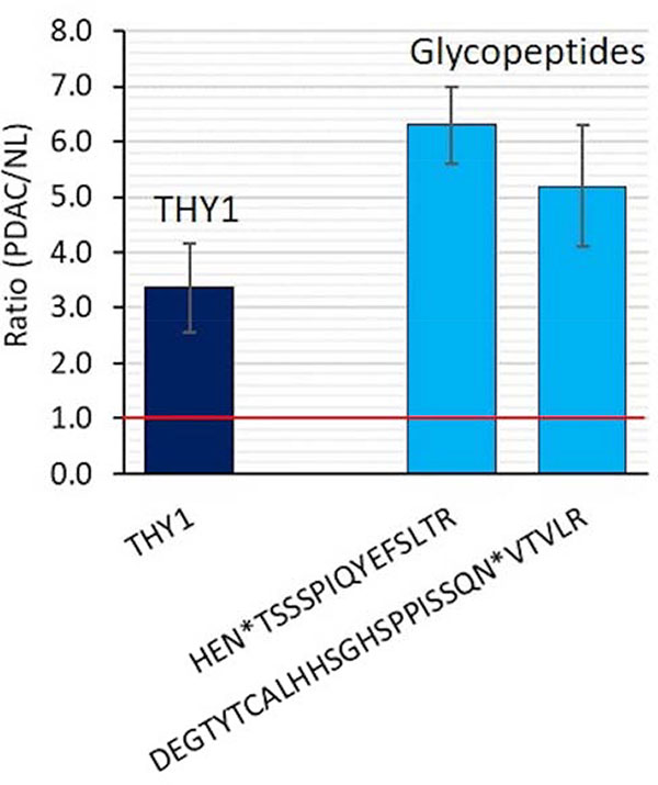
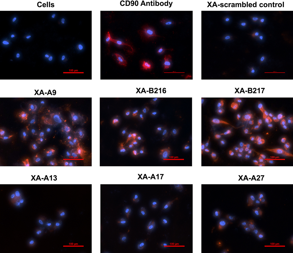
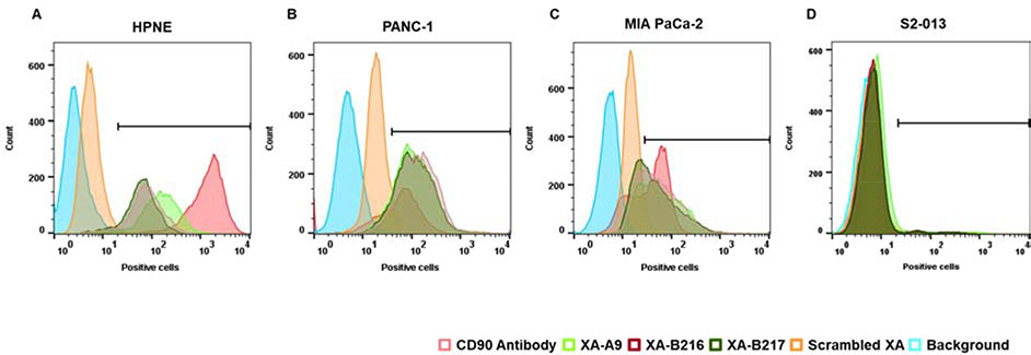
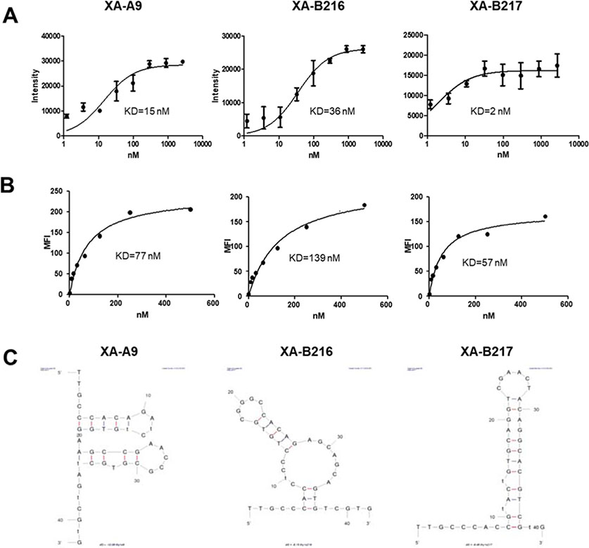
Modified DNA aptamers incorporated with amino-acid like side chains or drug-like ligands can offer unique advantages and enhance specificity as affinity ligands. Thy-1 membrane glycoprotein (THY1 or CD90) was previously identified as a biomarker candidate of neovasculature in pancreatic ductal adenocarcinoma (PDAC). The current study developed and evaluated modified DNA X-aptamers targeting THY1 in PDAC. The expression and glycosylation of THY1 in PDAC tumor tissues were assessed using immunohistochemistry and quantitative proteomics. Bead-based X-aptamer library that contains 108 different sequences was used to screen for high affinity THY1 X-aptamers. The sequences of the X-aptamers were analyzed with the next-generation sequencing. The affinities of the selected X-aptamers to THY1 were quantitatively evaluated with flow cytometry. Three high affinity THY1 X-aptamers, including XA-B217, XA-B216 and XA-A9, were selected after library screening and affinity binding evaluation. These three X-aptamers demonstrated a high binding affinity and specificity to THY1 protein and the THY1 expressing cell lines, using THY1 antibody as a comparison. The development of these X-aptamers provides highly specific and non-immunogenic affinity ligands for THY1 binding in the context of biomarker development and clinical applications. They could be further exploited to assist molecular imaging of PDAC targeting THY1.

Keywords: Aptamer; Pancreatic cancer; Proteomics; Thy-1 membrane glycoprotein (THY1 or CD90).


Conflict of interest statement

Declaration of competing interest The authors declare that they have no known competing financial interests or personal relationships that could have appeared to influence the work reported in this paper.

Figures

Figure 1.
Figure 1.
THY1 expression and N-linked glycosylation in PDAC tissues indicated by proteomics. THY1 was overexpressed (the left bar) and heavily glycosylated on N-linked glycosylation sites (the two right bars) in PDAC tissues compared to normal controls. The two blue bars on the right represent the changes in abundance of the glycopeptides derived from THY1 in PDAC tissue compared to normal pancreas.
Figure 2.
Figure 2.
THY1 expression in PDAC tissues (in comparison to KDR), PanIN lesions and PDBu-stimulated HUVEC cells. A) IHC staining of THY1 and KDR on 12 PDAC tissue slides. The vascular staining of THY1 was positive on 10 out of 12 PDAC samples, whereas the KDR staining was positive on half of the 12 PDAC samples. B) The vascular staining of THY1 was increased substantially in PanIN 2 & 3, compared to PanIN1. C) Florescence images of THY1 expression on HUVEC and PDBu-stimulated HUVEC cells. THY1 expression was visually increased after cancer stimulation by PDBu.
Figure 3.
Figure 3.
Level of THY1 expression on various human pancreatic cancer cell lines. Human pancreatic cancer cell lines (HPNE, PANC-1, MIA PaCa-2, and S2–013) were examined for level of THY1 expression using anti-human THY1 antibody. Percentage of positive cells from flow cytometric immunofluorescence histograms is presented.
Figure 4.
Figure 4.
Scheme of the X-aptamer selection method.
Figure 5.
Figure 5.
The structure and linkage chemistry of modifications X, Y, W.
Figure 6.
Figure 6.
Screening of binding affinity of individual synthesized XA-THY1s. Biotin conjugated XA-THY1s (500 nM) were incubated with human THY1 expressing HPNE cells. Anti-human THY1 antibody was used as positive control for specific expression of THY1 on HPNE cells. The relative extent of XA binding to the HPNE cells was assessed by fluorescence microscopy analysis using a Nikon Eclipse TE2000-E inverted microscope (Nikon Instruments Inc., Melville, NY). Both XA-THY1s and anti-human THY1 antibody were labeled with red fluorescence. Hoechst 33342 (Thermo Fisher Scientific) was used to counterstain nuclei (blue).
Figure 7.
Figure 7.
Binding affinity and specificity of selected XAs. Binding of selected XA-A9, XA-B216 and XA-B217 with various pancreatic cancer cell lines were analyzed by flow cytometry. All three XAs demonstrated high binding affinity and specificity to the THY1 expressing HPNE cells, using the binding activities of THY1 antibody (98%) and scrambled aptamers (1%) as positive and negative control, respectively (A). No binding was observed with THY1 negative pancreatic cancer S2–013 cells with those selected XAs (D). Further validation with other pancreatic cancer cell lines confirmed these selected XAs had similar binding affinity to PANC-1 (B) and MIA PaCa-2 cells (C) compared to THY1 antibody binding.
Figure 8.
Figure 8.
Binding affinity of selected XAs and their equilibrium dissociation constant. Filter-binding assays were performed with the biotinylated XAs and purified THY1 protein. Saturation binding curves were generated and the equilibrium dissociation constants, Kd, were calculated from the equation Y = Bmax X/(Kd + X), assuming a single binding site (A). Biotin-conjugated THY1 specific XAs were incubated with THY1 expressing HPNE cells at various concentrations and their binding affinity were analyzed by fluorescence intensity with flow cytometry analysis. The equilibrium dissociation constant (Kd) was obtained by fitting the dependence of fluorescence intensity of specific binding on the concentration of the X-aptamers to the equation (B). Secondary structures of the selected XA-A9, XA-B216 and XA-B217 were predicted with the MFold program (C).

References

    1. Saalbach A, et al., The Thy-1/Thy-1 ligand interaction is involved in binding of melanoma cells to activated Thy-1- positive microvascular endothelial cells. Microvasc Res, 2002. 64(1): p. 86–93. - PubMed
    1. Hermann PC, et al., Distinct populations of cancer stem cells determine tumor growth and metastatic activity in human pancreatic cancer. Cell Stem Cell, 2007. 1(3): p. 313–23. - PubMed
    1. Pan S, et al., Proteomics portrait of archival lesions of chronic pancreatitis. PLoS One, 2011. 6(11): p. e27574. - PMC - PubMed
    1. Zhu J, et al., Overexpression of CD90 (Thy-1) in pancreatic adenocarcinoma present in the tumor microenvironment. PLoS One, 2014. 9(12): p. e115507. - PMC - PubMed
    1. Foygel K, et al., Detection of pancreatic ductal adenocarcinoma in mice by ultrasound imaging of thymocyte differentiation antigen 1. Gastroenterology, 2013. 145(4): p. 885–894 e3. - PMC - PubMed

MeSH terms