. 2020 Nov 26;18(1):182.
doi: 10.1186/s12915-020-00918-w.

Paxillin mediates ATP-induced activation of P2X7 receptor and NLRP3 inflammasome

Affiliations

Paxillin mediates ATP-induced activation of P2X7 receptor and NLRP3 inflammasome

Wenbiao Wang et al. BMC Biol. .

Abstract

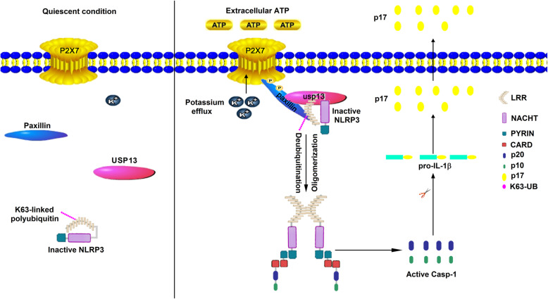
Background: Extracellular adenosine triphosphate (ATP), a key danger-associated molecular pattern (DAMP) molecule, is released to the extracellular medium during inflammation by injured parenchymal cells, dying leukocytes, and activated platelets. ATP directly activates the plasma membrane channel P2X7 receptor (P2X7R), leading to an intracellular influx of K+, a key trigger inducing NLRP3 inflammasome activation. However, the mechanism underlying P2X7R-mediated activation of NLRP3 inflammasome is poorly understood, and additional molecular mediators have not been identified. Here, we demonstrate that Paxillin is the molecule connecting the P2X7 receptor and NLRP3 inflammasome through protein interactions.

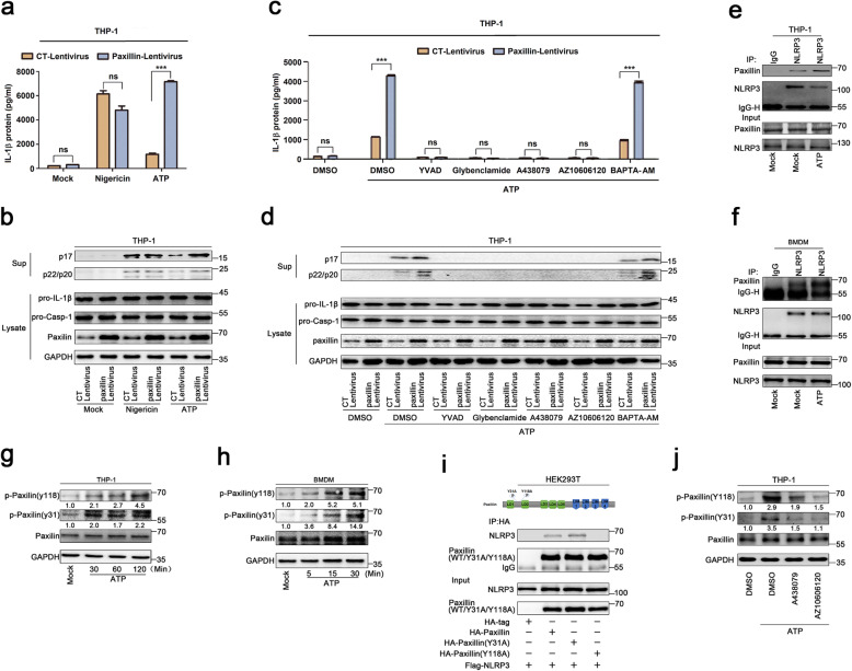
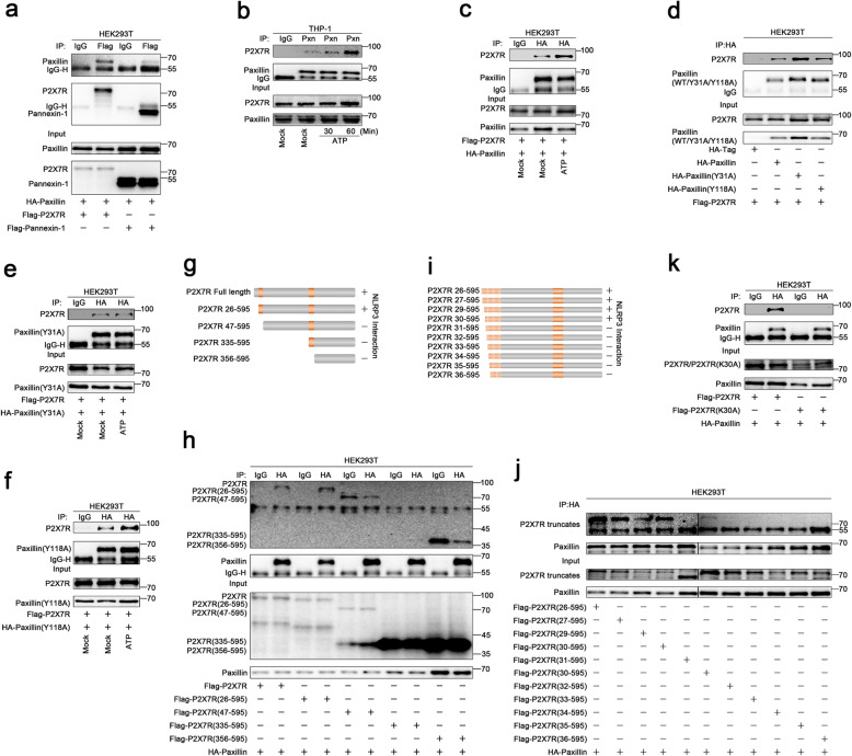
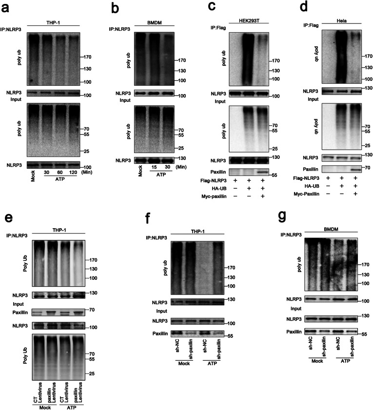
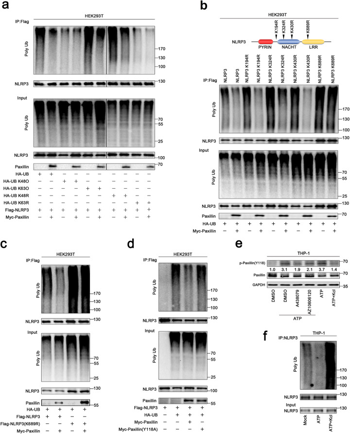
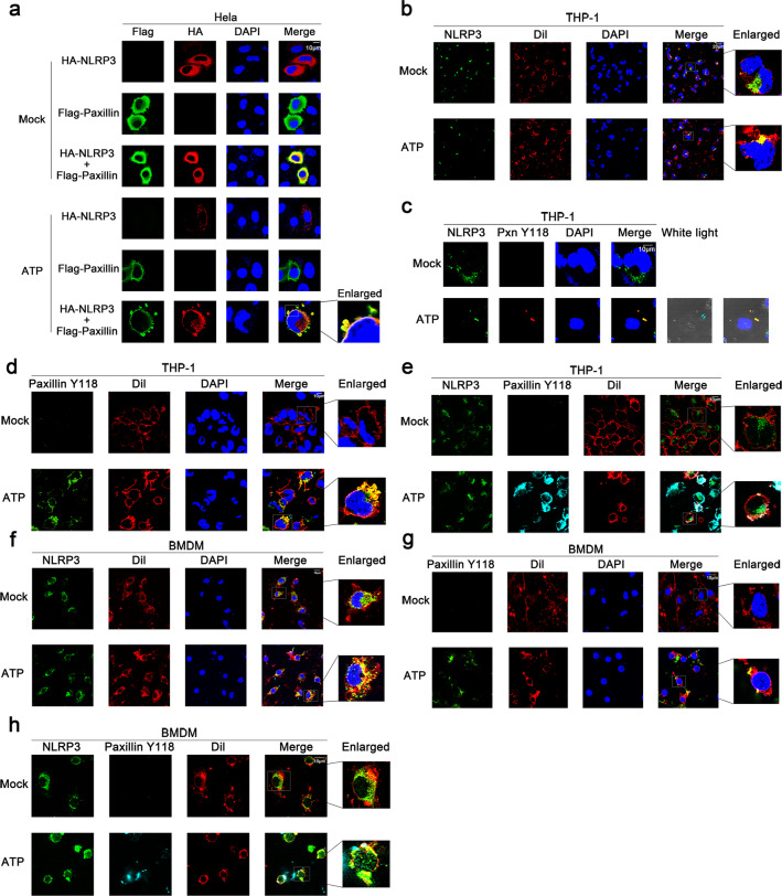
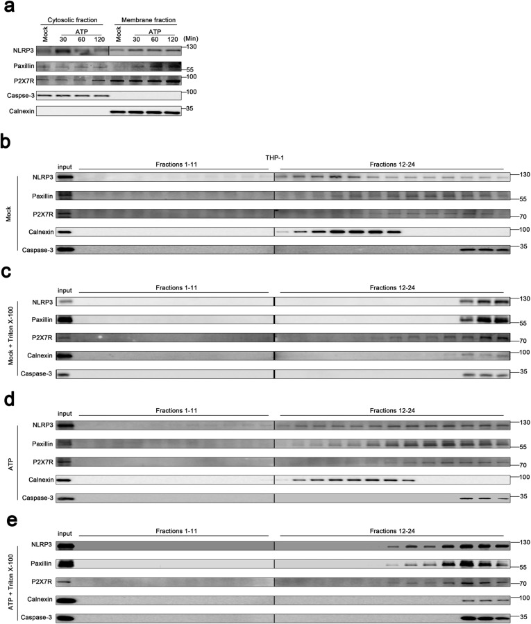
Results: We show a distinct mechanism by which Paxillin promotes ATP-induced activation of the P2X7 receptor and NLRP3 inflammasome. Extracellular ATP induces Paxillin phosphorylation and then facilitates Paxillin-NLRP3 interaction. Interestingly, Paxillin enhances NLRP3 deubiquitination and activates NLRP3 inflammasome upon ATP treatment and K+ efflux. Moreover, we demonstrated that USP13 is a key enzyme for Paxillin-mediated NLRP3 deubiquitination upon ATP treatment. Notably, extracellular ATP promotes Paxillin and NLRP3 migration from the cytosol to the plasma membrane and facilitates P2X7R-Paxillin interaction and PaxillinNLRP3 association, resulting in the formation of the P2X7R-Paxillin-NLRP3 complex. Functionally, Paxillin is essential for ATP-induced NLRP3 inflammasome activation in mouse BMDMs and BMDCs as well as in human PBMCs and THP-1-differentiated macrophages.

Conclusions: We have identified paxillin as a mediator of NLRP3 inflammasome activation. Paxillin plays key roles in ATP-induced activation of the P2X7 receptor and NLRP3 inflammasome by facilitating the formation of the P2X7R-Paxillin-NLRP3 complex.

Keywords: Adenosine triphosphate, ATP; P2X7 receptor; Paxillin; The NACHT, LRR, and PYD domain-containing protein 3, NLRP3; Ubiquitin-specific peptidase 13, USP13.


Conflict of interest statement

The authors have no conflicts of interest to disclose.

Figures

Fig. 1
Fig. 1
Paxillin binds directly to the LRR domain of NLRP3 in the cytosol. a Identification of NLRP3 LRR domain-Paxillin interaction by yeast two-hybrid analysis. Yeast strain AH109 cells were transformed with the combination of BD and AD plasmid, as indicated (1–3). Transformed yeast cells were first grown on the SD-minus Trp/Leu plates for 3 days. The colony of yeast was then streaked on SD-minus Trp/Leu/Ade/His plates (QDO). BD-p53 and AD-T (5) was used as a positive control and BD-Lam and AD-T (4) as a negative control. b HEK293T cells were co-transfected with pHA-Paxillin and pFlag-Vector, pFlag-NLRP3, pFlag-ASC, and pFlag-Casp-1. Lysates were prepared and subjected to IP using an anti-Flag antibody and analyzed by immunoblotting using an anti-HA or anti-Flag antibody (top) or subjected directly to Western blot using an anti-Flag or anti-HA antibody (as input) (bottom). c HEK293T cells were co-transfected with pHA-Paxillin and pFlag-Vector, pFlag-NLRP3, pFlag-PYRIN, pFlag-NACHT, and pFlag-LRR. Lysates were prepared and subjected to IP using an anti-Flag antibody and analyzed by immunoblotting using an anti-HA or anti-Flag antibody (top) or subjected directly to Western blot using an anti-Flag or anti-HA antibody (as input) (bottom). d Extracts from HEK293T cells transfected with Flag-Paxillin were incubated with 10 μg GST proteins or GST-LRR protein that was incubated with glutathione-Sepharose beads. The mixture was washed three times and then analyzed by immunoblotting using anti-Flag and anti-GST antibody (top). Lysates from transfected HEK293T cells were analyzed by immunoblotting using anti-Flag antibody (as input) (bottom). e Extracts from HEK293T cells transfected with Flag-NLRP3 were incubated with 10 μg GST proteins or GST-Paxillin protein which was incubated with glutathione-Sepharose beads. The mixture was washed three times and then analyzed by immunoblotting using anti-Flag and anti-GST antibody (top). Lysates from transfected HEK293T cells were analyzed by immunoblotting using anti-Flag antibody (as input) (bottom). f HeLa cells were transfected with pFlag-Paxillin and pHA-NLRP3 or co-transfected with pFlag-Paxillin and pHA-NLRP3. Subcellular localization of Flag-Paxillin (green), HA-NLRP3 (red), and the nucleus marker DAPI (blue) was examined by confocal microscopy
Fig. 2
Fig. 2
ATP promotes Paxillin phosphorylation and Paxillin-NLRP3 interaction. ad THP-1 cells stably expressing control lentivirus or Paxillin lentivirus were generated and differentiated into macrophages, which were then treated with ATP (5 mM) or Nigericin (2 μM) for 2 h (a, b) and treated with DMSO, YVAD (50 μM), Glybenclamide (25 μg/ml), A438079 (10 μM), AZ10606120 (10 μM), and BAPTA-AM (30 μM) for 1 h before the treatment with ATP (5 mM) for 2 h (c, d). IL-1β levels in supernatants were determined by ELISA (a, c). Mature IL-1β (p17) and cleaved Casp-1 (p22/p20) in supernatants or Paxillin, pro-IL-1β, and pro-Casp-1 in lysates were determined by Western blot (b, d). e TPA-differentiated THP-1 macrophages were treated with DMSO or ATP (5 mM) for 2 h. Lysates were prepared and subjected to IP (top) or subjected to Western blot (as input) (bottom). f BMDM cells prepared from C57BL/6 mice bone marrow were treated with LPS (1 μg/ml) for 6 h, and the primed BMDMs were stimulated by DMSO or ATP (5 mM) for 30 min. Lysates were prepared and subjected to IP (top) or subjected to Western blot (as input) (bottom). g, h TPA-differentiated THP-1 macrophages were treated with ATP (5 mM) for 30, 60, and 120 min (g). LPS-primed BMDMs were stimulated by ATP (5 mM) for 5, 15, and 30 min (h). The protein level of p-Paxillin(Y118), p-Paxillin(Y31), Paxillin, and GAPDH was determined by Western blot. i HEK293T cells were co-transfected with pFlag-NLRP3 and pHA-Vector, pHA-Paxillin, pHA-Paxillin-(Y31A), and pHA-Paxillin-(Y118A). Lysates were prepared and subjected to IP (top) or subjected to Western blot (as input) (bottom). j TPA-differentiated THP-1 macrophages were treated with DMSO, A438079 (10 μM), and AZ10606120 (10 μM) for 1 h before the treatment with ATP (5 mM) for 2 h. The protein level of p-Paxillin(Y118), p-Paxillin (Y31), Paxillin, and GAPDH was determined by Western blot. Data shown are means ± SEMs; ***p < 0.0001. ns, no significance. Densitometry of the blots was measured by ImageJ
Fig. 3
Fig. 3
ATP stimulates P2X7R-Paxillin interaction. a HEK293T cells were co-transfected with pFlag-P2X7R and pHA-Paxillin or pFlag-Pannexin-1 and pHA-Paxillin. b TPA-differentiated THP-1 macrophages were mock-treated or treated with ATP (5 mM) for 30 and 60 min. c HEK293T cells were co-transfected with pFlag-P2X7R and pHA-Paxillin and treated with DMSO or ATP (5 mM) for 2 h. d HEK293T cells were co-transfected with pFlag-P2X7R and pHA-Vector, pHA-Paxillin, pHA-Paxillin(Y31A), and pHA-Paxillin(Y118A). e HEK293T cells were co-transfected with pFlag-P2X7R and pHA-paxillin(Y31A) and treated with DMSO or ATP (5 mM) for 2 h. f HEK293T cells were co-transfected with pFlag-P2X7R and pHA-Paxillin(Y118A) and treated with DMSO or ATP (5 mM) for 2 h. g The diagrams of P2X7R, P2X7R(26–595), P2X7R(47–595), P2X7R(335–595), and P2X7R(356–595) (left). h HEK293T cells were co-transfected with pHA-Paxillin and pFlag-P2X7R, pFlag-P2X7R(26–595), pFlag-P2X7R(47–595), pFlag-P2X7R(335–595), and pFlag-P2X7R(356–595). i The diagrams of P2X7R(26/27/29/30/31/32/33/34/35/36–595). j HEK293T cells were co-transfected with 2 μg pHA-Paxillin and 6 μg pFlag-P2X7R(26–595), pFlag-P2X7R(27–595), pFlag-P2X7R(29–595), pFlag-P2X7R(30–595), pFlag-P2X7R(31–595), pFlag-P2X7R(30–595), pFlag-P2X7R(32–595), pFlag-P2X7R(33–595), pFlag-P2X7R(34–595), pFlag-P2X7R(35–595), and pFlag-P2X7R(36–595). k HEK293T cells were co-transfected with pFlag-P2X7R and pHA-Paxillin or pFlag-P2X7R(K30A) and pHA-Paxillin
Fig. 4
Fig. 4
Paxillin promotes NLRP3 deubiquitination depending on extracellular ATP. a TPA-differentiated THP-1 macrophages were treated with ATP (5 mM) for 30, 60, and 120 min. b BMDM cells prepared from C57BL/6 mice bone marrow were treated with LPS (1 μg/ml) for 6 h, and the primed BMDM cells were stimulated by ATP (5 mM) for 15 and 30 min. c, d HEK293T cells (c) and Hela cells (d) were co-transfected with pFlag-NLRP3, pHA-Ubiquitin, or pMyc-Paxillin. e THP-1 cells stably expressing control lentivirus or Paxillin lentivirus were generated and differentiated into macrophages, which were then treated with ATP (5 mM) for 2 h. f THP-1 cells stably expressing shRNA targeting Paxillin were generated and differentiated into macrophages, which were then treated with ATP (5 mM) for 2 h. g BMDMs prepared from C57BL/6 mice bone marrow were infected by lentivirus that express shRNA targeting Paxillin for 3 days. Before stimulation, the BMDMs were treated with LPS (1 μg/ml) for 6 h, and the primed BMDMs were stimulated by ATP (5 mM) for 30 min
Fig. 5
Fig. 5
Paxillin promotes NLRP3 K63-linked deubiquitination depending on extracellular ATP and K+ efflux. a HEK293T cells were co-transfected with pFlag-NLRP3 and pHA-UB, pHA-UB(K48O), pHA-UB(K63O), pHA-UB(K48R), pHA-UB(K63R), or pMyc-Paxillin. b HEK293T cells were co-transfected with pHA-UB and pFlag-NLRP3, pFlag-NLRP3(K194R), pFlag-NLRP3(K324R), pFlag-NLRP3(K403R), pFlag-NLRP3(K689R), or pMyc-Paxillin. c HEK293T cells were co-transfected with pHA-UB and pFlag-NLRP3, pFlag-NLRP3(K689R), or pMyc-Paxillin. d HEK293T cells were co-transfected with pFlag-NLRP3 and pHA-UB, pMyc-Paxillin, or pMyc-Paxillin(Y118A). e TPA-differentiated THP-1 macrophages were treated with DMSO, A438079 (10 μM), or AZ10606120 (10 μM) for 1 h before the treatment with ATP (5 mM) for 2 h or treated with ATP (5 mM) for 2 h and with/without 50 mM extracellular KCl. The protein levels of p-Paxillin-(y118), Paxillin, and GAPDH were determined by Western blotting. f TPA-differentiated THP-1 macrophages were treated with ATP (5 mM) for 2 h in the presence or absence of 50 mM extracellular KCl. Lysates were prepared and subjected to denature-IP (top) or subjected to Western blot (as input) (bottom) (a–m). Densitometry of the blots was measured by ImageJ
Fig. 6
Fig. 6
USP13 is essential for Paxillin-mediated NLRP3 deubiquitination upon ATP treatment. a HEK293T cells were co-transfected with pHA-Paxillin and pFlag-BRCC3, pFlag-EIF3S5, pFlag-USP13, or pFlag-OTUB1. Lysates were prepared and subjected to IP (top) or subjected to Western blot (as input) (bottom). b Hela cells stably expressing sh-USP13 or sh-EIF3S5 were generated and analyzed. c The stable Hela cells were co-transfected with pFlag-NLRP3, pHA-UB, or pMyc-Paxillin. Lysates were prepared and subjected to denature-IP (top) or subjected to Western blot (bottom). d, e HEK293T cells were transfected with pFlag-USP13 and pHA-Paxillin. f, g HEK293T cells were transfected with pFlag-USP13 and pHA-NLRP3. Lysates were prepared and subjected to IP (top) or subjected to Western blot (bottom) (dg). h HEK293T cells were co-transfected with pFlag-USP13 and pHA-NLRP3 or pHA-NLRP3 mutants. i The diagrams of USP13 and its mutants, as indicated. j HEK293T cells were co-transfected with pHA-NLRP3 and pFlag-USP13 or pFlag-USP13 mutants. k HEK293T cells were co-transfected with pHA-Paxillin and pFlag-USP13 or pFlag-USP13 mutants. l HEK293T cells were co-transfected with pFlag-USP13 and pHA-Paxillin or pHA-Paxillin mutants. Lysates were prepared and subjected to IP (top) or subjected to Western blot (bottom) (h, jl). m HEK293T cells stably expressing sh-USP13 were generated. The stable cells were co-transfected with pFlag-NLRP3 and pHA-UB. n The stable HEK293T cells were co-transfected with pFlag-NLRP3 or pHA-UB with or without Spautin-1 (20 μM) for 6 h. o The stable HEK293T cells were co-transfected with pFlag-NLRP3, pHA-UB, or pMyc-Paxillin with or without Spautin-1 (20 μM) for 6 h. Lysates were prepared and subjected to denature-IP (top) or subjected to Western blot (bottom) (mo). p, q TPA-differentiated THP-1 macrophages were treated with ATP (5 mM) for 2 h with/without Spautin-1 (5, 10, and 20 μM). IL-1β in supernatants was determined by ELISA (p). Mature IL-1β(p17) and cleaved Caspase-1(p22/p20) in supernatants and Paxillin, pro-IL-1β, and pro-Casp-1 in lysates were determined by Western blot (q). Data shown are means ± SEMs; ***p < 0.0001
Fig. 7
Fig. 7
ATP induces Paxillin and NLRP3 localization near the plasma membrane. a Hela cells were transfected with pFlag-Paxillin and pHA-NLRP3, or co-transfected with pFlag-Paxillin and pHA-NLRP3, and treated with ATP (5 mM) for 2 h. Subcellular localization of Flag-Paxillin (green), HA-NLRP3 (red), and the nucleus marker DAPI (blue) was examined by confocal microscopy. b TPA-differentiated THP-1 macrophages were treated with ATP (5 mM) for 2 h. Subcellular localization of NLRP3 (green), the membrane marker Dil (red), and DAPI was examined by confocal microscopy. c TPA-differentiated THP-1 macrophages were treated with ATP (5 mM) for 2 h. Subcellular localization of NLRP3 (green), Paxillin(Y118) (red), DAPI (blue), and white light was examined by confocal microscopy. d TPA-differentiated THP-1 macrophages were treated with ATP (5 mM) for 2 h. Subcellular localization of Paxillin(Y118) (green), the membrane marker Dil (red), and DAPI (blue) was examined by confocal microscopy. e TPA-differentiated THP-1 macrophages were treated with ATP (5 mM) for 2 h. Subcellular localization of NLRP3 (green), Paxillin(Y118) (cyan), and the membrane marker Dil (red) was examined by confocal microscopy. f LPS-primed BMDMs were stimulated by ATP (5 mM) for 30 min. Subcellular localization of NLRP3 (green), the membrane marker Dil (red), and DAPI (blue) was examined by confocal microscopy. g LPS-primed BMDMs were stimulated by ATP (5 mM) for 30 min. Subcellular localization of Paxillin(Y118) (green), the membrane marker Dil (red), and DAPI (blue) was examined by confocal microscopy. h LPS-primed BMDMs were stimulated by ATP (5 mM) for 30 min. Subcellular localization of NLRP3 (green), Paxillin(Y118) (cyan), and the membrane marker Dil (red) was examined by confocal microscopy
Fig. 8
Fig. 8
ATP induces Paxillin and NLRP3 membrane migration to facilitate the formation of the P2X7R-Paxillin-NLRP3 complex. a Subcellular fractionation of TPA-differentiated THP-1 macrophages in the presence of ATP (5 mM) for 30, 60, and 120 min. The protein levels of NLRP3, Paxillin, P2X7R, Caspase-3, and Calnexin in the cytosolic fraction (left) and membrane fraction (right) were determined by Western blot. be Membrane flotation assays of TPA-differentiated THP-1 macrophages post-nuclear lysates on a 10–45% Optiprep gradient under the treatment of ATP (5 mM) for 30 min in the presence or absence of 1% Triton X-100. The protein level of NLRP3, Paxillin, P2X7R, Caspase-3, and Calnexin was determined by Western blot in each 24 fractions and used as the input
Fig. 9
Fig. 9
Paxillin is required for ATP- and Nigericin-induced NLRP3 inflammasome activation. a, b BMDMs prepared from C57BL/6 mice bone marrow were infected by lentivirus that express sh-Paxillin for 3 days. LPS-primed BMDMs were stimulated by MDP (50 μg/ml) for 6 h, ATP (5 mM) for 30 min, poly (dA:dT) (5 μg/ml) for 6 h, or Salmonella for 4 h. c, d LPS-primed BMDMs were stimulated by ATP (5 mM) for 30 min, Nigericin (2 μM) for 2 h, MSU (100 μg/ml) for 6 h, or Alum crystals (200 mg/ml) for 6 h. e, f BMDCs prepared from C57BL/6 mice bone marrow were infected by lentivirus that express sh-Paxillin for 3 days. LPS-primed BMDCs were stimulated by ATP (5 mM) for 30 min, Nigericin (2 μM) for 2 h, or MSU (100 μg/ml) for 6 h. IL-1β levels in supernatants were determined by ELISA (a, c, e). Mature IL-1β (p17) and cleaved Casp-1 (p10) in supernatants as well as Paxillin, pro-IL-1β, and pro-Casp-1 in lysates were determined by Western blot (b, d, f). g, h PBMCs isolated from healthy individuals were infected by lentivirus that express sh-Paxillin for 2 days. Before stimulation, PBMCs were treated with LPS (1 μg/ml) for 6 h, and PBMCs were then stimulated by ATP (5 mM) for 30 min, Nigericin (2 μM) for 2 h, MSU (100 μg/ml) for 6 h, Alum crystals (200 mg/ml) for 6 h, or MDP (50 μg/ml) for 6 h. Paxillin in lysates was determined by Western blot (g). IL-1β levels in supernatants were determined by ELISA (h). i, j THP-1 cells stably expressing sh-NC or sh-Paxillin were generated and differentiated into macrophages, which were then treated with ATP (5 mM) for 2 h, Nigericin (2 μM) for 2 h, or MSU (100 μg/ml) for 6 h. IL-1β levels in supernatants were determined by ELISA (i). Mature IL-1β (p17) and cleaved Casp-1 (p22/p20) in supernatants and Paxillin, pro-IL-1β, and pro-Casp-1 in lysates were determined by Western blot (j). Data shown are means ± SEMs; ***p < 0.0001. ns, no significance
Fig. 10
Fig. 10
Paxillin regulates ATP-induced activation of P2X7R receptor and NLRP3 inflammasome by formatting the P2X7R-Paxillin-NLRP3 complex. In the quiescent condition, the inactive, Paxillin and USP13 distribute in the cytoplasm (left). However, in response to extracellular ATP, Paxillin is a molecular adaptor protein, which recruits usp13 and NLRP3 at the plasma membrane for the activation of the NLRP3 inflammasome for efficient processing of P2X7R (right)

References

    1. Schroder K, Tschopp J. The inflammasomes. Cell. 2010;140(6):821–832. doi: 10.1016/j.cell.2010.01.040. - DOI - PubMed
    1. Martinon F, Agostini L, Meylan E, Tschopp J. Identification of bacterial muramyl dipeptide as activator of the NALP3/cryopyrin inflammasome. Curr Biol. 2004;14(21):1929–1934. doi: 10.1016/j.cub.2004.10.027. - DOI - PubMed
    1. Martinon F, Mayor A, Tschopp J. The inflammasomes: guardians of the body. Annu Rev Immunol. 2009;27:229–265. doi: 10.1146/annurev.immunol.021908.132715. - DOI - PubMed
    1. Martinon F, Burns K, Tschopp J. The inflammasome: a molecular platform triggering activation of inflammatory caspases and processing of proIL-beta. Mol Cell. 2002;10(2):417–426. doi: 10.1016/S1097-2765(02)00599-3. - DOI - PubMed
    1. Di Virgilio F. Liaisons dangereuses: P2X(7) and the inflammasome. Trends Pharmacol Sci. 2007;28(9):465–472. doi: 10.1016/j.tips.2007.07.002. - DOI - PubMed

Publication types

LinkOut - more resources