. 2020 Nov 26;15(1):333.
doi: 10.1186/s13023-020-01613-3.

The genetic landscape of crystallins in congenital cataract

Affiliations

The genetic landscape of crystallins in congenital cataract

Vanita Berry et al. Orphanet J Rare Dis. .

Abstract

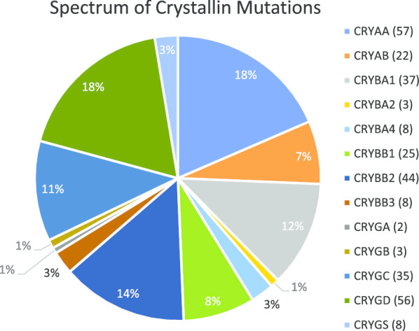
Background: The crystalline lens is mainly composed of a large family of soluble proteins called the crystallins, which are responsible for its development, growth, transparency and refractive index. Disease-causing sequence variants in the crystallins are responsible for nearly 50% of all non-syndromic inherited congenital cataracts, as well as causing cataract associated with other diseases, including myopathies. To date, more than 300 crystallin sequence variants causing cataract have been identified.

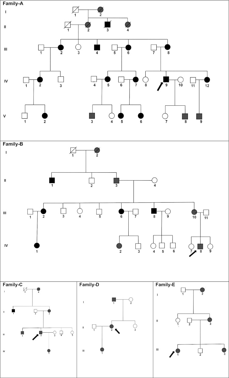
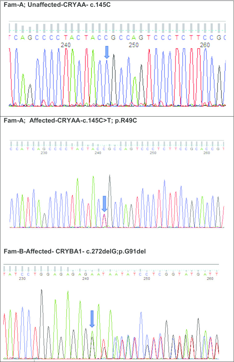
Methods: Here we aimed to identify the genetic basis of disease in five multi-generation British families and five sporadic cases with autosomal dominant congenital cataract using whole exome sequencing, with identified variants validated using Sanger sequencing. Following bioinformatics analysis, rare or novel variants with a moderate to damaging pathogenicity score, were filtered out and tested for segregation within the families.

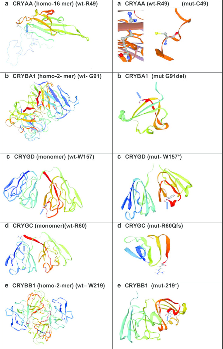
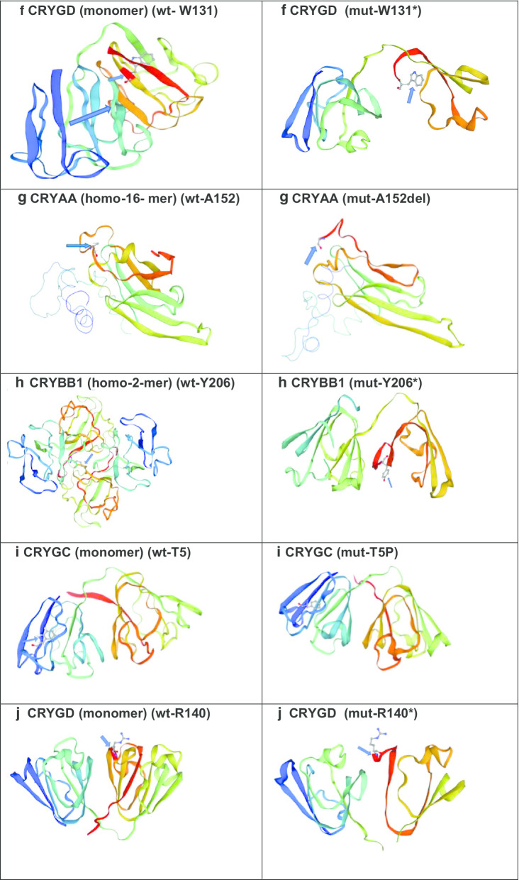
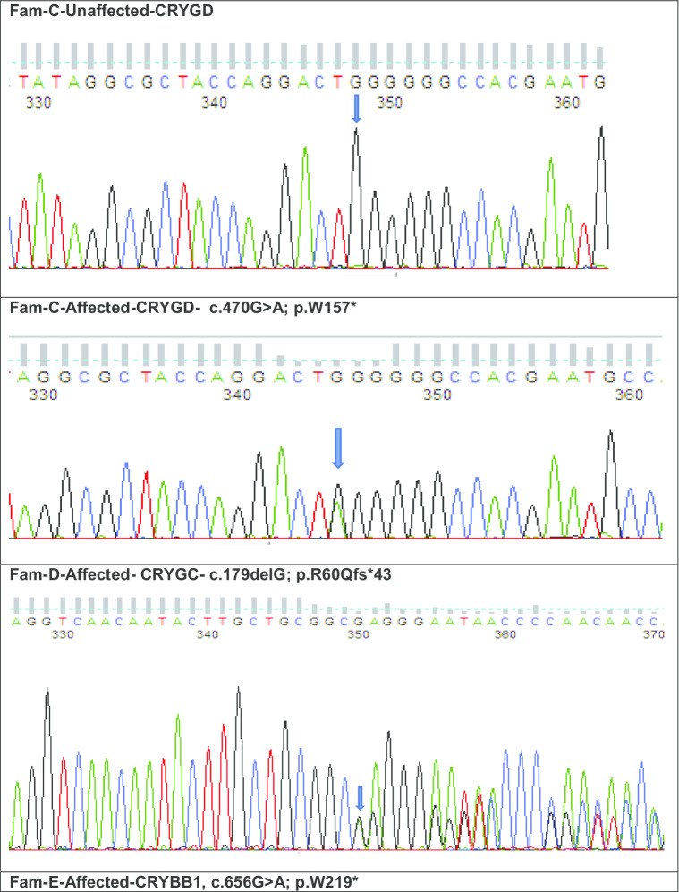
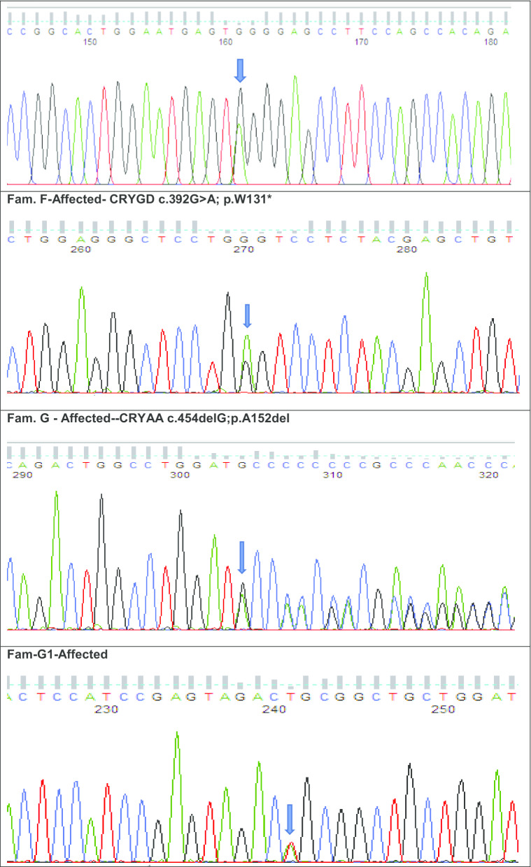
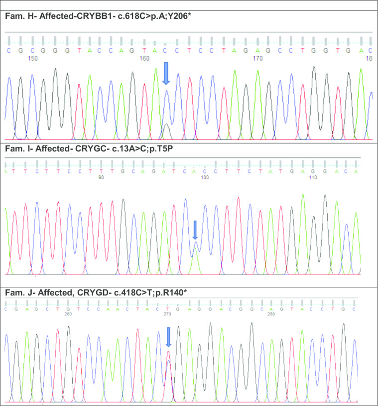
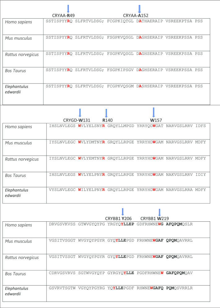
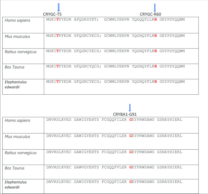
Results: We have identified 10 different heterozygous crystallin variants. Five recurrent variants were found: family-A, with a missense variant (c.145C>T; p.R49C) in CRYAA associated with nuclear cataract; family-B, with a deletion in CRYBA1 (c.272delGAG; p.G91del) associated with nuclear cataract; and family-C, with a truncating variant in CRYGD (c.470G>A; W157*) causing a lamellar phenotype; individuals I and J had variants in CRYGC (c.13A>C; T5P) and in CRYGD (c.418C>T; R140*) causing unspecified congenital cataract and nuclear cataract, respectively. Five novel disease-causing variants were also identified: family D harboured a variant in CRYGC (c.179delG; R60Qfs*) responsible for a nuclear phenotype; family E, harboured a variant in CRYBB1 (c.656G>A; W219*) associated with lamellar cataract; individual F had a variant in CRYGD (c.392G>A; W131*) associated with nuclear cataract; and individuals G and H had variants in CRYAA (c.454delGCC; A152del) and in CRYBB1 (c.618C>A; Y206*) respectively, associated with unspecified congenital cataract. All novel variants were predicted to be pathogenic and to be moderately or highly damaging.

Conclusions: We report five novel variants and five known variants. Some are rare variants that have been reported previously in small ethnic groups but here we extend this to the wider population and record a broader phenotypic spectrum for these variants.

Keywords: Autosomal dominant congenital cataract; Crystallins; Next generation sequencing.


Conflict of interest statement

The authors declare that they have no competing interests.

Figures

Fig. 1
Fig. 1
Frequency pie charts showing spectrum of cataract-causing crystallin variants. Total number of 308 disease-causing variants to date (novel and recurrent) are shown in 13 crystallins expressed in lens. (https://cat-map.wustl.edu/)
Fig. 2
Fig. 2
Structural view of Crystallins: (https://swissmodel.expasy.org/repository/uniprot/) a CRYAA—wild-type and missense mutant amino acid at position 49 (Arginine); b CRYBA1—wild-type and indel mutant amino acid at position 91(Glycine); cCRYGD wild-type and mutant stop codon at amino acid position 157 (Tryptophan); d CRYGC—wild-type and mutant frame-shift variant at amino acid position 60 (Arginine); e CRYBB1—wild-type and mutant stop codon amino acid at position 219 (Tryptophan); f CRYGD–wild-type and mutant stop codon amino acid at 131 (Tryptophan); g CRYAA—wild-type and mutant indel variant at amino acid position 152 (Alanine); h CRYBB1—wild-type and mutant stop codon amino acid at 206 (Tyrosine); i CRYGC—wild-type and missense mutant amino acid at position 5 (Threonine) and j CRYGD—wild-type and mutant stop codon amino acid at 140 (Arginine)
Fig. 2
Fig. 2
Structural view of Crystallins: (https://swissmodel.expasy.org/repository/uniprot/) a CRYAA—wild-type and missense mutant amino acid at position 49 (Arginine); b CRYBA1—wild-type and indel mutant amino acid at position 91(Glycine); cCRYGD wild-type and mutant stop codon at amino acid position 157 (Tryptophan); d CRYGC—wild-type and mutant frame-shift variant at amino acid position 60 (Arginine); e CRYBB1—wild-type and mutant stop codon amino acid at position 219 (Tryptophan); f CRYGD–wild-type and mutant stop codon amino acid at 131 (Tryptophan); g CRYAA—wild-type and mutant indel variant at amino acid position 152 (Alanine); h CRYBB1—wild-type and mutant stop codon amino acid at 206 (Tyrosine); i CRYGC—wild-type and missense mutant amino acid at position 5 (Threonine) and j CRYGD—wild-type and mutant stop codon amino acid at 140 (Arginine)
Fig. 3
Fig. 3
a Family A: Abridged pedigree with nuclear cataract; b Family B: Abridged pedigree with nuclear cataract; c Family C: Abridged pedigree with pulverulent cataract; d Family D: Abridged pedigree with nuclear cataract; e Family E: Abridged pedigree with lamellar cataract. The diagonal line indicates a deceased family member. Squares and circles symbolize males and females, respectively. Open and filled symbols indicate unaffected and affected individuals, respectively. The arrow indicates the family members who participated in the WES analysis. All the members available in the family were sequenced to show the segregation
Fig. 4
Fig. 4
Sequence analysis of Crystallin variants: a CRYAA –wild type and missense variant c.145C>T in unaffected and affected member of family—A with nuclear cataract; b CRYBA1—an indel variant at c.272delG in an affected member of family B with nuclear cataract; c CRYGD—wild type in unaffected and stop codon variant c.470G>A in affected member of family—C with pulverulent cataract; d CRYGC—a frameshift mutation at c.179delG is shown in the affected member of family-D with nuclear cataract; e CRYBB1—a stop codon variant c.656G>A in an affected member of family-E with lamellar cataract; f CRYGD– mutant stop codon amino acid at c.392G>A in an affected female with nuclear cataract; g CRYAA—a mutant indel variant at c.454delG in affected male with congenital cataract and (G1) CRYGA—another missense novel disease-causing variant of uncertain significance at c.118A>T in the same individual G; h CRYBB1—a stop codon mutation at c.618C>A in affected female with congenital cataract; i CRYGC—a missense variant at c.13A>C in an affected male with congenital cataract and j CRYGD a stop codon variant at c.418C>T in an affected female with nuclear cataract
Fig. 4
Fig. 4
Sequence analysis of Crystallin variants: a CRYAA –wild type and missense variant c.145C>T in unaffected and affected member of family—A with nuclear cataract; b CRYBA1—an indel variant at c.272delG in an affected member of family B with nuclear cataract; c CRYGD—wild type in unaffected and stop codon variant c.470G>A in affected member of family—C with pulverulent cataract; d CRYGC—a frameshift mutation at c.179delG is shown in the affected member of family-D with nuclear cataract; e CRYBB1—a stop codon variant c.656G>A in an affected member of family-E with lamellar cataract; f CRYGD– mutant stop codon amino acid at c.392G>A in an affected female with nuclear cataract; g CRYAA—a mutant indel variant at c.454delG in affected male with congenital cataract and (G1) CRYGA—another missense novel disease-causing variant of uncertain significance at c.118A>T in the same individual G; h CRYBB1—a stop codon mutation at c.618C>A in affected female with congenital cataract; i CRYGC—a missense variant at c.13A>C in an affected male with congenital cataract and j CRYGD a stop codon variant at c.418C>T in an affected female with nuclear cataract
Fig. 5
Fig. 5
a The multiple-sequence alignments from different vertebrate species. Arrows show conserved arginine at p.R49 and alanine at p.A152 in CRYAA protein (https://www.ncbi.nlm.nih.gov/nuccore/?term); b The multiple-sequence alignments from different vertebrate species. Arrows show conserved tryptophan at p.W131, p.W157 and arginine at p.R140. in CRYGD protein (https://www.ncbi.nlm.nih.gov/nuccore/?term=Homo+sapiens+CRYGD); c The multiple-sequence alignments from different vertebrate species. Arrows show conserved tyrosine at p,Y206 and tryptophan at p.W219 in CRYBB1 protein (https://www.ncbi.nlm.nih.gov/nuccore/?term=human+CRYBB1); d The multiple-sequence alignments from different vertebrate species. Arrows show conserved threonine at p,T5 and arginine at p.R60 in CRYGC protein (https://www.ncbi.nlm.nih.gov/nuccore/?term=human+CRYGC); e The multiple-sequence alignments from different vertebrate species. Arrows show conserved glycine at p. in CRYBA1 protein (https://www.ncbi.nlm.nih.gov/nuccore/?term=human+CRYBA1)
Fig. 5
Fig. 5
a The multiple-sequence alignments from different vertebrate species. Arrows show conserved arginine at p.R49 and alanine at p.A152 in CRYAA protein (https://www.ncbi.nlm.nih.gov/nuccore/?term); b The multiple-sequence alignments from different vertebrate species. Arrows show conserved tryptophan at p.W131, p.W157 and arginine at p.R140. in CRYGD protein (https://www.ncbi.nlm.nih.gov/nuccore/?term=Homo+sapiens+CRYGD); c The multiple-sequence alignments from different vertebrate species. Arrows show conserved tyrosine at p,Y206 and tryptophan at p.W219 in CRYBB1 protein (https://www.ncbi.nlm.nih.gov/nuccore/?term=human+CRYBB1); d The multiple-sequence alignments from different vertebrate species. Arrows show conserved threonine at p,T5 and arginine at p.R60 in CRYGC protein (https://www.ncbi.nlm.nih.gov/nuccore/?term=human+CRYGC); e The multiple-sequence alignments from different vertebrate species. Arrows show conserved glycine at p. in CRYBA1 protein (https://www.ncbi.nlm.nih.gov/nuccore/?term=human+CRYBA1)

References

    1. Sheeladevi S, Lawrenson JG, Fielder AR, Suttle CM. Global prevalence of childhood cataract: a systematic review. Eye. 2016;30:1160–1169. doi: 10.1038/eye.2016.156. - DOI - PMC - PubMed
    1. Gogate P, Kalua K, Courtright P. Blindness in childhood in developing countries: time for a reassessment? PLoS Med. 2009;6:e1000177. doi: 10.1371/journal.pmed.1000177. - DOI - PMC - PubMed
    1. Ionides A, Francis P, Berry V, Mackay D, Bhattacharya S, Shiels A, et al. Clinical and genetic heterogeneity in autosomal dominant cataract. Brit J Ophthalmol. 1999;83:802–808. doi: 10.1136/bjo.83.7.802. - DOI - PMC - PubMed
    1. Churchill A, Graw J. Clinical and experimental advances in congenital and paediatric cataracts. Philos Trans R Soc Lond B Biol Sci. 2011;366:1234–1249. doi: 10.1098/rstb.2010.0227. - DOI - PMC - PubMed
    1. Shiels A, Hejtmancik JF. Mutations and mechanisms in congenital and age-related cataracts. Exp Eye Res. 2017;156:95–102. doi: 10.1016/j.exer.2016.06.011. - DOI - PMC - PubMed

Publication types