Review
. 2021 Feb;54(1):11-23.
doi: 10.1016/j.otc.2020.09.024. Epub 2020 Sep 29.

Heads-up Surgery: Endoscopes and Exoscopes for Otology and Neurotology in the Era of the COVID-19 Pandemic

Affiliations
Review

Heads-up Surgery: Endoscopes and Exoscopes for Otology and Neurotology in the Era of the COVID-19 Pandemic

Sarah E Ridge et al. Otolaryngol Clin North Am. 2021 Feb.

Abstract

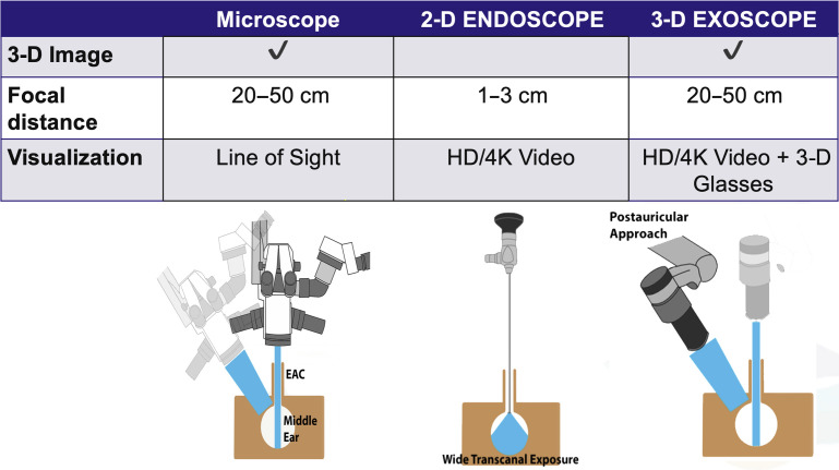
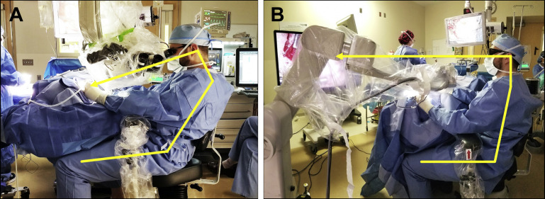
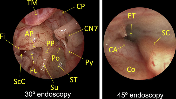
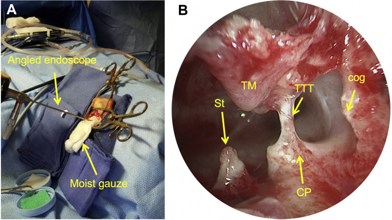
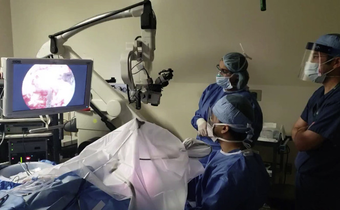
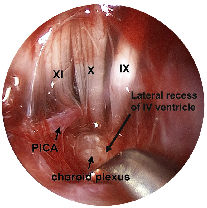
A new era of surgical visualization and magnification is poised to disrupt the field of otology and neurotology. The once revolutionary benefits of the binocular microscope now are shared with rigid endoscopes and exoscopes. These 2 modalities are complementary. The endoscope improves visualization of the hidden recesses through the external auditory canal or canal-up mastoidectomy. The exoscope provides an immersive visual experience and superior ergonomics compared with binocular microscopy. Endoscopes and exoscopes are poised to disrupt the standard of care for surgical visualization and magnification in otology and neurotology.

Keywords: Aerosol generating procedure; Endoscopic ear surgery; Ergonomics; Exoscopic ear surgery; Mastoidectomy; Microscope; Minimally-invasive; PPE.


Conflict of interest statement

Disclosure The senior author has financial relationships with 3NT Medical, Akouos, Frequency Therapeutics, Boston Pharmaceuticals, and Agilis.

Figures

Fig. 1
Fig. 1
General specifications for binocular microscope, 2-D Hopkins rod telescope, and 3-D exoscope. The microscope and exoscope are used with a line of sight approach, with similar focal distances and true depth perception, allowing the user to operate with both hands. In contrast, the 2-D endoscope has a much shorter focal distance but can be used in small corridors to provide a high contrast wide-angle view with enhanced depth of field. EAC, external auditory canal.
Fig. 2
Fig. 2
Traditional heads-down microscopic surgery is associated with unfavorable ergonomics. Arrows illustrate the posture of the surgeon and the corresponding line of sight when using each device. (A) Binocular microscopy, even with armrests, Trendelenburg positioning, and 250-mm focal length places strain on the neck, shoulders and back during prolonged dissection. The stack height of the microscope increases the distance between the end of the oculars to the ear, requiring outstretched arms. (B) Heads-up exoscopic surgery results in a relaxed posture and enhanced body mechanics.
Fig. 3
Fig. 3
Transcanal endoscopic tympanotomy, left ear. Endoscopes provide a wide-angle view and greater depth of field with minimal soft tissue and bony dissection compared with binocular microscopy. (Left panel) A 30° endoscopic view of the left ear mesotympanum and retrotympanum. (Right panel) A 45° endoscopic view of the protympanum. AP, anterior pillar; CA, carotid artery; CN7, facial nerve; Co, cochlea; CP, cochleariform process; ET, eEustachian tube; Fi, finiculus; Fu, fustis; Po, ponticulus; PP, posterior pillar; Py, pyramidal process. SC, semicanal; ScC, subcochlear canaliculus; ST, sinus tympani; Su, subiculum; TM, tympanic membrane.
Fig. 4
Fig. 4
TMEES, left ear. (A) Following a canal-up mastoidectomy for extensive cholesteatoma, a 30° or 45° endoscope (stabilized with a gauze sponge) is introduced into the aditus ad antrum to visualize residual disease. (B) Angled endoscopy directed anteroinferiorly reveals the posterosuperior surfaces of relevant anatomy that can be difficult to visualize transcanal. CP, cochleariform process. St, stapes; TM, tympanic membrane; TTT, tensor tympani tendon.
Fig. 5
Fig. 5
Endoscopic and exoscopic-assisted retrolabyrinthine craniotomy for removal of complex skull base neoplasm. The entire procedure was performed under a barrier drape (OtoTent) to reduce particle dispersion during an AGP in April 2020.
Fig. 6
Fig. 6
Endoscopic dissection of the lateral recess of the IV ventricle following retrolabyrinthine craniotomy, left ear. A 30° endoscope was oriented posteriorly and introduced into the posterior fossa, resulting in a wide-angle view with enhanced depth of field. In contrast, the foramen of Luschka and root entry zones of cranial nerves IX, X, and XI cannot be visualized microscopically using this same approach without sacrificing the posterior canal. Endoscopes enable less-invasive auditory brainstem implant surgery for deaf patients with cochlear nerve aplasia and other cochlear anomalies. PICA, posterior inferior cerebellar artery.

References

    1. Mudry A. The history of the microscope for use in ear surgery. Am J Otol. 2000;21(6):877–886. - PubMed
    1. Eivazi S., Afkari H., Bednarik R., et al. Analysis of disruptive events and precarious situations caused by interaction with neurosurgical microscope. Acta Neurochir (Wien) 2015 doi: 10.1007/s00701-015-2433-5. - DOI - PubMed
    1. Kapadiya M., Tarabichi M. An overview of endoscopic ear surgery in 2018. Laryngoscope Investig Otolaryngol. 2019 doi: 10.1002/lio2.276. - DOI - PMC - PubMed
    1. Kennedy D.W. Serious misconceptions regarding functional endoscopic sinus surgery. Laryngoscope. 1986;96:1170–1171. - PubMed
    1. Kennedy D.W. Functional endoscopic sinus surgery: technique. Arch Otolaryngol. 1985 doi: 10.1001/archotol.1985.00800120037003. - DOI - PubMed