

Development and validation of a deep learning algorithm detecting 10 common abnormalities on chest radiographs
- PMID: 33243843
- PMCID: PMC8134811
- DOI: 10.1183/13993003.03061-2020
Development and validation of a deep learning algorithm detecting 10 common abnormalities on chest radiographs
Abstract
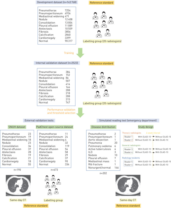
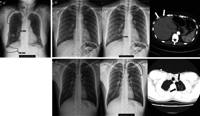
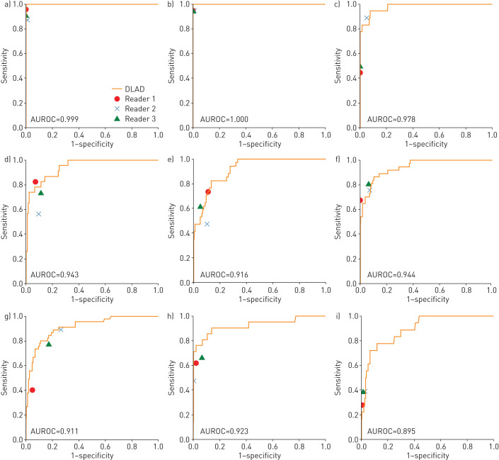
We aimed to develop a deep learning algorithm detecting 10 common abnormalities (DLAD-10) on chest radiographs, and to evaluate its impact in diagnostic accuracy, timeliness of reporting and workflow efficacy.DLAD-10 was trained with 146 717 radiographs from 108 053 patients using a ResNet34-based neural network with lesion-specific channels for 10 common radiological abnormalities (pneumothorax, mediastinal widening, pneumoperitoneum, nodule/mass, consolidation, pleural effusion, linear atelectasis, fibrosis, calcification and cardiomegaly). For external validation, the performance of DLAD-10 on a same-day computed tomography (CT)-confirmed dataset (normal:abnormal 53:147) and an open-source dataset (PadChest; normal:abnormal 339:334) was compared with that of three radiologists. Separate simulated reading tests were conducted on another dataset adjusted to real-world disease prevalence in the emergency department, consisting of four critical, 52 urgent and 146 nonurgent cases. Six radiologists participated in the simulated reading sessions with and without DLAD-10.DLAD-10 exhibited area under the receiver operating characteristic curve values of 0.895-1.00 in the CT-confirmed dataset and 0.913-0.997 in the PadChest dataset. DLAD-10 correctly classified significantly more critical abnormalities (95.0% (57/60)) than pooled radiologists (84.4% (152/180); p=0.01). In simulated reading tests for emergency department patients, pooled readers detected significantly more critical (70.8% (17/24) versus 29.2% (7/24); p=0.006) and urgent (82.7% (258/312) versus 78.2% (244/312); p=0.04) abnormalities when aided by DLAD-10. DLAD-10 assistance shortened the mean±sd time-to-report critical and urgent radiographs (640.5±466.3 versus 3371.0±1352.5 s and 1840.3±1141.1 versus 2127.1±1468.2 s, respectively; all p<0.01) and reduced the mean±sd interpretation time (20.5±22.8 versus 23.5±23.7 s; p<0.001).DLAD-10 showed excellent performance, improving radiologists' performance and shortening the reporting time for critical and urgent cases.
Copyright ©ERS 2021.
Conflict of interest statement
Conflict of interest: J.G. Nam reports grants from the National Research Foundation of Korea funded by the Ministry of Science and ICT (NRF-2018R1A5A1060031), and from Seoul National University Hospital Research Fund (03-2019-0190), during the conduct of the study. Conflict of interest: M. Kim is an employee of Lunit Inc., and was involved in the development of the algorithm and writing the corresponding part of the manuscript, but did not have control over any of the validation data submitted for publication. Conflict of interest: J. Park is an employee of Lunit Inc., and was involved in the development of the algorithm and writing the corresponding part of the manuscript, but did not have control over any of the validation data submitted for publication. Conflict of interest: E.J. Hwang has nothing to disclose. Conflict of interest: J.H. Lee has nothing to disclose. Conflict of interest: J.H. Hong has nothing to disclose. Conflict of interest: J.M. Goo has nothing to disclose. Conflict of interest: C.M. Park reports grants from the National Research Foundation of Korea funded by the Ministry of Science and ICT (NRF-2018R1A5A1060031), and from Seoul National University Hospital Research Fund (03-2019-0190), during the conduct of the study.
Figures
Comment in
-
Artificial intelligence for thoracic radiology: from research tool to clinical practice.Eur Respir J. 2021 May 20;57(5):2100625. doi: 10.1183/13993003.00625-2021. Print 2021 May. Eur Respir J. 2021. PMID: 34016606 No abstract available.
References
-
- United Nations Scientific Committee on the Effects of Atomic Radiation. Sources and Effects of Ionizing Radiation. Annex D. New York, United Nations, 2000.
Publication types
MeSH terms
LinkOut - more resources
Full Text Sources
Other Literature Sources
Medical