Review
. 2020 Nov;52(11):1787-1797.
doi: 10.1038/s12276-020-00522-6. Epub 2020 Nov 26.

Advances in transcriptome analysis of human brain aging

Affiliations
Review

Advances in transcriptome analysis of human brain aging

Seokjin Ham et al. Exp Mol Med. 2020 Nov.

Abstract

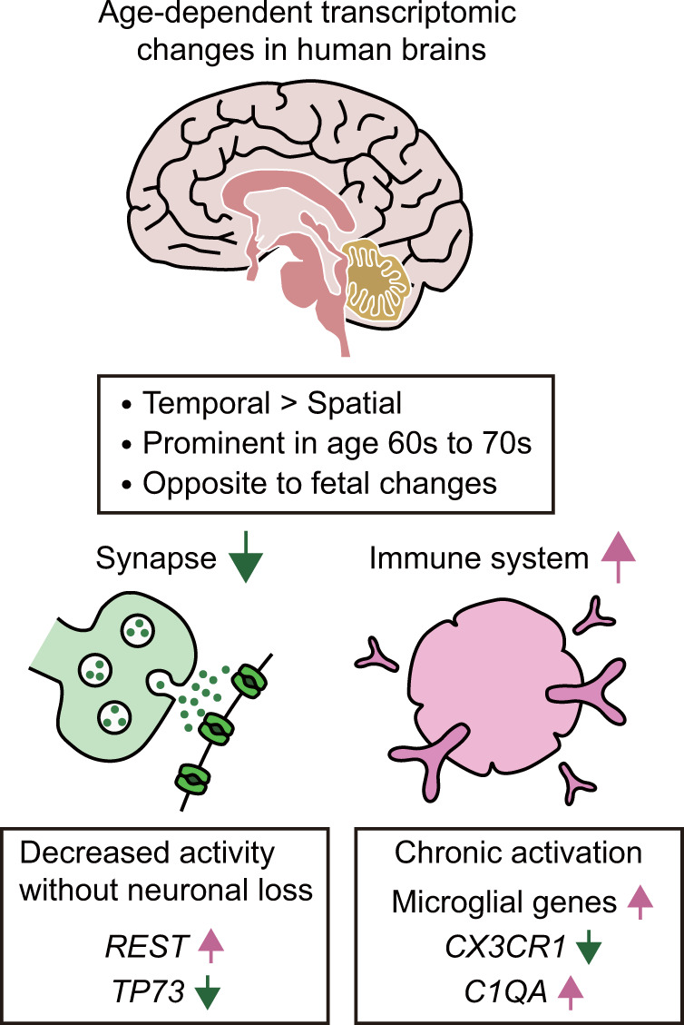
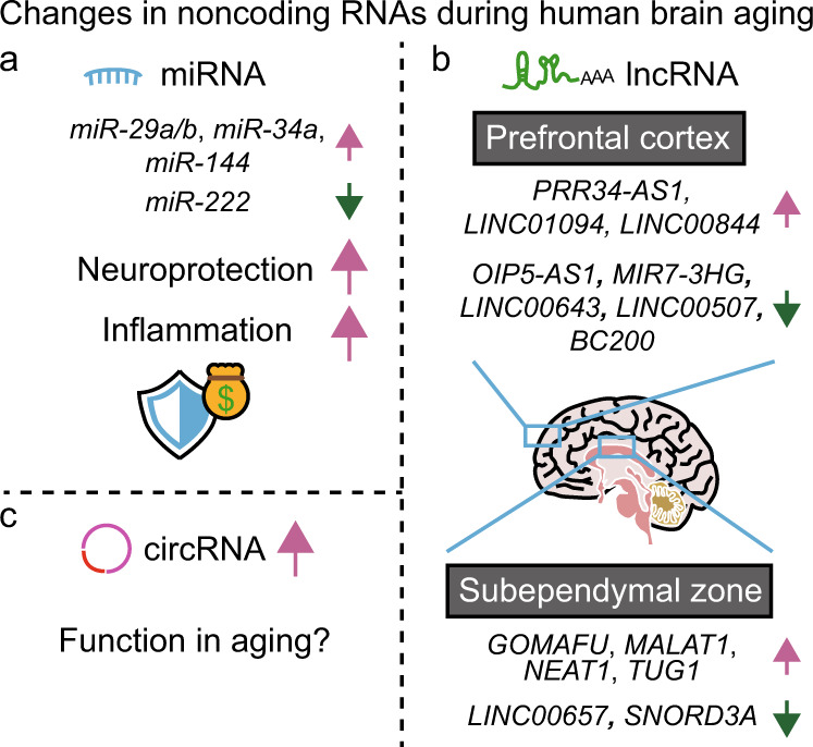
Aging is associated with gradual deterioration of physiological and biochemical functions, including cognitive decline. Transcriptome profiling of brain samples from individuals of varying ages has identified the whole-transcriptome changes that underlie age-associated cognitive declines. In this review, we discuss transcriptome-based research on human brain aging performed by using microarray and RNA sequencing analyses. Overall, decreased synaptic function and increased immune function are prevalent in most regions of the aged brain. Age-associated gene expression changes are also cell dependent and region dependent and are affected by genotype. In addition, the transcriptome changes that occur during brain aging include different splicing events, intersample heterogeneity, and altered levels of various types of noncoding RNAs. Establishing transcriptome-based hallmarks of human brain aging will improve the understanding of cognitive aging and neurodegenerative diseases and eventually lead to interventions that delay or prevent brain aging.


Conflict of interest statement

The authors declare that they have no conflict of interest.

Figures

Fig. 1
Fig. 1. Global age-dependent transcriptomic changes in human brains.
Expression changes are mainly temporal by relative preservation of their spatial identity. Substantial expression changes are detected in the sixth and seventh decades of human life. The directions of gene expression changes during adult aging tend to be opposite those occurring during fetal development. Throughout multiple brain regions, age-dependent transcriptomic changes include decreased synaptic function and increased immunity. It is worth noting that synaptic aging occurs even prior to neuronal loss. Decreased synaptic function underlies increased expression of repressor element-1-silencing transcription factor (REST) and decreased expression of the tumor protein 73 (TP73) gene. Increased immune responses are accompanied by upregulation of microglial genes and complement component 1q A (C1QA) and downregulation of genes encoding immunosuppressive factors, including C-X3-C motif chemokine receptor 1 (CX3CR1).
Fig. 2
Fig. 2. Targeted analysis of transcriptomic changes during human brain aging.
a Age-dependent upregulation of myelination-related genes, which is undetectable in global analysis, is detected via gene coexpression network analysis. Genes that exhibit highly positive correlations among themselves are connected to one another. Different colors represent different groups of genes related to specific functions. b Glia-specific genes lose their regional identity during brain aging, whereas neuron-specific genes preserve their regional identity. Glial markers include genes that are specifically expressed in astrocytes and oligodendrocytes. The dimensions of the transcriptome are reduced for visualization by principal component analysis or multidimensional scaling.
Fig. 3
Fig. 3. Genetic backgrounds that affect gene expression and alternative splicing in an age-dependent manner.
a. An example of expression quantitative trait loci (eQTLs) during brain aging. Genotypes of eQTL single-nucleotide polymorphisms (SNPs) are associated with the expression of target genes (eGenes) on the same chromosome (cis-eQTLs) or on different chromosomes (trans-eQTLs). Individuals with the GG allele in the SNP rs55675298 exhibit an age-dependent increase in the expression of the tumor protein-coding p53 family gene TP73, whereas individuals with the GT or TT allele do not. b. An example of a splicing quantitative trait locus (sQTL) in aged brains. Genotypes of sQTL SNPs are associated with alternative splicing of target genes. The number of G alleles in the SNP rs2439540 correlates positively with hnRNP splicing factors, including HNRNPA2B1, and correlates negatively with the intron usage level of TBC1 domain family member 7 (TBC1D7). Consistent with these observations, the mRNA level of HNRNPA2B1 correlates negatively with the intron usage level of TBC1D7.
Fig. 4
Fig. 4. Noncoding RNAs that participate in human brain aging.
a Several microRNAs (miRNAs) enhance neuroprotection in the elderly at the cost of inflammation. The expression of miR-29a/b, miR-34a, and miR-144 increases with age, whereas that of miR-222 decreases. b Long noncoding RNAs (lncRNAs) display age-dependent expression changes with preserved regional specificity. In the prefrontal cortex, the expression levels of the antisense RNA of the gene encoding proline-rich protein (PRR34-AS1), LINC01094, and LINC00844 correlate positively with age. In contrast, the expression levels of the antisense RNA of the gene encoding opacity‐associated (Opa) interacting protein 1 (OIP5-AS1), MIR7-3 host gene (MIR7-3HG), LINC00643, LINC00507, and brain cytoplasmic 200 (BC200) correlate negatively with age. In the subependymal zone, GOMAFU, metastasis-associated lung adenocarcinoma transcript 1 (MALAT1), nuclear paraspeckle assembly transcript 1 (NEAT1), and taurine-upregulated gene 1 (TUG1) display age-dependent upregulation, whereas the expression of LINC00657 and small nucleolar RNA, C/D box 3A (SNORD3A) is downregulated with age. c Circular RNAs (circRNAs) tend to accumulate during brain aging, but the function of their accumulation in aging remains unclear.

References

    1. Morrison JH, Baxter MG. The ageing cortical synapse: hallmarks and implications for cognitive decline. Nat. Rev. Neurosci. 2012;13:240–250. - PMC - PubMed
    1. Lopez-Otin C, Blasco MA, Partridge L, Serrano M, Kroemer G. The hallmarks of aging. Cell. 2013;153:1194–1217. - PMC - PubMed
    1. Kennedy BK, et al. Geroscience: linking aging to chronic disease. Cell. 2014;159:709–713. - PMC - PubMed
    1. Pakkenberg B, et al. Aging and the human neocortex. Exp. Gerontol. 2003;38:95–99. - PubMed
    1. Lu T, et al. Gene regulation and DNA damage in the ageing human brain. Nature. 2004;429:883–891. - PubMed

Publication types