Review
. 2021 Mar;14(2):305-316.
doi: 10.1038/s41385-020-00359-2. Epub 2020 Nov 26.

The central role of the nasal microenvironment in the transmission, modulation, and clinical progression of SARS-CoV-2 infection

Affiliations
Review

The central role of the nasal microenvironment in the transmission, modulation, and clinical progression of SARS-CoV-2 infection

Oreste Gallo et al. Mucosal Immunol. 2021 Mar.

Abstract

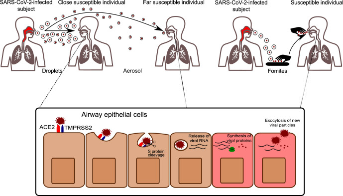
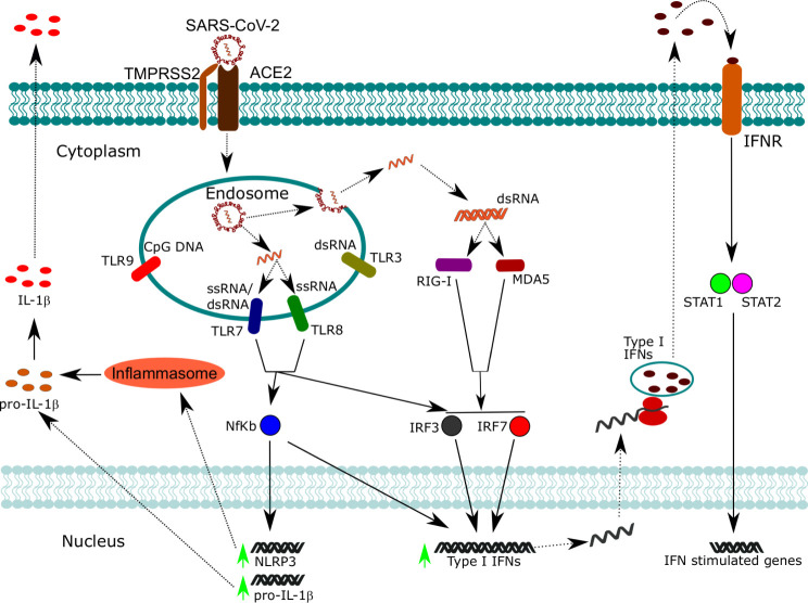
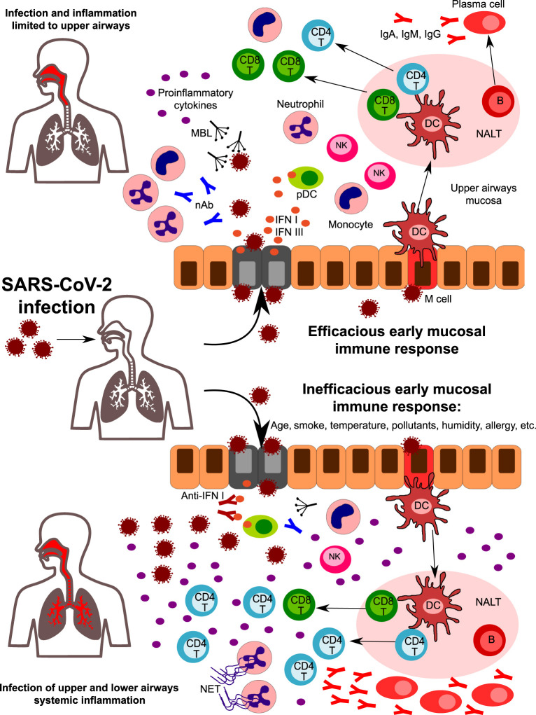
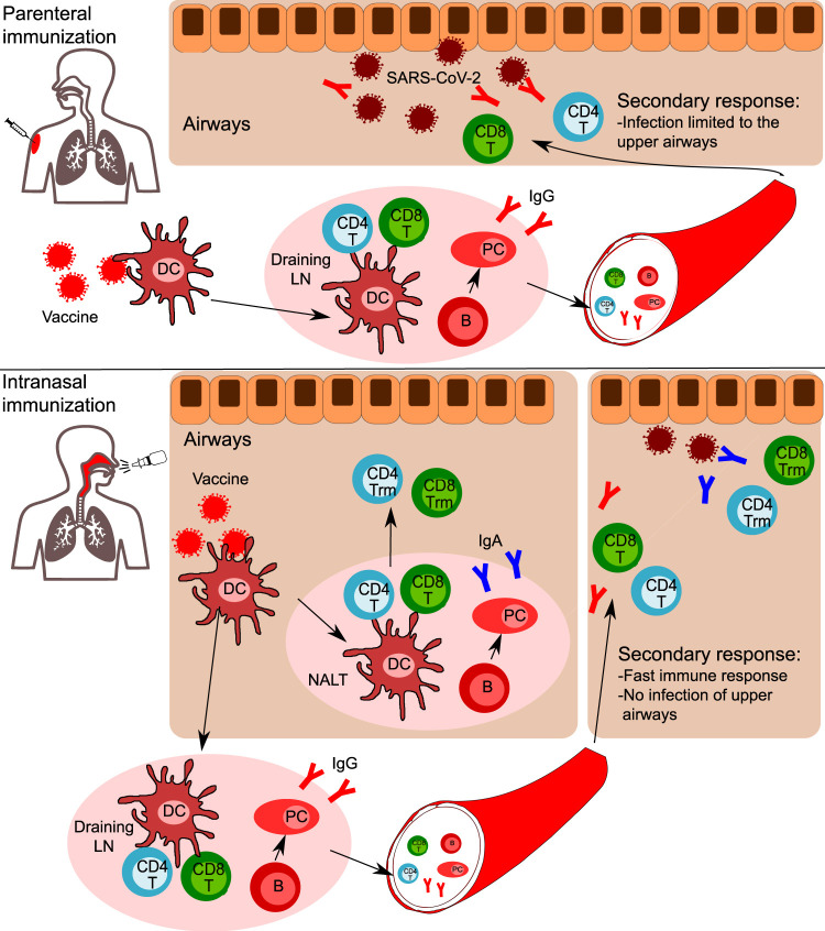
The novel coronavirus SARS-CoV-2 enters into the human body mainly through the ACE2 + TMPRSS2+ nasal epithelial cells. The initial host response to this pathogen occurs in a peculiar immune microenvironment that, starting from the Nasopharynx-Associated Lymphoid Tissue (NALT) system, is the product of a long evolutionary process that is aimed to first recognize exogenous airborne agents. In the present work, we want to critically review the latest molecular and cellular findings on the mucosal response to SARS-CoV-2 in the nasal cavity and in NALT, and to analyze its impact in the subsequent course of COVID-19. Finally, we want to explore the possibility that the regulation of the systemic inflammatory network against the virus can be modulated starting from the initial phases of the nasal and nasopharyngeal response and this may have several clinical and epidemiological implications starting from a mucosal vaccine development.


Conflict of interest statement

The authors declare no competing interests.

Figures

Fig. 1
Fig. 1
SARS-CoV-2 transmission and infection. SARS-CoV-2 can be transmitted by respiratory particles released from an infected subject. Droplets are large particles that commonly deposit within a few meters away from the emitting subject and are responsible for the infection of close individuals. Aerosol instead has a smaller diameter (<5 μm) and can infect subjects at higher distances. In addition to these routes, also contaminated objects (fomites) can be a source of transmission, especially if not practicing regular hand hygiene. Once entered in the airways, SARS-CoV-2 interacts with S protein to its receptor ACE2. The proteolytic cleavage of S, mediated by the cellular protease TMPRSS2, facilitates SARS-CoV-2 infection, which is followed by the release of viral nucleic acid, protein synthesis, and assembly of new viral particles.
Fig. 2
Fig. 2
SARS-CoV-2 recognition by nucleic acid sensors and activation of pro-inflammatory and antiviral responses. Following SARS-CoV-2 infection, viral particles are contained within endosomal compartments. Here, viral ssRNA is released and can be recognized by TLR7 and TLR8, which activate downstream signaling pathways converging to the activation of NF-kB, IRF3, and IRF7. NF-kB promotes the transcription of pro-inflammatory cytokines such as pro-IL-1β and the inflammasome component NLRP3. The inflammasome promotes the maturation of pro-IL-1β in mature IL-1β, which can be secreted. NF-kB cooperates also with IRF3 and IRF7 for the expression of type I IFNs. When the viral ssRNA is released in the cytoplasm, it is replicated by an RNA-dependent RNA polymerase which forms dsRNA intermediates. dsRNA can be recognized by cytoplasmic sensors such as RIG-I and MDA5, which converge to the activation of IRF3 and IRF7. Type I IFNs can act in an autocrine manner, activating STAT1 and STAT2 and thus promoting the expression of interferon-stimulated genes involved in anti-viral defense.
Fig. 3
Fig. 3
A hypothetical role for the early mucosal response in the upper airways in SARS-CoV-2 infection progression. Following SARS-CoV-2 entrance in the nasal or oral cavity, epithelial cells of the upper airways are the primary target site of infection. Innate immunity components in the upper airway mucosa are responsible for the first line of defense. Humoral components such as natural antibodies (nAb) and lectins (including mannose-binding lectin, MBL) can recognize glycoside structures of the virus, while epithelial infected cells and plasmacytoid DC release high amounts of type I IFNs, that are crucial in the initial antiviral response. Cells of innate immunity are activated by viral PAMPS and by the release of DAMPS from infected cells. An efficacious early mucosal innate response allows the development of adaptive immunity, with the expansion of CD4 + helper and CD8 + cytotoxic T cells and the differentiation of Ig-secreting plasma cells. Coordinated innate and adaptive responses contribute to the final elimination of the pathogen (upper part). However, factors such as age, smoke, pollutants, temperature, humidity, and genetics can affect the early response, leading to an inefficacious control of viral replication (lower part). Reduced levels of nAb and/or MBL, the presence of autoantibodies neutralizing type I IFN activity, and of impaired development of antigen-specific CD8 + T cells are some of the factors that can predispose to uncontrolled viral propagation and infection of lower airways. Viral escape is accompanied by a huge release of pro-inflammatory cytokines, with the recruitment and expansion of several subsets of innate and adaptive immunity. Neutrophils accumulate in the lungs and are massively activated, leading to NETs formation. All together, these mechanisms amplify the inflammatory response, leading to uncontrolled systemic inflammation.
Fig. 4
Fig. 4
Comparison of parenteral and intranasal vaccination strategies. Following parenteral immunization, dendritic cells (DC) rapidly uptake the vaccine's antigens and migrate to draining lymph nodes. Here, DC prime antigen-specific CD4 + and CD8 + T cells. Contemporary, antigen-specific B cells activate and differentiate in memory B cells and Ig-secreting plasma cells. CD4 + T cells, CD8 + T cells, B cells, and plasma cells egress the lymph node into the bloodstream. CD4 + T cells, CD8 + T cells, and B cells enter the pool of recirculating lymphocytes, while plasma cells will home to bone marrow niches, where they will continue to secrete antigen-specific IgG. In case of a secondary response (SARS-CoV-2 encounter), in addition to the presence of specific IgG, CD4 + and CD8 + T cells will be recruited to the airways. The infection is limited to upper airways, with no involvement of lower airways and no systemic inflammation. In the case of intranasal vaccination, in addition to the response occurring in draining lymph nodes, a mucosal response occurs in the NALT. B cells differentiate in plasma cells secreting IgA, while CD4 + and CD8 + T cells migrating in the airway mucosa develop a tissue-resident phenotype, thus do not recirculate but reside in tissues. In the case of SARS-CoV-2 encounter, specific T cells and IgA are immediately available in the upper airways to fight the virus and are subsequently assisted by IgG and the recruitment of T cells from the bloodstream. Thus, a faster immune response occurs, with rapid elimination of the virus and no infection of upper airways.

References

    1. Kiyono H, Fukuyama S. NALT-versus Peyer's-patch-mediated mucosal immunity. Nat. Rev. Immunol. 2004;4:699–710. 1:CAS:528:DC%2BD2cXntFCmtL0%3D, 7097243, 15343369. - PMC - PubMed
    1. Brandtzaeg P, Kiyono H, Pabst R, Russell MW. Terminology: nomenclature of mucosa-associated lymphoid tissue. Mucosal. Immunol. 2008;1:31–37. 1:CAS:528:DC%2BD1cXhtlWhuw%3D%3D, 19079158. - PubMed
    1. Tacchi L, et al. Nasal immunity is an ancient arm of the mucosal immune system of vertebrates. Nat. Commun. 2014;5:1–11. - PMC - PubMed
    1. Elad D, Wolf M, Keck T. Air-conditioning in the human nasal cavity. Respir. Physiol. Neurobiol. 2008;163:121–127. 18565805. - PubMed
    1. Newsome H, et al. Clinical importance of nasal air conditioning: a review of the literature. Am. J. Rhinol. Allergy. 2019;33:763–769. 31291132. - PubMed