Review
. 2020 Nov 10:11:565470.
doi: 10.3389/fimmu.2020.565470. eCollection 2020.

Much More Than IL-17A: Cytokines of the IL-17 Family Between Microbiota and Cancer

Affiliations
Review

Much More Than IL-17A: Cytokines of the IL-17 Family Between Microbiota and Cancer

Arianna Brevi et al. Front Immunol. .

Abstract

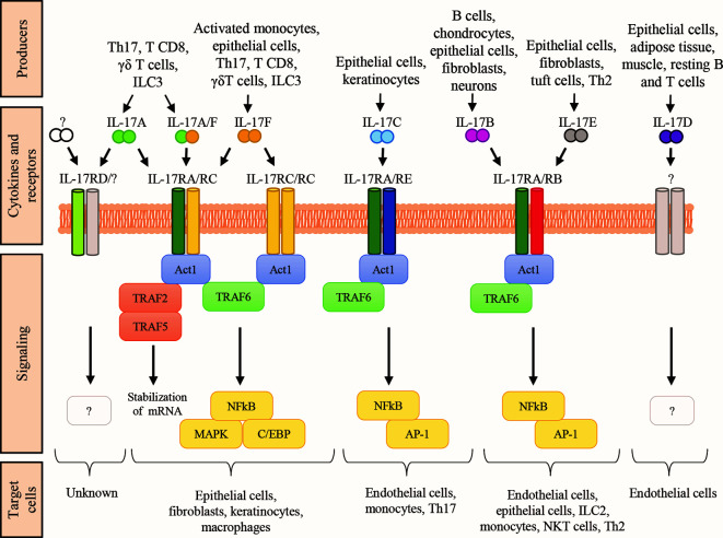
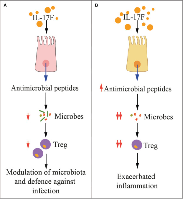
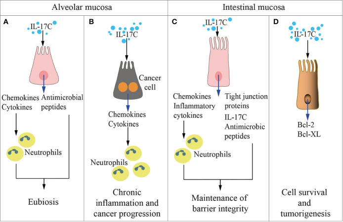
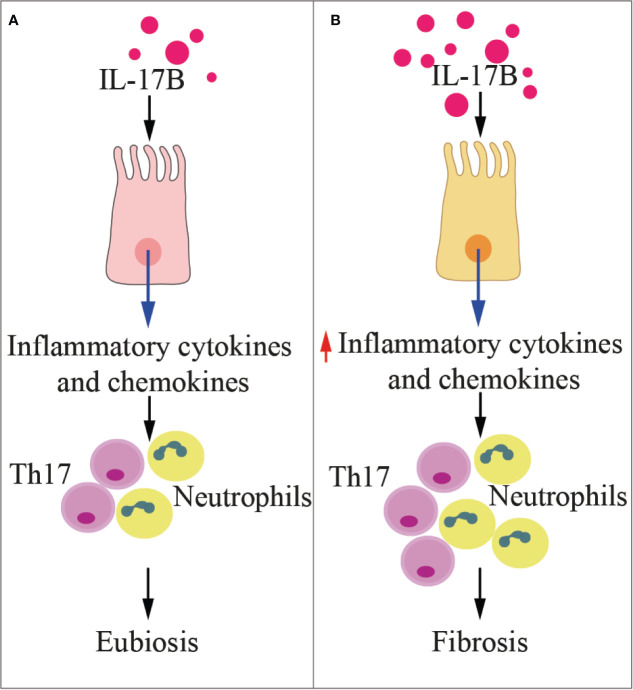
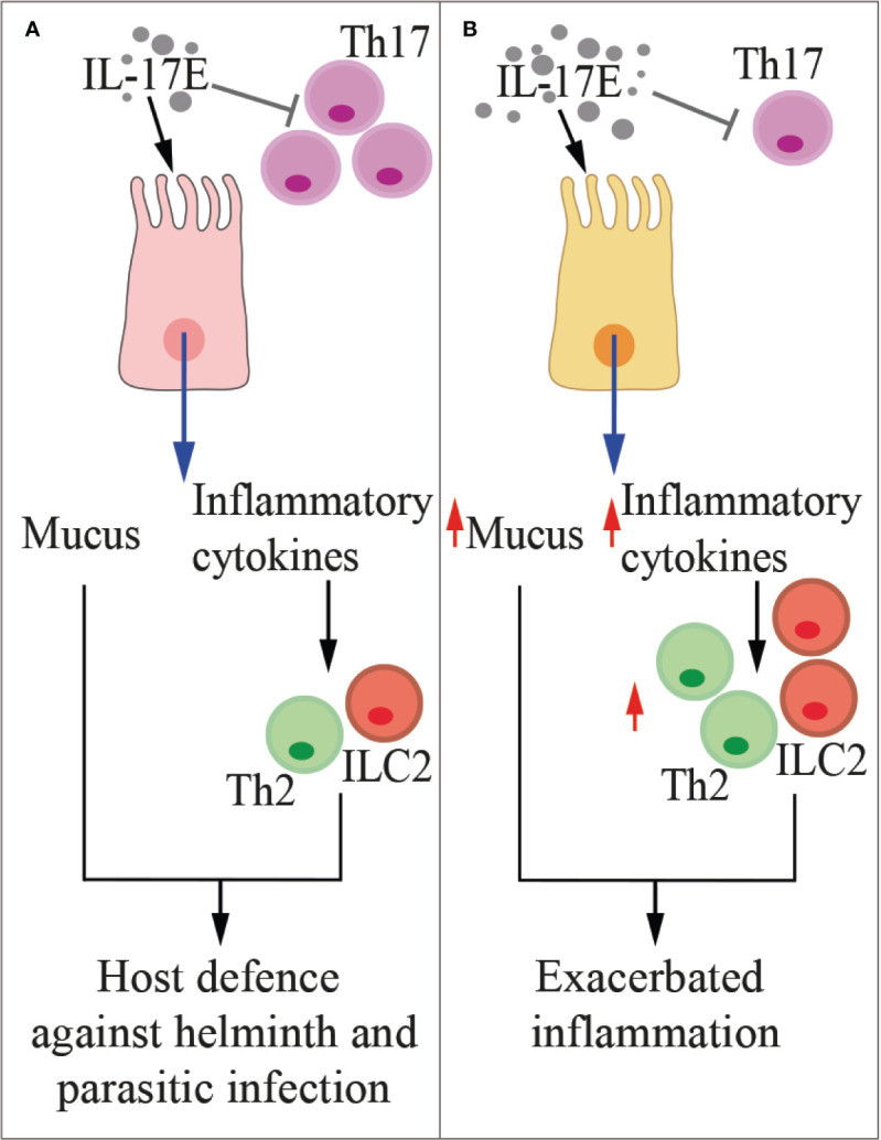
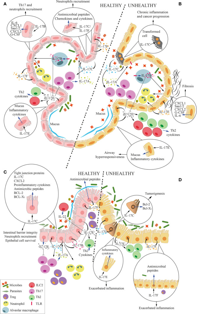
The interleukin-(IL-)17 family of cytokines is composed of six members named IL-17A, IL-17B, IL-17C, IL-17D, IL-17E, and IL-17F. IL-17A is the prototype of this family, and it was the first to be discovered and targeted in the clinic. IL-17A is essential for modulating the interplay between commensal microbes and epithelial cells at our borders (i.e., skin and mucosae), and yet, for protecting us from microbial invaders, thus preserving mucosal and skin integrity. Interactions between the microbiota and cells producing IL-17A have also been implicated in the pathogenesis of immune mediated inflammatory diseases and cancer. While interactions between microbiota and IL-17B-to-F have only partially been investigated, they are by no means less relevant. The cellular source of IL-17B-to-F, their main targets, and their function in homeostasis and disease distinguish IL-17B-to-F from IL-17A. Here, we intentionally overlook IL-17A, and we focus instead on the role of the other cytokines of the IL-17 family in the interplay between microbiota and epithelial cells that may contribute to cancer pathogenesis and immune surveillance. We also underscore differences and similarities between IL-17A and IL-17B-to-F in the microbiota-immunity-cancer axis, and we highlight therapeutic strategies that directly or indirectly target IL-17 cytokines in diseases.

Keywords: Th17; arthritis; autoimmunity; cancer; gut; immunotherapy; microbiome; microbiota.


Figures

Figure 1
Figure 1
The IL-17 family of cytokines. Schematic representation of the cytokines belonging to the IL-17 family, their respective receptor complexes coupled with intracellular signaling, and their target cells. Cytokines are reported in a mechanistic rather than alphabetic order. Producers each cytokine are also shown. AP-1, activator protein-1; C/EBP, CCAAT enhancer-binding protein; ILC, innate lymphoid cells; MAPK, mitogen-activated protein kinase; NKT natural killer T cells; Th2, T helper-2 cells; Th17, T helper-17 cells; TRAF, TNF-receptor associated factor; NF-kB, nuclear factor kB.
Figure 2
Figure 2
IL-17F in health and disease. The cartoon summarizes the main functions of IL-17F in physiologic conditions (A) and during inflammation (B). Treg, regulatory T cells.
Figure 3
Figure 3
IL-17C in health and disease. The cartoon summarizes the functions of IL-17 C in the alveolar (left panels) and the intestinal (right panels) mucosae in physiologic conditions (A, C) and during inflammation and cancer development (B, D).
Figure 4
Figure 4
IL-17B in health and disease. The cartoon summarizes the functions of IL-17B in physiologic conditions (A) and during inflammation (B). Th17, T helper 17 cells.
Figure 5
Figure 5
IL-17E in health and disease. The cartoon summarizes the functions of IL-17E in physiologic conditions (A) and during inflammation (B). Th2, type-2 T helper cells, ILC2, type-2 innate lymphoid cells.
Figure 6
Figure 6
Overall function of IL-17 cytokines at the microbiota-host interface in the lung and the gut. Cartoon summarizing the overall role of IL-17 cytokines at the interface between microbiota and alveolar (A, B) and intestinal mucosa (C, D) in health (A, C) and disease (B, D). Circles within the panels enlarge and focus on several effects of IL-17 cytokines on epithelial cells. Blue arrows represent the secretion of cytokines or the expression of genes by cells, whereas black arrows represent the stimulation of the cell by the cytokines. (A, C) In physiologic conditions, IL-17A, which is mostly released by Th17 cells, keep growth of commensal microbes residing in the lumen of the respiratory tract or the intestine under control. Mucosal epithelial cells secrete IL-17B, IL-17C, IL-17E and IL-17F in response to stimuli coming from the local microbiota. More in details, IL-17B, produced by alveolar macrophages (MØs) under TLR4-mediated stimulation, act on epithelial cells inducing release of several factors, among which IL-6, serum amyloid A1 and A2 (Saa1/2), CXCL1, CXCL2, and G-CSF. Some of these factors favor local recruitment of Th17 cells and neutrophils, which also contribute to maintain an adequate balance in the microbiota composition. Additionally, IL-17B induces monocytes to release TNF-α and IL-1β, which also favor neutrophil recruitment (not shown). IL-17C and IL-17F are released by epithelial cells, and act in autocrine and paracrine fashion inducing the production of antimicrobial peptides, but also chemokines and cytokines that favor neutrophil recruitment. IL-17C also promotes Th17 cell responses, and it supports barrier integrity through tight junction formation in epithelial cells. Also IL-17C induces expression of TNF-α and IL-1β in monocytes (not shown). IL-17E favors the induction of type 2 responses by Th2 cells and ILC2, whereas IL-17B blocks this action, thus avoiding excessive type 2 immune responses. While IL-17D activates NK-mediated immune surveillance (not shown), its relationship with lung and gut microbiota remains unknown. Therefore, IL-17D is not depicted in the figure. IL17E inhibits IL-23, IL-1β and IL-6 expression in activated dendritic cells (not shown), thus blocking the induction of pathogenic Th17 cells. Healthy alveolar epithelial cells also secrete mucus in response to IL-17E to protect the epithelium from bacterial adhesion. (B, D) In pathologic conditions, excessive IL-17A causes local inflammation. In response to the expansion of pathobionts, MØs release more IL-17B, which acts on epithelial cells to induce pro-inflammatory signals (IL-6, Saa1/2, CXCL1, CXCL2, and G-CSF), which may induce lung fibrosis. Stimuli from pathogenic bacteria unleash IL-17C hyperproduction, leading to chronic inflammation and tumorigenesis, also through the upregulation of Bcl-2 and Bcl-XL. Excessive IL-17E signaling is associated with stronger Type 2 immune reaction (Th2 and ILC2) that exacerbate airways hyperresponsiveness and gut inflammation. Unbalanced IL-17F in the gut induces the release of excessive antimicrobial peptides, which constrains Treg-inducing bacteria, therefore promoting gut inflammation.

References

    1. Lynch SV, Pedersen O. The Human Intestinal Microbiome in Health and Disease. N Engl J Med (2016) 375:2369–79. 10.1056/NEJMra1600266 - DOI - PubMed
    1. Sender R, Fuchs S, Milo R. Are We Really Vastly Outnumbered? Revisiting the Ratio of Bacterial to Host Cells in Humans. Cell (2016) 164(3):337–40. 10.1016/j.cell.2016.01.013 - DOI - PubMed
    1. Ley RE, Peterson DA, Gordon JI. Ecological and evolutionary forces shaping microbial diversity in the human intestine. Cell (2006) 124(4):837–48. 10.1016/j.cell.2006.02.017 - DOI - PubMed
    1. Gilbert JA, Blaser MJ, Caporaso JG, Jansson JK, Lynch SV, Knight R. Current understanding of the human microbiome. Nat Med (2018) 24(4):392–400. 10.1038/nm.4517 - DOI - PMC - PubMed
    1. Dominguez-Bello MG, Costello EK, Contreras M, Magris M, Hidalgo G, Fierer N, et al. Delivery mode shapes the acquisition and structure of the initial microbiota across multiple body habitats in newborns. Proc Natl Acad Sci U S A (2010) 107(26):11971–5. 10.1073/pnas.1002601107 - DOI - PMC - PubMed

Publication types

LinkOut - more resources