Review
. 2021 Apr;126(4):505-516.
doi: 10.1007/s11547-020-01315-7. Epub 2020 Nov 27.

Complicated pyelonephritis associated with chronic renal stone disease

Affiliations
Review

Complicated pyelonephritis associated with chronic renal stone disease

Federica Ciccarese et al. Radiol Med. 2021 Apr.

Abstract

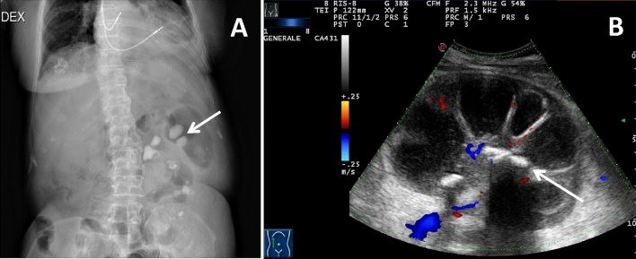
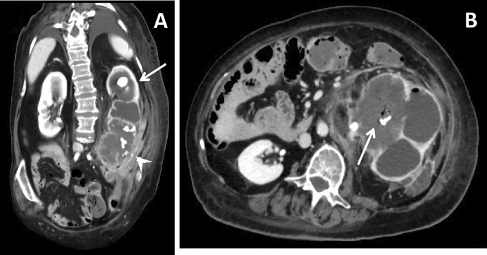
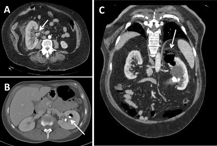
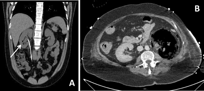
Purpose: This article reviews imaging manifestations of complicated pyelonephritis associated with chronic renal stones disease, in particular xanthogranulomatous pyelonephritis (XGP) and emphysematous pyelonephritis (EPN), as potential mimics of other renal diseases and malignances and provides helpful tips and differentiating features that may alert the radiologist to suspect a diagnosis of infection.

Materials and methods: A retrospective review of the records from 6 adult patients (5 females and 1 male, mean age 72,3 years) with diagnosis of XGP associated with chronic nephrolithiasis and 7 adult patients (6 females and 1 male, mean age 59,3 years) with diagnosis of EPN associated with chronic nephrolithiasis from January 2010 to January 2020 was carried out. Computed tomography urography (CTU) with at least an unenhanced scan, and the parenchymal and excretory phases after contrast medium administration performed at our Teaching Hospital were included. When available images related to conventional radiography, ultrasound (US) and magnetic resonance imaging of the same patients, the comparison with CTU images was carried out.

Conclusion: A possible diagnosis of XGP or EPN must always be taken into account when a pyelonephritis is associated with untreated kidney stones, especially whenever clinical presentation is atypical, current therapy is not effective and imaging shows features of dubious interpretation. Due to their rarity and atypical presentation, a multidisciplinary approach is required and an expert radiologist represents a key figure in the multidisciplinary team as he can help to differentiate between benign and malignant lesions and thus avoid unnecessary radical surgical procedures.

Keywords: Complicated pyelonephritis; Emphysematous pyelonephritis; Nephrolithiasis; Renal stone disease; Xanthogranulomatous pyelonephritis.


Conflict of interest statement

All authors certify that they have no affiliations with or involvement in any organization or entity with any financial interest or non-financial interest in the subject matter or materials discussed in this manuscript.

Figures

Fig. 1
Fig. 1
Radiographic and ultrasonographic (US) appearance of xanthogranulomatous Pyelonephritis. A 69-year-old female patient presenting with right flank pain. Conventional radiography (a) shows the presence of several big radiopaque staghorn calculus in the left kidney (arrow) and an enlargement of the renal outline. US (b) shows an enlarged left kidney with a loss of the normal renal architecture due to the cortical thinning and the presence of multiple hypoechoic areas in the parenchyma, corresponding to dilated calyxes; the renal pelvis is occupied by multiple amorphous echogenicities with acoustic shadowing corresponding to a staghorn calculus (arrow); Doppler US examination demonstrates no particular alterations in the vascularization of the organ
Fig. 2
Fig. 2
CT staging of xanthogranulomatous pyelonephritis. a A 76-year-old female patient presenting with mild fever and stage I XGP with multiple voluminous high-density staghorn calculi in the contracted renal pelvis and inferior calyxes (arrows), concomitant calyx dilatation and cortical thinning. b A 68-year-old male patient presenting with weight loss and mild fever and stage II XGP where, besides the presence of multiple hypodense areas of xanthogranulomatous tissue inside an inhomogeneous parenchyma, is evident a thickening of the Gerota fascia with streaks of perirenal fat (arrow) and enlarged lymph nodes at the renal hilum and in the para-aortic region. c A 82-year-old female patient presenting with pain, fever and hematuria and severe stage III XGP with a voluminous fluid collection bounded by vascularized tissue in the retroperitoneal space involving the psoas muscle (arrow)
Fig. 3
Fig. 3
MRI appearance of xanthogranulomatous pyelonephritis. A 75-year-old female patient presenting with flank pain and fever. a Coronal T1-weighted images shows hypointense parenchymal cavities (arrow). b Coronal T2-weighted sequence shows hyperintense cavities (arrow) surrounded by a solid isointense rim and containing fluid levels, with upper more hyperintense component and lower more hypointense component, due to inflammatory debris and necrotic-purulent material; the combination of contracted renal pelvis, calyx dilatation and cortical thinning together constitutes the bear’s sign
Fig. 4
Fig. 4
CT of the xanthogranulomatous pyelonephritis associated with renal tumor. A 64-year-old female patient presenting with abdominal pain, fever and hematuria. Coronal oblique MPR reconstruction (a) and axial scan (b) of the parenchymal phase of an unusual and rare case of XGP associated with high-grade squamous cell carcinoma (G3, pT4), histopathologically confirmed. The upper portion of the kidney is characterized by multiple hypodense areas of xanthogranulomatous tissue and voluminous high-density staghorn calculi (arrow in A); the lower pole shows a complete and severe distortion of the parenchyma, with several calcifications (arrowhead in A and arrow in B), that turned out to be with a carcinomatous degeneration. Several adjacent structures are invaded by the pathological process
Fig. 5
Fig. 5
CT staging of emphysematous pyelonephritis. a Axial oblique MPR reconstruction of the parenchymal phase of a 53-year-old diabetic female patient presenting with mild fever shows a class I EPN, characterized by gas only inside collecting system, in association with the presence of a calculus in the renal pelvis (arrow). b Axial scan of the parenchymal phase of a 73-year-old diabetic female patient presenting with right flank discomfort shows two calyceal calculi with initial parenchyma spread of gas (arrow) in a class II EPN. c Coronal oblique MPR reconstruction of a 66-year-old diabetic female patient presenting with abdominal pain shows a class III EPN with a voluminous hypodense fluid-gaseous abscess filled with gas bubbles, dissecting the interstitial space and invading the perinephric space (arrow)
Fig. 6
Fig. 6
Pitfalls in renal gas collections diagnosis. a Coronal oblique MPR reconstruction of unenhanced phase of a 60-year-old male asymptomatic patient shows gas bubbles in the inferior calyces of right kidney (arrow) in patients with ureteral stent. b Axial scan of the parenchymal phase of a 56-year-old female patient presenting with abdominal pain, fever and hematuria shows unusual and severe case of an extensive renal infarction, where the left kidney is characterized by a widespread subversion of the parenchyma, which appears to have a mainly gaseous density, with concomitant signs of free air in the retroperitoneum

References

    1. Brikowski TH, Lotan Y, Pearle MS. Climate-related increase in the prevalence of urolithiasis in the United States. Proc Natl Acad Sci U S A. 2008;105(28):9841–9846. doi: 10.1073/pnas.0709652105. - DOI - PMC - PubMed
    1. Morgan MS, Pearle MS. Medical management of renal stones. BMJ. 2016;14(352):i52. doi: 10.1136/bmj.i52. - DOI - PubMed
    1. Johri N, Cooper B, Robertson W, Choong S, Rickards D, Unwin R. An update and practical guide to renal stone management. Nephron Clin Pract. 2010;116(3):c159-71. doi: 10.1159/000317196. - DOI - PubMed
    1. Vargas AD, Bragin SD, Mendez R. Staghorn calculis: its clinical presentation, complications and management. J Urol. 1982;127(5):860–862. doi: 10.1016/S0022-5347(17)54109-9. - DOI - PubMed
    1. Lin SY, Lin CL, Chang CH, Wu HC, Chen WC, Wang IK, Liu YL, Sung FC, Chang YJ, Kao CH. Comparative risk of chronic kidney diseases in patients with urolithiasis and urological interventions: a longitudinal population-based study. Urolithiasis. 2017;45(5):465–472. doi: 10.1007/s00240-016-0929-y. - DOI - PubMed