. 2021 Feb:96:1-17.
doi: 10.1016/j.matbio.2020.11.002. Epub 2020 Nov 25.

Decorin regulates cartilage pericellular matrix micromechanobiology

Affiliations

Decorin regulates cartilage pericellular matrix micromechanobiology

Daphney R Chery et al. Matrix Biol. 2021 Feb.

Abstract

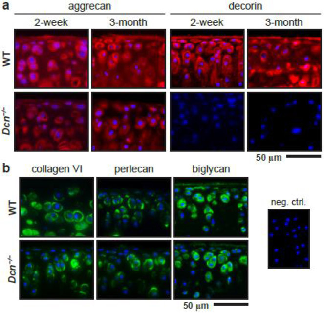
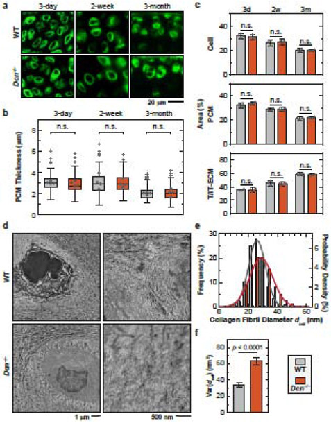
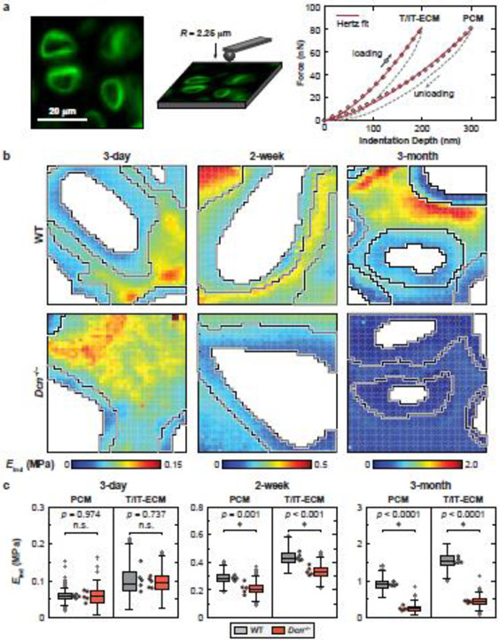
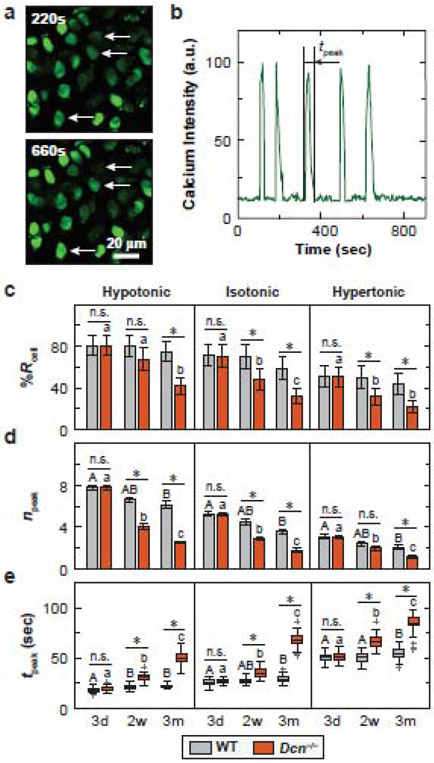
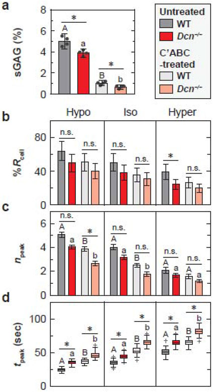
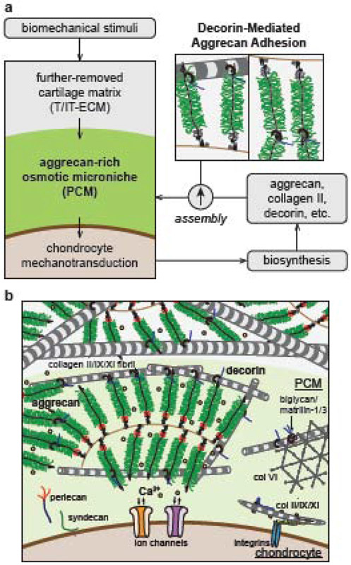
In cartilage tissue engineering, one key challenge is for regenerative tissue to recapitulate the biomechanical functions of native cartilage while maintaining normal mechanosensitive activities of chondrocytes. Thus, it is imperative to discern the micromechanobiological functions of the pericellular matrix, the ~ 2-4 µm-thick domain that is in immediate contact with chondrocytes. In this study, we discovered that decorin, a small leucine-rich proteoglycan, is a key determinant of cartilage pericellular matrix micromechanics and chondrocyte mechanotransduction in vivo. The pericellular matrix of decorin-null murine cartilage developed reduced content of aggrecan, the major chondroitin sulfate proteoglycan of cartilage and a mild increase in collagen II fibril diameter vis-à-vis wild-type controls. As a result, decorin-null pericellular matrix showed a significant reduction in micromodulus, which became progressively more pronounced with maturation. In alignment with the defects of pericellular matrix, decorin-null chondrocytes exhibited decreased intracellular calcium activities, [Ca2+]i, in both physiologic and osmotically evoked fluidic environments in situ, illustrating impaired chondrocyte mechanotransduction. Next, we compared [Ca2+]i activities of wild-type and decorin-null chondrocytes following enzymatic removal of chondroitin sulfate glycosaminoglycans. The results showed that decorin mediates chondrocyte mechanotransduction primarily through regulating the integrity of aggrecan network, and thus, aggrecan-endowed negative charge microenvironment in the pericellular matrix. Collectively, our results provide robust genetic and biomechanical evidence that decorin is an essential constituent of the native cartilage matrix, and suggest that modulating decorin activities could improve cartilage regeneration.

Keywords: Chondrocyte; Decorin; Mechanotransduction; Nanomechanics; Pericellular matrix.


Conflict of interest statement

Declaration of Competing Interest None.

Figures

Figure 1.
Figure 1.
Distribution of extracellular matrix biomolecules in wild-type (WT) and decorin-null (Dcn−/−) cartilage via immunofluorescence (IF) imaging. a) IF images illustrate the reduced staining of aggrecan core protein and the absence of decorin in Dcn−/− cartilage at 2 weeks and 3 months of ages. b) IF images show similar distribution of pericellular matrix (PCM) biomarkers, type VI collagen, perlecan and biglycan, in WT and Dcn−/− cartilage at 2 weeks of age (blue: DAPI; shown together was the negative control).
Figure 2.
Figure 2.
Decorin-null (Dcn−/−) cartilage develops normal PCM morphology and moderate changes of collagen fibrillar nanostructure in the PCM. a) Representative immunofluorescence (IF) images on fresh, unfixed cartilage cryo-sections immunolabeled with type VI collagen at 3 days, 2 weeks and 3 months of ages. b) Box-and-whisker plot of the distribution of cartilage PCM thickness (≥ 120 cells from n = 4 animals for each genotype). c) Comparison of the proportions of areas occupied by the cell, PCM and T/IT-ECM in WT and Dcn−/− cartilage (n = 4). See Table S1 for the complete list of descriptive statistics and adjusted p-values. d) Representative TEM images of collagen fibril structure on the sagittal sections of 3-month-old wild-type (WT) and Dcn−/− cartilage PCM. e) Histogram of fibril diameter distribution (> 1,400 fibrils from n = 4 animals). Shown together were the normal distribution, N(μ, σ2), fits to fibril diameters (for each fit, values of μ and σ correspond to the mean and standard deviation of fibril diameters shown in Table S2). f) Comparison of fibril diameter heterogeneity (variance) between the two genotypes (mean ± 95% CI).
Figure 3.
Figure 3.
Decorin-null (Dcn−/− cartilage exhibits reduced micromodulus in the PCM and T/IT-ECM. a) Left panel: Schematic illustration of immunofluorescence (IF)-guided AFM nanomechanical mapping on the cryo-section of 2-week-old wild-type (WT) murine cartilage using a microspherical tip (R ≈ 2.25 μm); the PCM is immunolabeled with collagen VI. Right panel: Two representative indentation force versus depth (F-D) curves obtained on the 2-week-old WT cartilage cryo-section (measured in PBS, 10 μm/s rate), solid line: finite thickness-corrected Hertz model fit to the entire loading portion of the F-D curve. b) Representative indentation modulus maps of the PCM and T/IT-ECM partitioned for WT and Dcn−/− cartilage at 3 days, 2 weeks and 3 months of ages. Moduli corresponding to cell remnants were removed (white voids). c) Box-and-whisker plots of the PCM and T/IT-ECM micromodulus for WT versus Dcn−/− cartilage at each age (> 1,100 locations for each region, n = 5 animals). Each circle represents the average modulus of one animal, *: p < 0.05 between WT and Dcn−/− cartilage; n.s.: not significant. See Table S3 for the complete list of descriptive statistics and adjusted p-values.
Figure 4.
Figure 4.
Decorin-null (Dcn−/−) chondrocytes exhibit decreased chondrocyte intracellular Ca2+ activities in situ. a) Representative confocal images of [Ca2+]i signaling in isotonic DMEM for 2-week-old wild-type (WT) cartilage explants. Chondrocytes were labeled with Ca-520™ AM and time series images were recorded using a confocal microscope with a 20× objective submerged in DMEM at 37 °C. b) Representative [Ca2+]i oscillation intensity curve of a single cell over a 15-min time frame illustrating the definition of tpeak, the duration of each peak. c-e) Comparison of [Ca2+]i signaling characteristics between WT and Dcn−/− chondrocytes at each age and osmolarity: c) percentage of responding cells, %Rcell (mean ± 95% CI), d) number of peaks within the 15-min testing time frame, npeak (mean ± 95% CI), e) duration of each peak, tpeak (box-and-whisker plot). Data represent > 40 responding cells pooled from n = 4 animals for each group. *: p < 0.05 between genotypes; n.s.: not significant. For longitudinal comparisons, in WT cartilage, there was no significant difference among all three ages for %Rcell in all three osmolarities. Different letters indicate significant differences between ages within each genotype. See Tables S4-S6 for the complete list of descriptive statistics and adjusted p-values.
Figure 5.
Figure 5.
Impact of enzymatic removal of chondroitin-sulfate glycosaminoglycans (CS-GAGs) on chondrocyte intracellular Ca2+ activities in situ. a) Comparison of the amount of sGAGs in chondroitinase ABC (C’ABC)-treated and untreated cartilage explants from wild-type (WT) and decorin-null (Dcn−/−) mice subjected to 12 hours C’ABC treatment at 37°C. b-d) Comparison of [Ca2+]i signaling characteristics between the untreated and C’ABC-treated cartilage at 2 weeks of age in DMEM: b) percentage of responding cells, %Rcell (mean ± 95% CI), c) number of peaks within the 15-min testing timeframe, npeak (mean ± 95% CI), and d) duration of each peak, tpeak (box-and-whisker plot). Data represent > 40 responding cells pooled from n = 4 animals for each group. *: p < 0.05 between genotype within each treatment group (n.s.: not significant). For both WT and Dcn−/− cartilage, there was no significant difference between treatment groups for %Rcell in all three osmolarities. Different letters indicate significant differences between treatment groups within each genotype. See Tables S7-S8 for the complete list of descriptive statistics and adjusted p-values.
Figure 6.
Figure 6.
Schematic illustration of the working hypothesis on the structural role of decorin in cartilage pericellular matrix (PCM). a) Decorin regulates the structural integrity of PCM by mediating the molecular adhesion of aggrecan-aggrecan and aggrecan-collagen II fibrils therein. As a result, decorin regulates the PCM-mediated transmission of biomechanical stimuli from the extracellular matrix to chondrocytes, and thus, the mechanotransduction of chondrocytes. b) Schematic illustration of cartilage matrix molecular constituents, highlighting the crucial structural role of decorin in regulating both the PCM and T/IT-ECM. The schematics are inspired by Ref. [5, 19, 39].

Similar articles

Cited by

References

    1. Wang T, Lai JH, Yang F, Effects of hydrogel stiffness and extracellular compositions on modulating cartilage regeneration by mixed populations of stem cells and chondrocytes in vivo, Tissue Eng. A 22 (23-24) (2016) 1348–1356. - PMC - PubMed
    1. Maroudas A, Physicochemical properties of articular cartilage, in: Freeman MAR (Ed.), Adult Articular Cartilage, Pitman, England, 1979, pp. 215–290.
    1. Huey DJ, Hu JC, Athanasiou KA, Unlike bone, cartilage regeneration remains elusive, Science 338 (6109) (2012) 917–921. - PMC - PubMed
    1. Wilusz RE, Sanchez-Adams J, Guilak F, The structure and function of the pericellular matrix of articular cartilage, Matrix Biol. 39 (2014) 25–32. - PMC - PubMed
    1. Guilak F, Nims RJ, Dicks A, Wu CL, Meulenbelt I, Osteoarthritis as a disease of the cartilage pericellular matrix, Matrix Biol. 71-72 (2018) 40–50. - PMC - PubMed

Publication types

LinkOut - more resources