. 2021 Feb 11;104(2):325-335.
doi: 10.1093/biolre/ioaa215.

ZC3H4-a novel Cys-Cys-Cys-His-type zinc finger protein-is essential for early embryogenesis in mice†

Affiliations

ZC3H4-a novel Cys-Cys-Cys-His-type zinc finger protein-is essential for early embryogenesis in mice†

Jianmin Su et al. Biol Reprod. .

Abstract

Zinc finger domains of the Cys-Cys-Cys-His (CCCH) class are evolutionarily conserved proteins that bind nucleic acids and are involved in various biological processes. Nearly 60 CCCH-type zinc finger proteins have been identified in humans and mice, most have not been functionally characterized. Here, we provide the first in vivo functional characterization of ZC3H4-a novel CCCH-type zinc finger protein. Our results show that although Zc3h4 mutant embryos exhibit normal morphology at E3.5 blastocyst stage, they cannot be recovered at E7.5 early post-gastrulation stage, suggesting implantation failure. Outgrowth assays reveal that mutant blastocysts either fail to hatch from the zona pellucida, or can hatch but do not form a typical inner cell mass colony, the source of embryonic stem cells (ESCs). Although there is no change in levels of reactive oxygen species, Zc3h4 mutants display severe DNA breaks and reduced cell proliferation. Analysis of lineage specification reveals that both epiblast and primitive endoderm lineages are compromised with severe reductions in cell number and/or specification in the mutant blastocysts. In summary, these findings demonstrate the essential role of ZC3H4 during early mammalian embryogenesis.

Keywords: Blastocyst embryo; DNA break; cell lineage specification; epiblast; inner cell mass.


Figures

Figure 1
Figure 1
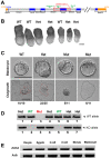
Zc3h4 knock-out allele generation, embryo collection and genotyping, and expression analysis. (A) Schematic of Zc3h4 knock-out allele generation and genotyping primers for WT allele and Mut allele. F, forward; R, reverse. (B) Representative genotyped embryos at E7.5. (C) E3.5 blastocysts from heterozygous intercrosses were imaged and subjected to 3-day-outgrowth assay before knowing the genotypes. After outgrowth culture, outgrowths were imaged again and then lysed for genotyping. Although mutant blastocysts were indistinguishable from littermates at E3.5, they do not form successful outgrowths. Outgrowths from WT and Het blastocysts displayed a distinctive ICM colony (red dashed line) surrounded by trophoblast cells (black dashed line). Mutant exhibited 2 distinct phenotypes, either failing to hatch from the zona pellucida with all blastomeres trapped in the zona (5 from 11) or they hatched but resulted in an outgrowth completely devoid of typical ICM colony (6 from 11). Scale bars, 100 μm. (D) Representative genotyping PCR of individual outgrowth. (E) RT-PCR to identify Zc3h4 expression in WT preimplantation embryos. Actb was used as loading control.
Figure 2
Figure 2
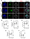
Decreased total cell number and altered lineage allocation was found in Zc3h4 mutant embryos. (A) IF of OCT4 (ICM marker), CDX2 (TE marker), and active TRP53 (apoptosis marker) in blastocysts of different genotypes. (B) Total cell number per blastocyst in Mut embryos was significantly decreased. (C) The number of OCT4 positive cells per blastocyst was severely reduced in Mut embryos. (D) The percentage of ICM cells (OCT4 positive) in Mut embryos was significantly decreased. (E) The number of CDX2 positive cells per blastocyst was severely reduced in Zc3h4 mutants. (F) The percentage of CDX2-positive TE cells was significantly increased in Zc3h4 mutants. Scale bar, 50 μm. *, P < 0.05.
Figure 3
Figure 3
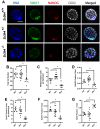
Both PE and EPI are defective in Zc3h4 mutant embryos. (A) IF of SOX17 (PE marker), NANOG (EPI marker), and CDX2 (TE marker) in blastocysts of different genotypes. (B) The total cell number per blastocyst in Zc3h4 mutant embryos is decreased, as well as a significantly reduced number (C) and percentage (D) of NANOG-positive EPI cells when compared with WT and Het littermates. A significantly reduced number (E) and percentage (F) was found for SOX17-positive PE cells when compared with WT and Het littermates. (G) Concordant with decreased PE and EPI ratios, a significant increase in the percent of CDX2-positive TE cells was detected in Zc3h4 mutants. Scale bar, 50 μm. *, P < 0.05.
Figure 4
Figure 4
Increased DNA breaks and decreased cell proliferation were found in Zc3h4 mutant embryos. (A) Fluorescent whole-mount TUNEL assay and EdU labeling assay were performed to see if ZC3H4 is involved in DNA breaks and cell proliferation, respectively. (B) Zc3h4 Mut blastocysts exhibited a significantly higher percentage of TUNEL-positive nuclei when compared with WT and Het littermates. (C) Percentage of EdU-positive nuclei was markedly decreased in Zc3h4 mutant blastocysts when compared with WT and Het littermates. Scale bar, 50 μm. *, P < 0.05.
Figure 5
Figure 5
IF of ROS (marker for oxidative homeostasis and general cell viability/health) in blastocysts of different genotypes. All genotypes displayed similarly low ROS levels, suggesting that ZC3H4 does not significantly regulate or alter oxidative homeostasis during early embryo development in mice. Scale bar, 50 μm.

References

    1. Arny M, Nachtigall L, Quagliarello J. The effect of preimplantation culture conditions on murine embryo implantation and fetal development. Fertil Steril 1987; 48:861–865. - PubMed
    1. Shi L, Wu J. Epigenetic regulation in mammalian preimplantation embryo development. Reprod Biol Endocrinol 2009; 7:59. - PMC - PubMed
    1. Sutherland AE, Calarco-Gillam PG. Analysis of compaction in the preimplantation mouse embryo. Dev Biol 1983; 100:328–338. - PubMed
    1. Houliston E, Maro B. Posttranslational modification of distinct microtubule subpopulations during cell polarization and differentiation in the mouse preimplantation embryo. J Cell Biol 1989; 108:543–551. - PMC - PubMed
    1. Latham KE, Solter D, Schultz RM. Activation of a two-cell stage-specific gene following transfer of heterologous nuclei into enucleated mouse embryos. Mol Reprod Dev 1991; 30:182–186. - PubMed

Publication types