Review
. 2020 Nov 27;18(1):449.
doi: 10.1186/s12967-020-02622-3.

Mesenchymal stem cell derived-exosomes: a modern approach in translational medicine

Affiliations
Review

Mesenchymal stem cell derived-exosomes: a modern approach in translational medicine

Sepideh Nikfarjam et al. J Transl Med. .

Abstract

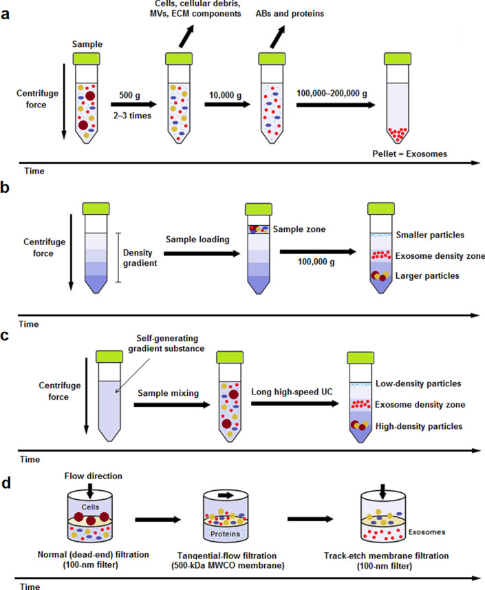
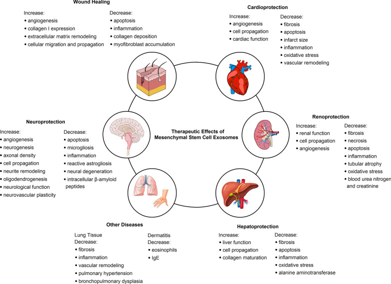
Mesenchymal stem cells (MSCs) have captured great attention in regenerative medicine for over a few decades by virtue of their differentiation capacity, potent immunomodulatory properties, and their ability to be favorably cultured and manipulated. Recent investigations implied that the pleiotropic effects of MSCs is not associated to their ability of differentiation, but rather is mediated by the secretion of soluble paracrine factors. Exosomes, nanoscale extracellular vesicles, are one of these paracrine mediators. Exosomes transfer functional cargos like miRNA and mRNA molecules, peptides, proteins, cytokines and lipids from MSCs to the recipient cells. Exosomes participate in intercellular communication events and contribute to the healing of injured or diseased tissues and organs. Studies reported that exosomes alone are responsible for the therapeutic effects of MSCs in numerous experimental models. Therefore, MSC-derived exosomes can be manipulated and applied to establish a novel cell-free therapeutic approach for treatment of a variety of diseases including heart, kidney, liver, immune and neurological diseases, and cutaneous wound healing. In comparison with their donor cells, MSC-derived exosomes offer more stable entities and diminished safety risks regarding the administration of live cells, e.g. microvasculature occlusion risk. This review discusses the exosome isolation methods invented and utilized in the clinical setting thus far and presents a summary of current information on MSC exosomes in translational medicine.

Keywords: Exosome; Exosome isolation; Extracellular vesicle; Mesenchymal stem cell; Regenerative medicine.


Conflict of interest statement

The authors declare that they have no competing interests.

Figures

Fig. 1
Fig. 1
Schematic representation of most frequently utilized exosome isolation methods for therapeutic purpose. a Differential ultracentrifugation (DUC): Sample is subjected to 2‒3 steps of low-speed (500 g) centrifugation to pellet out cells, microvesicles (MVs), extracellular matrix (ECM) components, and cellular debris. The supernatant is then centrifuged at 10,000 g for removal of apoptotic bodies (ABs) and contaminating proteins. Finally, exosomes are retrievd by a long (60–120 min) ultracentrifugation (UC) step at 100,000–200,000 g and subsequent washing of the pellet in PBS; b rate-zonal ultracentrifugation (RZUC): RZUC is a type of density gradient UC (DGUC) where sample is placed at the surface of a gradient density medium such as sucrose, and following a step of UC at 100,000 g, sample components migrate through the gradient density and separate according to their size and shape; c isopycnic ultracentrifugation (IPUC): IPUC is another type of DGUC that separates particles based on their density. Sample is usually mixed with a self-generating gradient substance such as CsCl, and is then subjected to a long UC step. In the end, distributed components form bands, so-called the isopycnic position, where the buoyant density of the collected particles matches with the gradient density of the surrounding solution. The banded exosomes can be retrieved from the density zone between 1.10 and 1.21 g/mL by fractionation; d sequential filtration (SF): Sample is first subjected to a 100-nm dead-end (normal) filteration process to separate cells and larger particles. Then, contaminating proteins are excluded via tangential flow filtration using a 500-kDa MWCO membrane. Lastly, the filtrate is once more passed through a track-etch membrane filter (with pore size of 100 nm) at very low pressure in order to inhibit passing of flexible nonexosomal EVs into the filtrate while allowing for passage of exosomes
Fig. 2
Fig. 2
Regenerative effects of mesenchymal stem cell-derived exosomes in different diseases in preclinical experimental models

References

    1. Teixeira FG, Carvalho MM, Sousa N, Salgado AJ. Mesenchymal stem cells secretome: a new paradigm for central nervous system regeneration? Cell Mol Life Sci. 2013;70(20):3871–3882. doi: 10.1007/s00018-013-1290-8. - DOI - PMC - PubMed
    1. Akbari A, Jabbari N, Sharifi R, Ahmadi M, Vahhabi A, Seyedzadeh SJ, et al. Free and hydrogel encapsulated exosome-based therapies in regenerative medicine. Life Sci. 2020;249:117447. doi: 10.1016/j.lfs.2020.117447. - DOI - PubMed
    1. Lai RC, Yeo RW, Lim SK. Mesenchymal stem cell exosomes. Semin Cell Dev Biol. 2015;40:82–88. doi: 10.1016/j.semcdb.2015.03.001. - DOI - PubMed
    1. Smirnov SV, Harbacheuski R, Lewis-Antes A, Zhu H, Rameshwar P, Kotenko SV. Bone-marrow-derived mesenchymal stem cells as a target for cytomegalovirus infection: implications for hematopoiesis, self-renewal and differentiation potential. Virology. 2007;360(1):6–16. doi: 10.1016/j.virol.2006.09.017. - DOI - PMC - PubMed
    1. Crapnell K, Blaesius R, Hastings A, Lennon DP, Caplan AI, Bruder SP. Growth, differentiation capacity, and function of mesenchymal stem cells expanded in serum-free medium developed via combinatorial screening. Exp Cell Res. 2013;319(10):1409–1418. doi: 10.1016/j.yexcr.2013.04.004. - DOI - PubMed

LinkOut - more resources