. 2020 Nov 27;17(1):363.
doi: 10.1186/s12974-020-02042-6.

Oleanolic acid ameliorates intestinal alterations associated with EAE

Affiliations

Oleanolic acid ameliorates intestinal alterations associated with EAE

Beatriz Gutierrez et al. J Neuroinflammation. .

Abstract

Background: Multiple sclerosis (MS) is a chronic demyelinating autoimmune disease affecting the CNS. Recent studies have indicated that intestinal alterations play key pathogenic roles in the development of autoimmune diseases, including MS. The triterpene oleanolic acid (OA), due to its anti-inflammatory properties, has shown to beneficially influence the severity of the experimental autoimmune encephalomyelitis (EAE), a preclinical model of MS. We herein investigate EAE-associated gut intestinal dysfunction and the effect of OA treatment.

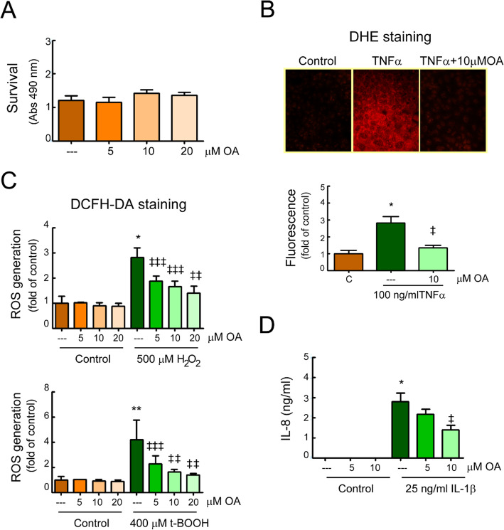
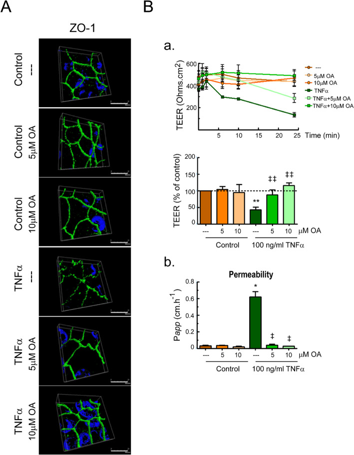
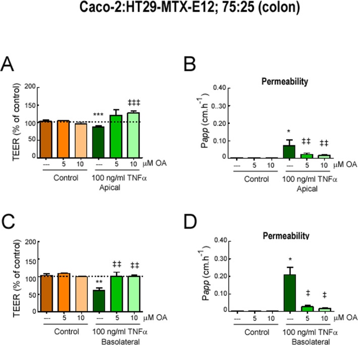
Methods: Mice with MOG35-55-induced EAE were treated with OA or vehicle from immunization day and were daily analyzed for clinical deficit. We performed molecular and histological analysis in serum and intestinal tissues to measure oxidative and inflammatory responses. We used Caco-2 and HT29-MTX-E12 cells to elucidate OA in vitro effects.

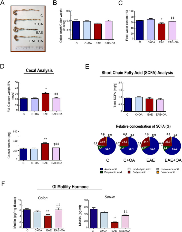
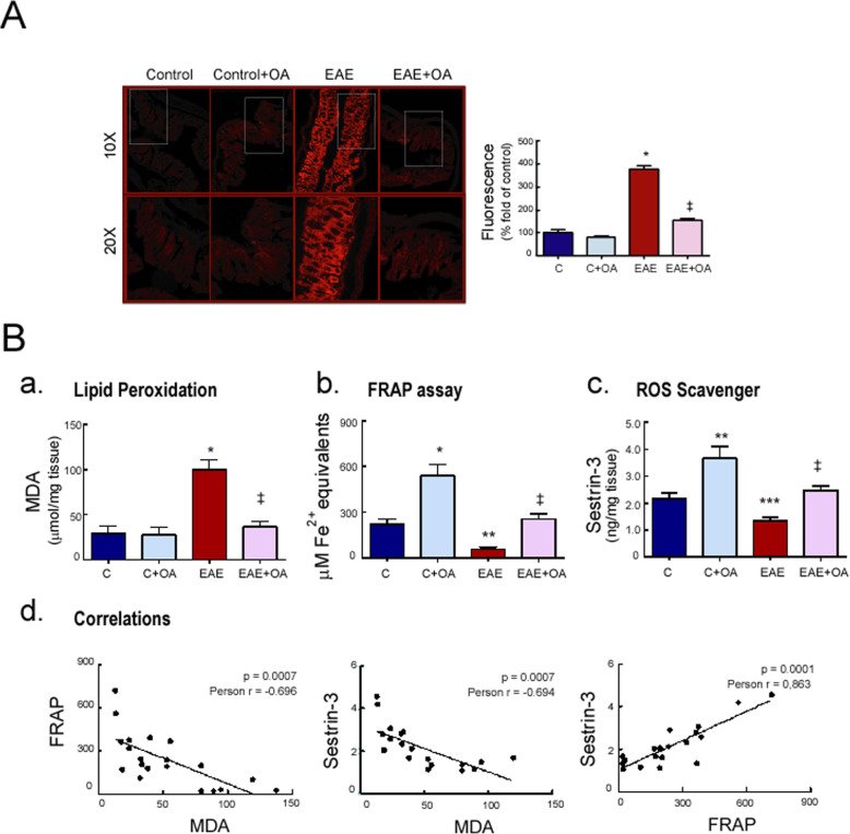
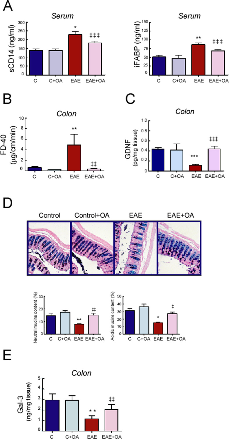
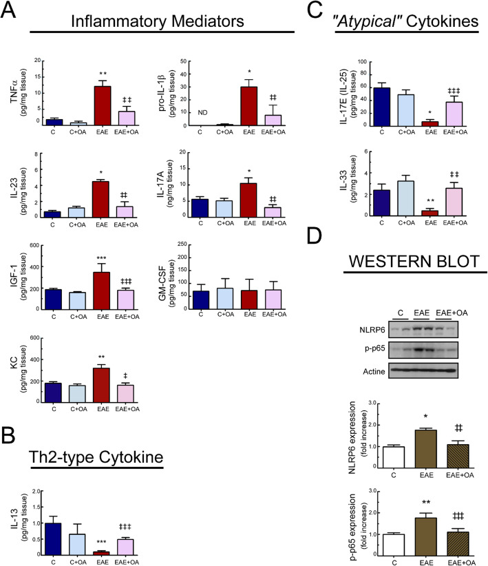
Results: We found that OA protected from EAE-induced changes in intestinal permeability and preserved the mucin-containing goblet cells along the intestinal tract. Serum levels of the markers for intestinal barrier damage iFABP and monocyte activation sCD14 were consistently and significantly reduced in OA-treated EAE mice. Beneficial OA effects also included a decrease of pro-inflammatory mediators both in serum and colonic tissue of treated-EAE mice. Moreover, the levels of some immunoregulatory cytokines, the neurotrophic factor GDNF, and the gastrointestinal hormone motilin were preserved in OA-treated EAE mice. Regarding oxidative stress, OA treatment prevented lipid peroxidation and superoxide anion accumulation in intestinal tissue, while inducing the expression of the ROS scavenger Sestrin-3. Furthermore, short-chain fatty acids (SCFA) quantification in the cecal content showed that OA reduced the high iso-valeric acid concentrations detected in EAE-mice. Lastly, using in vitro cell models which mimic the intestinal epithelium, we verified that OA protected against intestinal barrier dysfunction induced by injurious agents produced in both EAE and MS.

Conclusion: These findings reveal that OA ameliorates the gut dysfunction found in EAE mice. OA normalizes the levels of gut mucosal dysfunction markers, as well as the pro- and anti-inflammatory immune bias during EAE, thus reinforcing the idea that OA is a beneficial compound for treating EAE and suggesting that OA may be an interesting candidate to be explored for the treatment of human MS.

Keywords: Cytokines; EAE; Immune markers; Inflammation; Intestinal dysfunction; Mucins; Multiple sclerosis; Oleanolic acid; Oxidative stress; Triterpenes.


Conflict of interest statement

The authors declare that they have no competing interests.

Figures

Fig. 1
Fig. 1
Effect of OA treatment on colon length, fecal parameters and Histopathology in EAE mice. a Representative image of colons. b Colon length/weight ratio (colon were excised from anus to cecum, measured, and emptied before being weighed). c Percentage of water in fecal pellets collected from mice over one hour period. d Full cecum weight/body weight ratio and weight of cecal content. e Total and relative short-chain fatty acid (SCFA) concentrations in cecal contents. f Motillin protein concentration measured in colon extracts and serum samples, from mice of the indicated groups at day 23 post-immunization. Results were expressed as the mean ± SEM, n = 5–7 per group. *p < 0.001 and **p < 0.01 vs control; and ‡‡p < 0.01 and ‡‡‡p < 0.05 vs untreated-EAE. C, healthy mice. C + OA, healthy mice treated with OA. EAE, induced mice. EAE + OA, induced-mice treated with OA
Fig. 2
Fig. 2
OA treatment reduces oxidative stress in colon tissue from EAE mice. Representative photomicrographs of a colon tissue stained with DHE. Histological analysis by fluorescence microscopy and quantification. Objective lens × 10 and × 20. b Expression levels in colon of (a) malondialdehyde, MDA, (b) ferric reducing/antioxidant power, FRAP, and (c) the ROS disruptor Sestrin-3. (c, d) Scatter plots of the correlation between oxidative parameters. Results were expressed as the mean ± SEM, n = 5–7 per group. *p < 0.001, **p < 0.01, and ***p < 0.05 vs control; and p < 0.001 vs untreated-EAE. C, healthy mice. C + OA, healthy mice treated with OA. EAE, induced mice. EAE + OA, induced-mice treated with OA
Fig. 3.
Fig. 3.
OA treatment protects from intestinal permeability and mucus alteration in EAE mice. a Serum levels of soluble CD14 and iFABP were quantified by ELISA. b Intestinal sacs prepared from colon to assess intestinal permeability. Sacs were loaded with FITC-labeled Dextran (FD-40) and placed in a bath. After 120 min, the FD-40 concentration from the bath solutions was quantified. c Expression of GDNF in colon tissue quantified by ELISA. d Histological analysis of colon mucins stained with Alcian Blue/Periodic acid-Schiff (AB/PAS). Objective lens × 10, × 20, and × 20, respectively. Representative photomicrographs (left), quantification graphs (right). e Expression of galectin-3 (Gal-3) in colon tissue quantified by ELISA. Results were expressed as the mean ± SEM, n = 5–7 per group. *p < 0.001 and **p < 0.01 vs control; and p < 0.001, ‡‡p < 0.01, and ‡‡‡p < 0.05 vs untreated-EAE. C, healthy mice. C + OA, healthy mice treated with OA. EAE, induced mice. EAE + OA, induced-mice treated with OA
Fig. 4
Fig. 4
OA treatment modulates inflammatory parameters in colon tissue from EAE mice. Levels in colon of a the inflammatory mediators TNFα, IL-1β, IL-23, IL-17, IGF-1, GM-SCF, and KC. b The Th2-type cytokine IL-23 and c the atypical cytokines IL-25 and IL-33 quantified by ELISAs. d Protein expression of NLRP6 and p-P65-NFκB in colon assessed by western blot. Bar graphs represent the mean ± SEM of 5–7 animals. *p < 0.001, **p < 0.01, and ***p < 0.05 vs control; and p < 0.001, ‡‡p < 0.01, and ‡‡‡p < 0.05 vs untreated-EAE. C, healthy mice. C + OA, healthy mice treated with OA. EAE, induced mice. EAE + OA, induced-mice treated with OA
Fig. 5.
Fig. 5.
OA treatment inhibits responses of activated intestinal epithelial cells. Caco-2 monolayers were treated for 24 h with the indicated doses of OA and viability was measured (a). Caco-2 monolayers, pretreated for 30 min with different doses of OA, were incubated with the indicated stimuli. b After 24 h, cells were stained with DHE and superoxide anion accumulation was analyzed by fluorescence microscopy. Representative microphotographs (objective lens × 40) and quantification. c After 1 h, cells were stained with DCFH-DA and intracellular ROS-production was analyzed in a microplate reader. d After 24 h, IL-8 concentration in the cell-culture supernatant was measured by commercial ELISA. The assays were performed in duplicates, n = 3. Results were expressed as the mean ± SEM. *p < 0.001 and **p < 0.01 vs control; and p < 0.001, ‡‡p < 0.01, and ‡‡‡p < 0.05 vs stimuli without OA
Fig. 6
Fig. 6
OA treatment modulated intestinal barrier function in differentiated Caco-2 cell monolayers subjected to an inflammatory stimulus. Caco-2 cells were pretreated for 30 min with different doses of OA, and then stimulated with 100 ng/ml of TNFα for 24 h. a Representative immunofluorescence images of ZO-1. The three-dimensional projections show the ZO-1 profiles under the indicated treatments. b (a) Transepithelial electrical resistance (TEER) was tested at different time points, up to 24 h. TEER values at 24 h normalized to the untreated control (100%) are also showed. b (b) Transportation of FITC-dextran (FD-40) from apical to basolateral wells. The assay were performed in triplicates, n = 3. Results were expressed as the mean ± SEM. *p < 0.001 and **p < 0.01 vs control; and p < 0.001 and ‡‡p < 0.01 vs stimuli without OA
Fig. 7
Fig. 7
OA treatment modulated intestinal barrier function in differentiated Caco-2:HT29-MTX-E12 co-cultures subjected to an inflammatory stimulus. Differentiated Caco-2:HT29-MTX-E12 co-cultures at 75:25 proportions were stimulated with apical 100 ng/ml of TNFα (a, b) or basolateral 100 ng/ml of TNFα (c, d) for 24 h. a, d TEER values normalized to the untreated control; b, d FITC-dextran (FD-40) transport. The assays were performed in triplicates, n = 3. Results were expressed as the mean ± SEM. *p < 0.001 and **p < 0.01 vs control; and p < 0.001, ‡‡p < 0.01, and ‡‡‡p < 0.05 vs stimuli without OA

References

    1. Compston A, Coles A. Multiple sclerosis. Lancet. 2008;372:1502–1517. doi: 10.1016/S0140-6736(08)61620-7. - DOI - PubMed
    1. Calabresi PA. Diagnosis and management of multiple sclerosis. Am Fam Physician. 2004;70:1935–1944. - PubMed
    1. Levinthal DJ, Rahman A, Nusrat S, O’Leary M, Heyman R, Bielefeldt K. Adding to the burden: gastrointestinal symptoms and syndromes in multiple sclerosis. Mult Scler Int. 2013;2013:319201. - PMC - PubMed
    1. Yacyshyn B, Meddings J, Sadowski D, Bowen-Yacyshyn MB. Multiple sclerosis patients have peripheral blood CD45RO+ B cells and increased intestinal permeability. Dig Dis Sci. 1996;41:2493–2498. doi: 10.1007/BF02100148. - DOI - PubMed
    1. Buscarinu MC, Cerasoli B, Annibali V, Policano C, Lionetto L, Capi M, Mechelli R, Romano S, Fornasiero A, Mattei G, Piras E, Angelini DF, Battistini L, Simmaco M, Umeton R, Salvetti M, Ristori G. Altered intestinal permeability in patients with relapsing-remitting multiple sclerosis: A pilot study. Mult Scler. 2017;23:442–446. doi: 10.1177/1352458516652498. - DOI - PubMed

LinkOut - more resources