. 2020 Nov 27;11(1):511.
doi: 10.1186/s13287-020-02032-8.

Comprehensive proteomic analysis of exosomes derived from human bone marrow, adipose tissue, and umbilical cord mesenchymal stem cells

Affiliations

Comprehensive proteomic analysis of exosomes derived from human bone marrow, adipose tissue, and umbilical cord mesenchymal stem cells

Zheng-Gang Wang et al. Stem Cell Res Ther. .

Abstract

Background: Mesenchymal stem cell (MSC)-derived exosomes have shown comprehensive application prospects over the years. Despite performing similar functions, exosomes from different origins present heterogeneous characteristics and components; however, the relative study remains scarce. Lacking of a valuable reference, researchers select source cells for exosome studies mainly based on accessibility and personal preference.

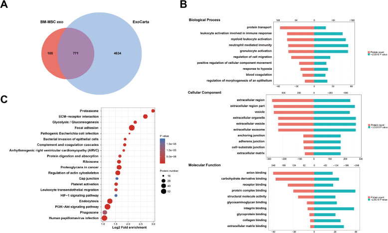
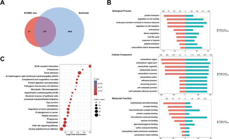
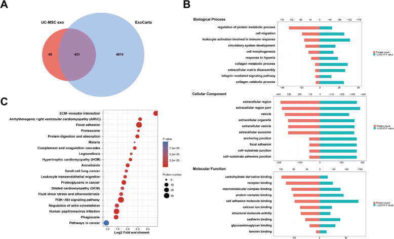
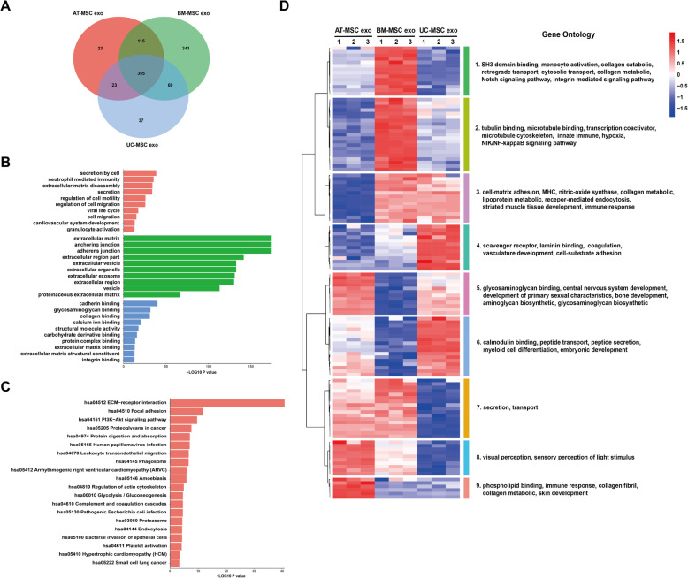
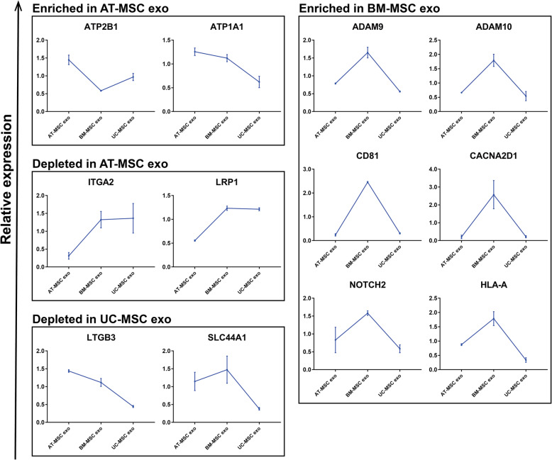
Methods: In this study, exosomes secreted by MSCs derived from different tissues were isolated, by ultracentrifugation, and proteomics analysis was performed. A total of 1014 proteins were detected using a label-free method.

Results: Bioinformatics analysis revealed their shared function in the extracellular matrix receptor. Bone marrow MSC-derived exosomes showed superior regeneration ability, and adipose tissue MSC-derived exosomes played a significant role in immune regulation, whereas umbilical cord MSC-derived exosomes were more prominent in tissue damage repair.

Conclusions: This study systematically and comprehensively analyzes the human MSC-derived exosomes via proteomics, which reveals their potential applications in different fields, so as to provide a reference for researchers to select optimal source cells in future exosome-related studies.

Keywords: Exosomes; Extracellular vesicles; Mesenchymal stem cells; Proteomics; Stem cell-based therapy.


Conflict of interest statement

The authors declare no conflict of interest.

Figures

Fig. 1
Fig. 1
Quality control of MSCs and exosomes. a MSCs were stained with Alizarin red, Oil red O, and Alcian blue to confirm their differentiation into osteogenesis, adipogenesis, and chondrogenesis. (Scale bar 50 μm and 100 μm). b Size distribution of exosomes measured by NTA. c The MSCs and exosomes expression levels of Calnexin, CD9, CD81, and TSG101 were detected via western blot analysis. d TEM images of exosomes derived from MSCs. (Scale bar 100 nm, 500 nm)
Fig. 2
Fig. 2
Bioinformatics analysis of BM-MSC exo. a Venn diagram of BM-MSC-derived exosomes against ExoCarta. b GO analysis of BM-MSC exo. c KEGG analysis of BM-MSC exo
Fig. 3
Fig. 3
Bioinformatics analysis of AT-MSC exo. a Venn diagram of AT-MSC-derived exosomes against ExoCarta. b GO analysis of AT-MSC exo. c KEGG analysis of AT-MSC exo
Fig. 4
Fig. 4
Bioinformatics analysis of UC-MSC exo. a Venn diagram of UC-MSC-derived exosomes against ExoCarta. b GO analysis of UC-MSC exo. c KEGG analysis of UC-MSC exo
Fig. 5
Fig. 5
Horizontal bioinformatics analysis of three sources of exosome proteins. a Venn diagrams of detected proteins in exosomes derived from human MSCs. b GO analysis of the shared proteins among three sources of MSCs. (Red: biological process, green: cellular component, blue: molecular function.) c KEGG analysis of the shared proteins among three sources of MSCs. d Heat map of the protein level of shared proteins among three sources of MSCs. Clusters are assembled by GO analysis. The significantly enriched items are shown on the right
Fig. 6
Fig. 6
Bioinformatics analysis of the specific proteins detected in each source of exosomes. a The volcanogram of differential proteins for BM-MSC exo vs AT-MSC exo, BM-MSC exo vs UC-MSC exo, and AT-MSC exo vs UC-MSC exo. b The cytolocalization of differential proteins for BM-MSC exo vs AT-MSC exo, BM-MSC exo vs UC-MSC exo, and AT-MSC exo vs UC-MSC exo. c GO analysis of specific proteins for each source of exosomes. (Red: biological process, green: cellular component, blue: molecular function). d KEGG analysis of specific proteins for each source of exosomes
Fig. 7
Fig. 7
Membrane protein enrichment in exosomes depends on source cell type. Relative expression of each membrane protein within a sample is depicted on slop charts and membrane protein grouped according to their enrichment or depletion in AT-MSC exo, enrichment in BM-MSC exo, and depletion in UC-MSC exo

References

    1. Zellner J, Johnstone B, Barry F. Mesenchymal stem cell based regenerative treatment of the knee: from basic science to clinics. Stem Cells Int. 2019;2019:7608718. doi: 10.1155/2019/7608718. - DOI - PMC - PubMed
    1. Mao XY, Pan T, Shen H. The rescue effect of mesenchymal stem cell on sodium iodate-induced retinal pigment epithelial cell death through deactivation of NF-kappaB-mediated NLRP3 inflammasome. Biomed Pharmacother. 2018;103:517–523. doi: 10.1016/j.biopha.2018.04.038. - DOI - PubMed
    1. Uccelli A, Moretta L, Pistoia V. Mesenchymal stem cells in health and disease. Nat Rev Immunol. 2008;8(9):726–736. doi: 10.1038/nri2395. - DOI - PubMed
    1. Piard C, Jeyaram A, Liu Y. 3D printed HUVECs/MSCs cocultures impact cellular interactions and angiogenesis depending on cell-cell distance. Biomaterials. 2019;222:119423. doi: 10.1016/j.biomaterials.2019.119423. - DOI - PMC - PubMed
    1. Gazdic M, Arsenijevic A, Markovic BS. Mesenchymal stem cell-dependent modulation of liver diseases. Int J Biol Sci. 2017;13(9):1109–1117. doi: 10.7150/ijbs.20240. - DOI - PMC - PubMed

Publication types