. 2021 Feb:269:120527.
doi: 10.1016/j.biomaterials.2020.120527. Epub 2020 Nov 16.

Driving Hierarchical Collagen Fiber Formation for Functional Tendon, Ligament, and Meniscus Replacement

Affiliations

Driving Hierarchical Collagen Fiber Formation for Functional Tendon, Ligament, and Meniscus Replacement

Jennifer L Puetzer et al. Biomaterials. 2021 Feb.

Abstract

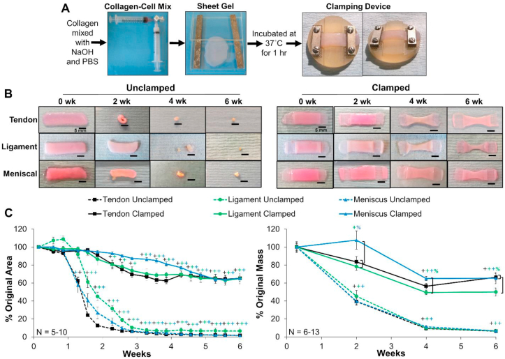
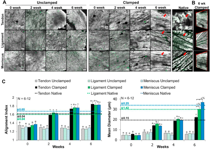
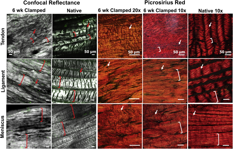
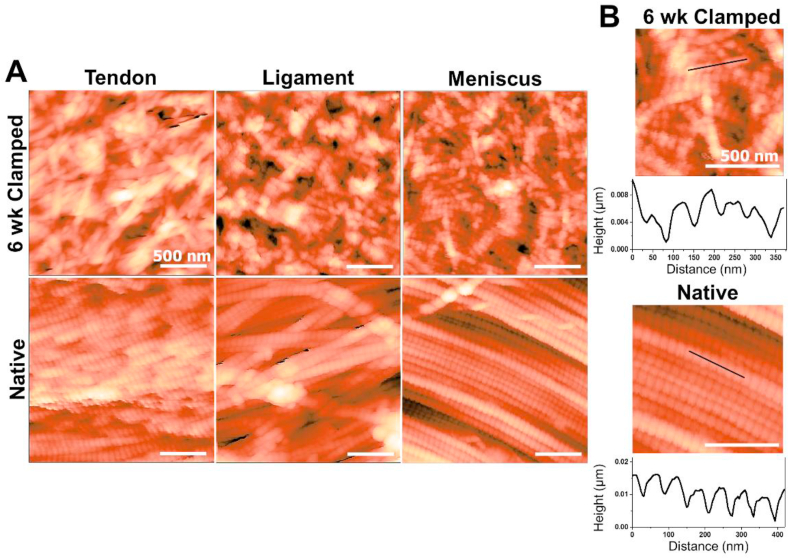
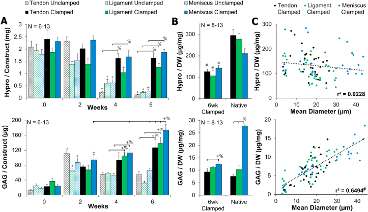
Hierarchical collagen fibers are the primary source of strength in musculoskeletal tendons, ligaments, and menisci. It has remained a challenge to develop these large fibers in engineered replacements or in vivo after injury. The objective of this study was to investigate the ability of restrained cell-seeded high density collagen gels to drive hierarchical fiber formation for multiple musculoskeletal tissues. We found boundary conditions applied to high density collagen gels were capable of driving tenocytes, ligament fibroblasts, and meniscal fibrochondrocytes to develop native-sized hierarchical collagen fibers 20-40 μm in diameter. The fibers organize similar to bovine juvenile collagen with native fibril banding patterns and hierarchical fiber bundles 50-350 μm in diameter by 6 weeks. Mirroring fiber organization, tensile properties of restrained samples improved significantly with time, reaching ~1 MPa. Additionally, tendon, ligament, and meniscal cells produced significantly different sized fibers, different degrees of crimp, and different GAG concentrations, which corresponded with respective juvenile tissue. To our knowledge, these are some of the largest, most organized fibers produced to date in vitro. Further, cells produced tissue specific hierarchical fibers, suggesting this system is a promising tool to better understand cellular regulation of fiber formation to better stimulate it in vivo after injury.

Keywords: Collagen; Fibrillogenesis; Hierarchical; Ligament; Meniscus; Tendon.


Conflict of interest statement

The authors declare that they have no known competing financial interests or personal relationships that could have appeared to influence the work reported in this paper.

Figures

Image 1
Graphical abstract
Fig. 1
Fig. 1
Hierarchical collagen organization with relative diameters of sub-level organization.
Fig. 2
Fig. 2
A) Cell-seeded collagen sheet gel formation and application of mechanical constraints of clamped constructs. B) Photographs of unclamped and clamped samples throughout culture and C) percentage of original area and mass of constructs. Unclamped samples significantly contracted from day 9 on, while clamped samples maintained their size through ~2–3 weeks. Scale bar = 5 mm, Data shown as mean ± S.E.M., Significance compared to +0 week and %bracket group (p < 0.05). (For interpretation of the references to color in this figure legend, the reader is referred to the Web version of this article.)
Fig. 3
Fig. 3
Confocal reflectance of A) collagen fiber development, B) 3D reconstruction of 6 week clamped tissues. Grey = collagen, green = auto-fluorescence of cells, scale bar = 50 μm. C) FFT based image analysis [25] of alignment (1 unorganized, 4.5 completely aligned) and mean collagen fiber diameter. Unclamped samples remained unorganized while clamped samples develop aligned fibrils by 2 weeks, which grew to significantly distinct collagen fibers (arrows) that matched respective juvenile native tissue diameters by 6 weeks. 3 to 16 images per construct were averaged, with 6–12 constructs analyzed per time point. Data shown as mean ± S.E.M., significance compared to *native, +0 week, %bracket group (p < 0.05). (For interpretation of the references to color in this figure legend, the reader is referred to the Web version of this article.)
Fig. 4
Fig. 4
Low magnification confocal reflectance 3D reconstructions and picrosirius red stained sections imaged with polarized light with 20x and 10× objectives, indicate that engineered samples develop hierarchical organization with fiber bundles 50–350 μm in diameter (brackets). Further, tenocytes develop strong crimp-like morphologies by 6 weeks, with similar morphologies appearing in ligament fibroblast and meniscal fibrochondrocyte cultures as well (arrows). Native tissue obtained from juvenile 2–6 week old bovine. (For interpretation of the references to color in this figure legend, the reader is referred to the Web version of this article.)
Fig. 5
Fig. 5
Atomic force microscopy analysis of 6 week clamped constructs and juvenile 2–6 week old bovine native tissue. A) Representative 2 μm × 2 μm images, B) topographical scans to determine d-period lengths. Engineered tissue developed average d-period lengths similar to native tissue by 6 weeks, however the banding pattern was less regular and well defined than native tissue. (For interpretation of the references to color in this figure legend, the reader is referred to the Web version of this article.)
Fig. 6
Fig. 6
A) Clamped collagen (hydroxyproline) content was largely unchanged, while clamped GAG content significantly increased with time. B) 6 week clamped samples had 40–70% collagen/dry weight (DW) and 50–100% GAG/DW of juvenile native tissue. Data shown as mean ± S.E.M. C) Clamped GAG/DW significantly correlated with mean collagen fiber diameter, while clamped collagen/DW did not correlate. Significance compared to +0 week, %bracket group,*native, and #significance determined by Pearson's correlation (p < 0.05). (For interpretation of the references to color in this figure legend, the reader is referred to the Web version of this article.)
Fig. 7
Fig. 7
Clamped samples improved tensile moduli to ~1 MPa by 6 week and all 6 week clamped samples had similar yield strength, ultimate tensile strength (UTS), and strain response when tested to failure at 0.75% strain/s. Tendon 6 week clamped constructs trended toward higher strains for the toe region and yielding compared to ligament. Data shown as mean ± S.E.M., significance compared to +0 week, %bracket group (p < 0.05), & denotes samples too small for mechanical analysis, ^ denotes trending (p < 0.1). (For interpretation of the references to color in this figure legend, the reader is referred to the Web version of this article.)

References

    1. Buehler M.J. Nature designs tough collagen: explaining the nanostructure of collagen fibrils. Proc. Natl. Acad. Sci. Unit. States Am. 2006;103:12285–12290. doi: 10.1073/pnas.0603216103. - DOI - PMC - PubMed
    1. Zhang G., Young B.B., Ezura Y., Favata M., Soslowsky L.J., Chakravarti S., Birk D.E. Development of tendon structure and function: regulation of collagen fibrillogenesis. J. Musculoskelet. Neuronal Interact. 2005;5:5–21. http://www.ncbi.nlm.nih.gov/pubmed/15788867 - PubMed
    1. Connizzo B.K., Yannascoli S.M., Soslowsky L.J. Structure-function relationships of postnatal tendon development: a parallel to healing. Matrix Biol. 2013;32:106–116. doi: 10.1016/j.matbio.2013.01.007. - DOI - PMC - PubMed
    1. Goh K.L., Listrat A., Béchet D. Hierarchical mechanics of connective tissues: integrating insights from nano to macroscopic studies. J. Biomed. Nanotechnol. 2014:2464–2507. doi: 10.1166/jbn.2014.1960. - DOI - PubMed
    1. Rodrigues M.T., Reis R.L., Gomes M.E. Engineering tendon and ligament tissues: present developments towards successful clinical products. J. Tissue Eng. Regen. Med. 2013:673–686. doi: 10.1002/term.1459. - DOI - PubMed

Publication types

LinkOut - more resources