. 2021 Jan 7;148(1):dev196568.
doi: 10.1242/dev.196568.

I-KCKT allows dissection-free RNA profiling of adult Drosophila intestinal progenitor cells

Affiliations

I-KCKT allows dissection-free RNA profiling of adult Drosophila intestinal progenitor cells

Kasun Buddika et al. Development. .

Abstract

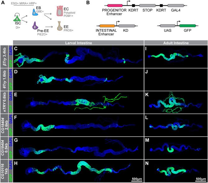
The adult Drosophila intestinal epithelium is a model system for stem cell biology, but its utility is limited by current biochemical methods that lack cell type resolution. Here, we describe a new proximity-based profiling method that relies upon a GAL4 driver, termed intestinal-kickout-GAL4 (I-KCKT-GAL4), that is exclusively expressed in intestinal progenitor cells. This method uses UV crosslinked whole animal frozen powder as its starting material to immunoprecipitate the RNA cargoes of transgenic epitope-tagged RNA binding proteins driven by I-KCKT-GAL4 When applied to the general mRNA-binder, poly(A)-binding protein, the RNA profile obtained by this method identifies 98.8% of transcripts found after progenitor cell sorting, and has low background noise despite being derived from whole animal lysate. We also mapped the targets of the more selective RNA binder, Fragile X mental retardation protein (FMRP), using enhanced crosslinking and immunoprecipitation (eCLIP), and report for the first time its binding motif in Drosophila cells. This method will therefore enable the RNA profiling of wild-type and mutant intestinal progenitor cells from intact flies exposed to normal and altered environments, as well as the identification of RNA-protein interactions crucial for stem cell function.

Keywords: CLIP; FMRP; Intestinal stem cell; PABP; eCLIP.


Conflict of interest statement

Competing interestsThe authors declare no competing or financial interests.

Figures

Fig. 1.
Fig. 1.
KD driver analysis identifies a pan-intestinal enhancer in adults. (A) Schematic of the intestinal stem cell lineage, which includes intestinal stem cells (ISCs) and enteroblasts (EBs), collectively referred to as progenitor cells, as well as differentiated enterocytes (ECs) and enteroendocrine cells (EEs). Cell type markers are shown in gray. (B) Schematic of the I-KCKT system, which involves three transgenes: a KD-expressing transgene under the control of an intestinal enhancer, a transgene in which a KDRT-flanked stop cassette separates GAL4 from a progenitor enhancer and a UAS responder, in this case, controlling GFP expression. (C-N) KD recombinase-mediated labeling of larval (C-H) and adult (I-N) intestinal cells. KD lines contain 3.4 kb βTry (C,I), 1.6 kb θTry (D,J), 0.6 kb κTry (E,K), 2.6 kb CG18404 (F,L), 0.5 kb CG18404 (G,M) or 1 kb CG10116 (H,N) enhancer sequences. KD expression pattern was detected based on KDRT-mediated activation of a tubulin-based GAL4.p65 driver in combination with a UAS-6XGFP responder. Full genotypes are listed in Table S3.
Fig. 2.
Fig. 2.
I-KCKT-GAL4.p65 and I-KCKT-GAL4 label most intestinal progenitor cells and ISC-KCKT-GAL4TS labels most ISCs. (A-J) Micrographs of five intestinal regions (R1-R5) stained for stinger-GFP (green), the intestinal progenitor marker mira-His2A.mCherry.HA (red) and the DAPI DNA marker (blue). GFP expression is driven by either I-KCKT-GAL4.p65 (A-E) or I-KCKT-GAL4 (F-J). (K-O) Micrographs of five intestinal regions (R1-R5) stained for stinger-GFP (green), the intestinal progenitor marker mira-His2A.mCherry.HA (red), the EB marker 3Xgbe-smGFP.V5.nls (white) and the DAPI DNA marker (blue). GFP is driven by ISC-KCKT-GAL4TS. Because of weak staining in either the red or green channel in A-O, not all cells co-expressing GFP and mCherry appear yellow. Single channel images showing this weak staining are included in Fig. S1. (P,Q) Histograms showing the percentage of mira-His2A.mCherry.HA-labeled intestinal progenitor (IP) cells per intestinal region (R1-R5) that are labeled with stinger-GFP driven by either I-KCKT-GAL4.p65 (P) or I-KCKT-GAL4 (Q). (R,S) Histograms showing the percentage of ISCs (R) and EBs (S) labeled with stinger-GFP driven by ISC-KCKT-GAL4TS. Graphs show mean±s.d. (n=5 for each intestinal region of each genotype). Full genotypes are listed in Table S3.
Fig. 3.
Fig. 3.
I-KCKT-GAL4 drivers are intestine-specific. (A-G) Pictures of adult female flies showing 6XGFP expression patterns driven by I-KCKT-GAL4.p65 (A), I-KCKT-GAL4TS at 30°C (B), EB-KCKT-GAL4TS at 30°C (C), ISC-KCKT-GAL4TS at 30°C (D), esgP{GawB}NP5130-GAL4 (E), Dl05151-G-GAL4 (F) or Su(H)GBE-GAL4 (G). Insets show enlargements of abdominal regions displaying intestinal GFP expression. (H) Western blot analysis of tissue extracts from total animals (T), dissected intestines with Malphigian tubules (I), or dissected carcasses (C) probed with either anti-GFP (top) or anti-tubulin (bottom) antibodies. Lysates were generated from adults harboring UAS-6XGFP driven by I-KCKT-GAL4.p65, I-KCKT-GAL4, esgP{GawB}NP5130-GAL4, Dl05151-G-GAL4, Su(H)GBE-GAL4, I-KCKT-GAL4TS at 18°C, I-KCKT-GAL4TS at 30°C or ISC-KCKT-GAL4TS at 30°C. Longer exposures of 18°C samples were also blank, indicating low background at the nonpermissive temperature. (I,J) Micrographs of intestines showing 6XGFP expression patterns driven by I-KCKT-GAL4TS from adults kept either at 18°C (I) or 30°C (J) and counterstained for the DAPI DNA marker (blue). Yellow arrowheads indicate GFP expression at the base of the Malphigian tubules. Full genotypes are listed in Table S3.
Fig. 4.
Fig. 4.
CLIP-seq of PABP driven by I-KCKT-GAL4 identifies progenitor expressed genes. (A) Representation of conventional FACS (left) and I-KCKT-based (right) intestinal progenitor RNA sequencing. (B) Volcano plot of differentially expressed genes in PABP IP versus total input. Each dot represents a single gene. Yellow indicates a false discovery rate adjusted P-value (FDR)<0.05 and a log2 fold change <1 or >−1. Green indicates log2 fold change >1 or <−1 and FDR≥0.05. Red indicates FDR<0.05 and log2 fold change >1 or <−1. A selected set of significantly changed genes are shown in blue. (C,D) Genome browser tracks of normalized total input and PABP IP at the esg (C) or osk (D) loci. Note that the two replicates of the total input (green and yellow) and the two replicates of the PABP IP (red and blue) have been overlaid. (E) Upset plot showing the overlap of genes identified by PABP CLIP-seq versus RNA-seq of FACS-isolated progenitor cells reported in Korzelius, Dutta, Buddika and Fast databases. Numbers above each bar show the size of each intersection. Only a select set of meaningful overlaps are shown. (F) Correlogram of Spearman's rho values for pairwise comparisons of progenitor expressed genes in PABP CLIP-seq; progenitor RNA-seq reported in Buddika, Dutta, Fast or Korzelius; or RNA-seq from Drosophila female head. (G) Heatmaps of odds ratio (green) and Jaccard index (gray) values for pairwise comparisons of PABP IP CLIP-seq, RNA-seq of total input or RNA-seq of dissected female heads against FACS-based progenitor RNA-seq. (H,I) Venn diagrams showing genes significantly enriched in PABP-CLIP that overlap with the top 10% of input genes based on normalized expression values (H) or genes present in input but not in any of the FACS-based RNA-seq gene lists (I). (J) Heatmaps of odds ratio (red) and Jaccard index (blue) values of pairwise comparisons of PABP IP enriched or PABP IP depleted genes with six other gene sets: fat body, head, ovary or testis enriched genes (identified relative to midgut genes) as well as midgut or progenitor enriched genes from Buddika (identified relative to whole animal input). **P<0.01, ***P<0.001, ****P<0.0001 based on Fisher's exact test.
Fig. 5.
Fig. 5.
eCLIP of FMRP driven by I-KCKT-GAL4 identifies intestinal target mRNAs. (A,B) Pie chart representing the percentage of target gene types (A) and binding site distribution (B) in mRNAs identified in FMRP eCLIP. (C) The top FMRP binding motif identified using DREME. (D) Venn diagram showing overlap between FMRP target genes with the cumulative progenitor transcriptome (identified by PABP IP or RNA-seq of FACS isolated progenitors). (E) Genome browser tracks of normalized size-matched input (SMI) and FMRP IP at the esg locus of the Drosophila genome. Note that the two replicates of FMRP IP were merged prior to genome browser visualization. Locations of four FMRP binding regions are indicated with gray bars. (F) Bar plot showing fold enrichment of 12 mRNAs in FMRP IP compared with whole intestinal input as determined by qPCR. Negative controls are separated from the other genes by a dotted line. (G) Box and whiskers plot showing the log value of mean transcript length of FMRP-bound versus unbound transcripts that are expressed in intestinal progenitors. Note that the length (bp) of the longest transcript isoform was used for this analysis whenever a gene had multiple transcript isoforms. *P<0.05, ****P<0.0001.

References

    1. Andrä I., Ulrich H., Dürr S., Soll D., Henkel L., Angerpointner C., Ritter J., Przibilla S., Stadler H., Effenberger M. et al. (2020). An evaluation of T-cell functionality after flow cytometry sorting revealed p38 MAPK activation. Cytometry A 97, 171-183. 10.1002/cyto.a.23964 - DOI - PubMed
    1. Andriatsilavo M., Stefanutti M., Siudeja K., Perdigoto C. N., Boumard B., Gervais L., Gillet-Markowska A., Al Zouabi L., Schweisguth F. and Bardin A. J. (2018). Spen limits intestinal stem cell self-renewal. PLoS Genet. 14, e1007773 10.1371/journal.pgen.1007773 - DOI - PMC - PubMed
    1. Antonello Z. A., Reiff T., Ballesta-Illan E. and Dominguez M. (2015). Robust intestinal homeostasis relies on cellular plasticity in enteroblasts mediated by miR-8-Escargot switch. EMBO J. 34, 2025-2041. 10.15252/embj.201591517 - DOI - PMC - PubMed
    1. Bailey T. L. (2011). DREME: motif discovery in transcription factor ChIP-seq data. Bioinformatics 27, 1653-1659. 10.1093/bioinformatics/btr261 - DOI - PMC - PubMed
    1. Bardin A. J., Perdigoto C. N., Southall T. D., Brand A. H. and Schweisguth F. (2010). Transcriptional control of stem cell maintenance in the Drosophila intestine. Development 137, 705-714. 10.1242/dev.039404 - DOI - PMC - PubMed

Publication types