. 2020 Nov 27;11(1):6029.
doi: 10.1038/s41467-020-19912-5.

Two-dimensional electronic spectroscopy of bacteriochlorophyll a with synchronized dual mode-locked lasers

Affiliations

Two-dimensional electronic spectroscopy of bacteriochlorophyll a with synchronized dual mode-locked lasers

JunWoo Kim et al. Nat Commun. .

Abstract

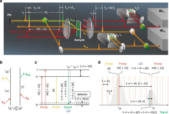
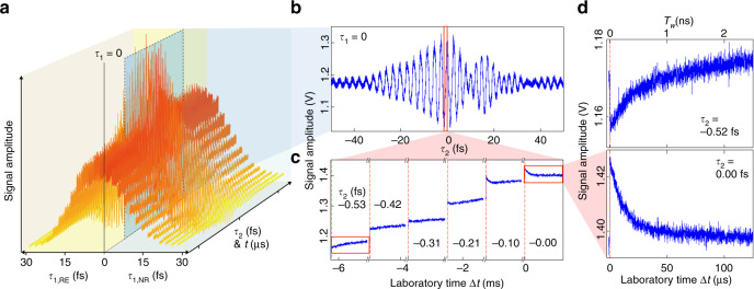
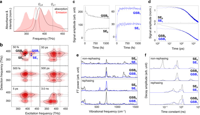
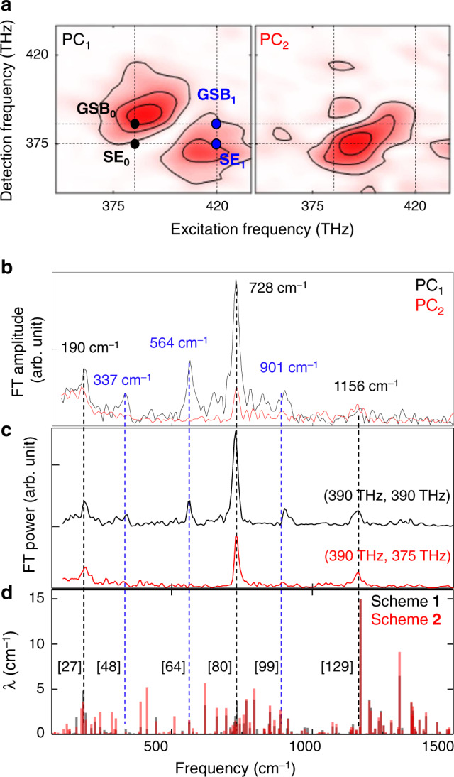
How atoms and electrons in a molecule move during a chemical reaction and how rapidly energy is transferred to or from the surroundings can be studied with flashes of laser light. However, despite prolonged efforts to develop various coherent spectroscopic techniques, the lack of an all-encompassing method capable of both femtosecond time resolution and nanosecond relaxation measurement has hampered various applications of studying correlated electron dynamics and vibrational coherences in functional materials and biological systems. Here, we demonstrate that two broadband (>300 nm) synchronized mode-locked lasers enable two-dimensional electronic spectroscopy (2DES) study of chromophores such as bacteriochlorophyll a in condensed phases to measure both high-resolution coherent vibrational spectrum and nanosecond electronic relaxation. We thus anticipate that the dual mode-locked laser-based 2DES developed and demonstrated here would be of use for unveiling the correlation between the quantum coherence and exciton dynamics in light-harvesting protein complexes and semiconducting materials.


Conflict of interest statement

The authors declare no competing interests.

Figures

Fig. 1
Fig. 1. Synchronized mode-locked laser-based two-dimensional electronic spectroscopy.
a Schematic representation of the SM-2DES experimental setup. Green circular plates represent beam splitters. The ML1 beam is split into two beams with wave vectors kA and kB, and the time delay τ1 between the two pulsed beams is scanned using a translational stage. The pulses propagating along the directions determined by kA and kB are used as a pair of pump pulses to excite optical chromophores in solution. The ML2 beam is split into two beams with wave vectors kC and kLO. The time delay τ2 between the kC and kLO pulses is scanned using another translational stage. The transient grating generated by kA and kB beams diffracts the probe with wave vector kC into the direction satisfying the desired phase-patching condition ksig = –kA (ML1) + kB (ML1) + kC (ML2), where the 2DES signal under detection propagates along the direction determined by ksig. The repetition frequencies of the pump and probe beams are fr + Δfr and fr, respectively. The 2DES signal is combined with the local oscillator field with a wave vector kLO that is collinear with the propagation direction of the 2DES signal electric field, and the interference signal is detected by a photodetector (PD). b Double-sided Feynman diagram describing one of the various terms contributing to the 2DES signal. c Energy-level diagram of a three-level system. |g〉 is the ground state, and |e〉 and |f〉 represent two vibrational states in the electronically excited state; n, m, and p are the relevant frequency comb mode numbers. νge (νgf) indicates the optical transition frequency between |g〉 and |e〉 (|f〉) states. The vibrational quantum beat with a frequency of νgfνfe ≈ (–n + m) fr is measured through the interferometric detection of the 2DES signal with LO from ML2. The beat signal contributing to the time-domain interferogram oscillates with a frequency of (–n + mfr, which is in the RF domain. d Comb structures of the pump, probe, and LO fields in the frequency domain. The line-broadened absorption bands of the |g〉−|e〉 and |g〉−|f〉 transitions are shown in grey.
Fig. 2
Fig. 2. Time-domain interferometric 2DES signal.
a Experimentally measured SM-2DES raw data of IR125/ethanol solution with respect to τ1 and τ2. Here, the repetition frequency detuning factor Δfr is 1.6 kHz. b SM-2DES interference signal with respect to τ2 when τ1 = 0 fs. The Tw-dependent decaying signals are hidden in the prominent τ2-dependent interference signal. c Zoomed-in signal taken from the red box in b. d Zoomed-in Tw-dependent signals τ2 = –0.52 fs (top) and τ2 = 0.00 fs (bottom) at τ1 = 0 fs.
Fig. 3
Fig. 3. Time-resolved SM-2DES spectra of BChla.
a The absorption (black) and emission (red) spectra of BChla/1-propanol solution. The power spectrum of the light source (pink area) is presented for the sake of direct comparison. b Absorptive (rephasing + non-rephasing) SM-2DES spectra of the BChla solution at Tw = 50, 500 fs, 5 ps (Δfr = 38.4 Hz), 50 ps, 500 ps and 3 ns (Δfr = 3.2 kHz). Each spectrum is normalized by its maximum amplitude. c, d The time traces of the rephasing 2DES signals at four different positions in the SM-2DES spectra, which are SE0 (387 THz, 375 THz), GSB0 (387 THz, 387 THz), SE1 (420 THz, 375 THz), and GSB1 (420 THz, 387 THz). e Fourier transforms of the oscillating components in the time traces plotted in c. f The distribution of decay time constants associated with the time traces shown in d.
Fig. 4
Fig. 4. Principal components analysis of BChla 2DES.
a The 2D optical frequency spectra of two major principal components of the 2D coherent vibrational spectra for rephasing interaction pathway. b The vibrational spectra of a multiplied by their score. Relatively intense six peaks at 190, 337, 564, 728, 901, and 1156 cm–1 are marked with their centre frequencies. c Coherent vibrational spectra obtained by Fourier-transforming the Tw-dependent 2DES signals at (390 THz, 390 THz) and (390 THz, 375 THz). The amplitudes of the three vibrational modes at 337, 564, and 901 cm−1 are significantly small in the CVS from the 2DES signal close to the SE0 position. d Theoretically calculated coherent vibrational spectra of BChla with respect to Qy excitation, where λ indicates vibrational reorganization energy. The numbers in square brackets are the indices of the vibrational modes. See Supplementary Note 6 for more details on the simulation schemes 1 and 2.
Fig. 5
Fig. 5. Symmetry-breaking modes and non-Condon effect.
HOMO (a) and LUMO (b) of BChla. The thick red arrow represents the Qy transition dipole moment (μeg). Blue and yellow balls represent nitrogen and magnesium atoms, while the grey balls do carbon atoms. x- and y-axes are defined from two orthogonal N–Mg–N lines. Thin red arrows emphasizes the nuclear motions of N atoms in the BChla centre. The key eigenvector elements of the D4h-to-D2h (c) and D4h-to-Cs (d) symmetry-breaking vibrational modes at the BChla centre are shown here. The full vibrational eigenvectors of these six modes are presented in Supplementary Fig. 11. The red arrows in c and d show the directions (eigenvector elements) of nitrogen atoms. e Schematic diagram describing a non-Condon effect on the reduced vibrational amplitudes of the three symmetry-breaking modes found in the CVS from the 2DES signal at (390 THz, 375 THz). Due to the destructive interference between the Franck–Condon factor and the Herzberg–Teller vibronic coupling, the oscillator strength associated with the Qy,0-S0 transition at the equilibrium coordinate Qsb in the excited state is smaller than that at the equilibrium coordinate Q0sb in the ground state. Such a non-Condon effect is also manifest in the broken mirror symmetry between the steady-state absorption and emission spectra in Fig. 3a.

Similar articles

Cited by

References

    1. Brixner T, et al. Two-dimensional spectroscopy of electronic couplings in photosynthesis. Nature. 2005;434:625–628. doi: 10.1038/nature03429. - DOI - PubMed
    1. Collini E, et al. Coherently wired light-harvesting in photosynthetic marine algae at ambient temperature. Nature. 2010;463:644. doi: 10.1038/nature08811. - DOI - PubMed
    1. Jang SJ, Mennucci B. Delocalized excitons in natural light-harvesting complexes. Rev. Mod. Phys. 2018;90:035003. doi: 10.1103/RevModPhys.90.035003. - DOI
    1. Blankenship, R. E. Molecular Mechanisms of Photosynthesis (John Wiley & Sons, 2014).
    1. van Amerongen, H., van Grondelle, R. & Valkunas, L. Photosynthetic Excitons (Wold Scientific, 2000).

Publication types