. 2020 Nov 27;3(1):724.
doi: 10.1038/s42003-020-01434-x.

Aberrant (pro)renin receptor expression induces genomic instability in pancreatic ductal adenocarcinoma through upregulation of SMARCA5/SNF2H

Affiliations

Aberrant (pro)renin receptor expression induces genomic instability in pancreatic ductal adenocarcinoma through upregulation of SMARCA5/SNF2H

Yuki Shibayama et al. Commun Biol. .

Abstract

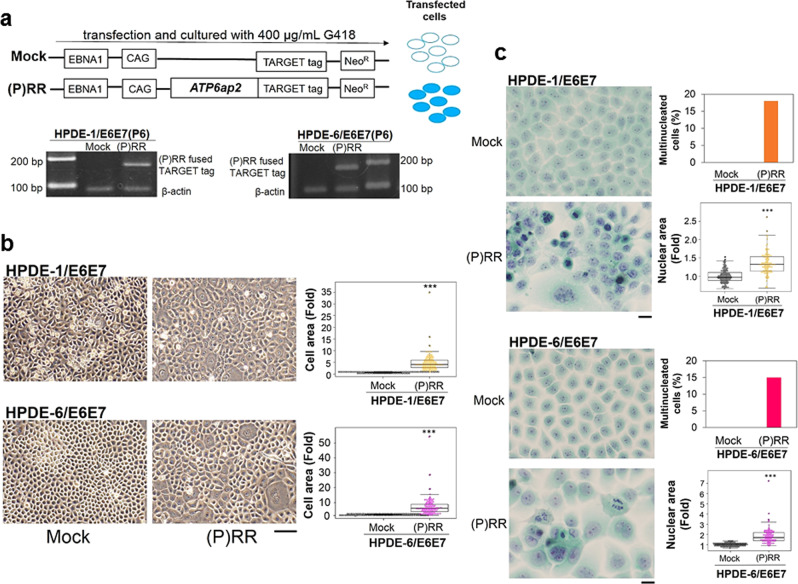
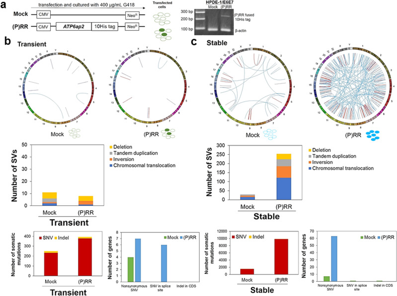
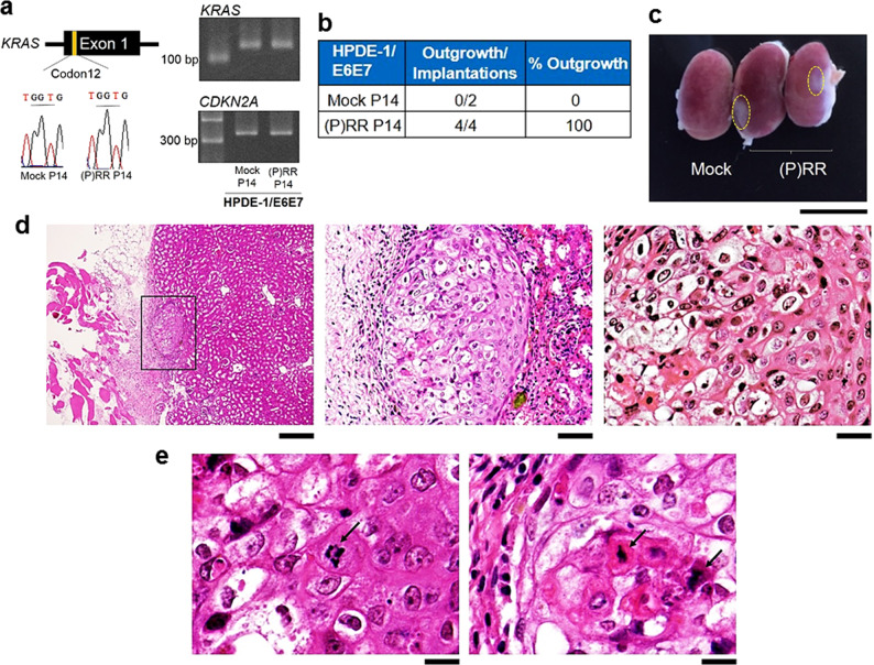
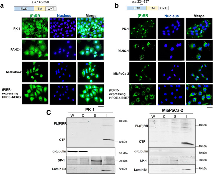
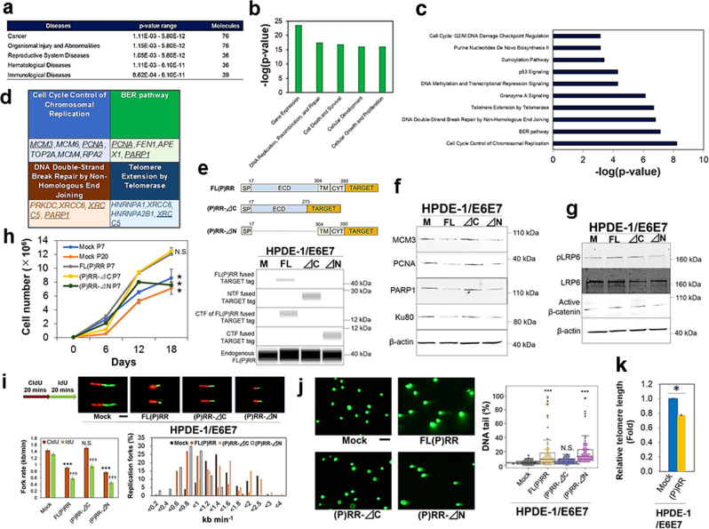
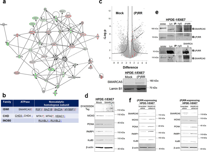
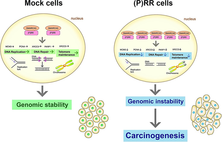
(Pro)renin receptor [(P)RR] has a role in various diseases, such as cardiovascular and renal disorders and cancer. Aberrant (P)RR expression is prevalent in pancreatic ductal adenocarcinoma (PDAC) which is the most common pancreatic cancer. Here we show whether aberrant expression of (P)RR directly leads to genomic instability in human pancreatic ductal epithelial (HPDE) cells. (P)RR-expressing HPDE cells show obvious cellular atypia. Whole genome sequencing reveals that aberrant (P)RR expression induces large numbers of point mutations and structural variations at the genome level. A (P)RR-expressing cell population exhibits tumour-forming ability, showing both atypical nuclei characterised by distinctive nuclear bodies and chromosomal abnormalities. (P)RR overexpression upregulates SWItch/Sucrose Non-Fermentable (SWI/SNF)-related, matrix-associated, actin-dependent regulator of chromatin, subfamily a, member 5 (SMARCA5) through a direct molecular interaction, which results in the failure of several genomic stability pathways. These data reveal that aberrant (P)RR expression contributes to the early carcinogenesis of PDAC.


Conflict of interest statement

The authors declare no competing interests.

Figures

Fig. 1
Fig. 1. (P)RR-expressing HPDE-1/E6E7 and HPDE-6/E6E7 cell population exhibited cellular atypia.
a Upper: Stably maintained vector containing ATP6ap2, which encodes (pro)renin receptor [(P)RR]. The inclusion of the EBNA1 gene enables this vector to be transferred to daughter cells at each round of cell division. Lower: Detection of (P)RR fused TARGET tag in human pancreatic ductal epithelial (HPDE)-1/E6E7 and HPDE-6 /E6E7 cells at six-passage. b Upper left: Representative image of HPDE-1/E6E7 cells expressing either Mock or (P)RR at six-passage under a phase-contrast microscope (×50). Upper right: The cell area in HPDE-1/E6E7 cells expressing either Mock or (P)RR. Averaged value of Mock cells is considered as 1 (N = 100 for each, ***P < 0.0001 vs. Mock). Lower left: Representative image of HPDE-6/E6E7 cells expressing either Mock or (P)RR at six-passage. Lower right: The cell area in HPDE-6/E6E7 cells expressing either Mock or (P)RR (N = 100 for each, ***P < 0.0001 vs. Mock). c Papanicolaou stain (×400). Upper left: Representative image of atypical nuclei in HPDE-1/E6E7 cells expressing either Mock or (P)RR at six-passage. Upper right: The percentage of multinucleated cells and the nuclear area in HPDE-1/E6E7 cells expressing either Mock or (P)RR. Averaged value of Mock cells is considered as 1 (N = 100 for each, ***P < 0.0001 vs. Mock). Lower left: Representative image of atypical nuclei in HPDE-6/E6E7 cells expressing either Mock or (P)RR at six-passage. Lower right: The percentage of multinucleated cells and the nuclear area in HPDE-6/E6E7 cells expressing either Mock or (P)RR. Averaged value of Mock cells is considered as 1 (N = 100 for each, ***P < 0.0001 vs. Mock). The horizontal line inside the box plot is the median and the vertical lines protruding the box extend to the minimum and the maximum values, respectively. The vertical width of the central box shows the inter-quartile deviation.
Fig. 2
Fig. 2. Genomic instability of HPDE-1/E6E7 cell population with transient and stable (P)RR expression.
a Left: Vector constructs for stable and non-replicative transient ATP6ap2 encoding (pro)renin receptor [(P)RR] expression. Transfected cells were cultured with G418 for 21 days and analyzed after one passage. Right: Detection of (P)RR fused 10His tag in HPDE-1/E6E7 cells. b Upper: Circos plot showing distribution of SVs in transient Mock- and (P)RR-expressing cell population. Middle: Number of each SV in transient Mock-and (P)RR-expressing cell population. Lower: Total number of somatic mutations and mutated genes of the exome in transient Mock- and (P)RR-expressing cell population. c Upper: Circos plot showing distribution of SVs in stable Mock- and (P)RR-expressing cell population. Middle: Number of each SV in stable Mock- and (P)RR-expressing cell population. Lower: Total number of somatic mutations and mutated genes of the exome in stable Mock- and (P)RR-expressing cell population. SNV Single nucleotide variant; Indel Insertion/deletion; CDS Coding sequence.
Fig. 3
Fig. 3. Tumour-forming ability in (P)RR-expressing HPDE-1/E6E7 cell population.
a The DNA sequence obtained by Sanger sequencing of codon 12 of KRAS (Left) and PCR products of KRAS and CDKN2A (Right) in 14-passage HPDE-1/E6E7 cells expressing either Mock or (P)RR. b Summary of the implantation and outgrowth of the HPDE-1/E6E7 cell population expressing either Mock or (P)RR. c Representative image showing the outgrowth of the HPDE-1/E6E7 cell population expressing (P)RR after implantation into kidney of immunodeficient mice (×2.7). Dotted line shows the area occupied by the HPDE-1/E6E7 cell populations expressing (P)RR. d Left: Tissue formed by (P)RR-expressing HPDE-1/E6E7 cell population engrafted into renal subcapsules of immunodeficient mice (×42). Middle: Tissue formed by the (P)RR-expressing HPDE-1/E6E7 cell population was composed of cells with different nuclear types and sizes (×200). Right: Atypical nuclei were characterized by the presence of distinctive nuclear bodies in (P)RR-expressing HPDE-1/E6E7 cells (×400). e Representative images indicating chromosomal abnormalities (×800), namely, the formation of a bridge between chromosomes (Left) and a fused chromosome (Right), as shown by arrows.
Fig. 4
Fig. 4. Intracellular localization of domains of (P)RR.
a (P)RR is expressed in both the cytoplasm and the nucleus in human PDAC cell lines and (P)RR-expressing HPDE-1/E6E7 cells. Immunofluorescence (IF) with anti-ATP6AP2 antibody recognizing a.a. 146–350, which covers from the extracellular to the cytoplasmic domains (×500). ECD Extracellular domain; TM Transmembrane domain; CYT Cytoplasmic domain. b (P)RR is expressed in the cytoplasm, as determined by IF with anti-ATP6AP2 antibody recognizing a.a. 224–237, which is localized in the extracellular domain (×500). c CTF of (P)RR is dominantly expressed in the insoluble nuclear fraction in human PDAC cell lines. Detection of each domain of (P)RR was performed using anti-ATP6AP2 antibody recognizing a.a. 146–350. Consistent results are obtained in three independent experiments. W Whole-cell lysates; C Cytoplasmic fraction; S Soluble nuclear fraction; I Insoluble nuclear fraction.
Fig. 5
Fig. 5. Dysfunction of genomic stability pathways by aberrant (P)RR expression in HPDE-1/E6E7 cells.
a Diseases expected from molecules downregulated by aberrant (P)RR expression. b Ingenuity Pathway Analysis (IPA) for molecular functions of the downregulated molecules under (P)RR overexpression. c Canonical pathways downregulated by (P)RR overexpression. d Canonical pathways identified with high IPA statistical confidence. Underline indicates molecules confirmed by western blot. e Constructs of deletion mutants in human (P)RR and confirmation of gene transfection in the vectors with each of Mock (M), FL(P)RR (FL), NTF of (P)RR (⊿C) and CTF of (P)RR (⊿N). Endogenous (P)RR was used as a loading control. f Expression of molecules involved in genomic stability pathways in cells with the deletion of each domain of (P)RR. Consistent results are obtained in three independent experiments. g Activation of Wnt components. Consistent results are obtained in three independent experiments. h Cell proliferative ability (mean ± SEM, N = 3 for each, *P < 0.05 vs. FL(P)RR, N.S., not significant). i DNA fibre assay. Cells were incubated sequentially with 5-chlorodeoxyuridine (CldU) and 5-iododeoxyuridine (IdU) for 20 min each. Upper: Representative images of DNA replication fork in cells (×800). Lower left: Quantitative evaluation of replication fork rates in cells (mean ± SEM, N = 100 for each, ***P < 0.0001 vs. Mock in CldU, N.S., not significant, †††P < 0.0001 vs. Mock in IdU). Lower right: Distribution of replication fork rate with CldU. j Single-cell gel electrophoresis in cells with the deletion of each domain of (P)RR at three-passage. Left: Representative images of DNA tail in cells (×100). Right: Quantification of the cells with DNA tails (N = 100 for each, ***P < 0.001 vs. Mock, N.S., not significant). The horizontal line inside the box plot is the median and the vertical lines protruding the box extend to the minimum and the maximum values, respectively. The vertical width of the central box shows the inter-quartile deviation. k Telomere length evaluated by Flow-FISH analyses (mean ± SEM, N = 3 for each, *P < 0.05).
Fig. 6
Fig. 6. Direct molecular binding of (P)RR with SWI/SNF-related, matrix-associated, actin-dependent regulator of chromatin, subfamily a, member 5 (SMARCA5).
a Molecular interaction network affected by aberrant (P)RR expression in HPDE-1/E6E7 cell population. Red shapes: upregulated molecules; Green shapes: downregulated molecules. Straight line: direct interaction. Dotted line: indirect interaction. The components of the imitation switch (ISWI) chromatin remodelling complex are dominant in this network. b The components of several chromatin remodelling complexes affected by aberrant (P)RR expression. Underline: molecules interacting with SMARCA5. Red arrows: upregulated molecules; blue arrows: downregulated molecules. c Upper: Volcano plot of SMARCA5. Arrows show the significant upregulation of SMARCA5 under (P)RR overexpression in HPDE-1/E6E7 cells. Lower: Representative image showing the upregulation of SMARCA5 with western blot. d Representative image for the expression of components of DNA replication, DNA repair and telomere maintenance machinery in HPDE-1/E6E7 cells with SMARCA5 overexpression. e Binding of CTF of (P)RR with SMARCA5 under coimmunoprecipitation using insoluble nucleus. MWM Molecular Weight Marker; Lys Lysates; rIgG rabbit IgG; IP Immunoprecipitation; IB Immunoblot. f Expression of molecules responsible for genomic stability in (P)RR-expressing HPDE-1/E6E7 cells transfected with two different SMARCA5 siRNAs. Consistent results are obtained in three independent experiments for western blot and coimmunoprecipitation.
Fig. 7
Fig. 7. Possible molecular mechanism of genomic instability induced by aberrant (P)RR expression.
Aberrant (P)RR expression enhances SMARCA5 expression through a direct molecular interaction, which results in the failure of several genomic stability pathways. In short, aberrant (P)RR expression induces genomic instability and contributes to early carcinogenesis of PDAC.

Similar articles

Cited by

References

    1. Rawla P, Sunkara T, Gaduputi V. Epidemiology of pancreatic cancer: global trends, etiology and risk factors. World J. Oncol. 2019;10:10–27. doi: 10.14740/wjon1166. - DOI - PMC - PubMed
    1. Witkiewicz AK, et al. Whole-exome sequencing of pancreatic cancer defines genetic diversity and therapeutic targets. Nat. Commun. 2015;6:6744. doi: 10.1038/ncomms7744. - DOI - PMC - PubMed
    1. Bailey P, et al. Genomic analyses identify molecular subtypes of pancreatic cancer. Nature. 2016;531:47–52. doi: 10.1038/nature16965. - DOI - PubMed
    1. Waddell N, et al. Whole genomes redefine the mutational landscape of pancreatic cancer. Nature. 2015;518:495–501. doi: 10.1038/nature14169. - DOI - PMC - PubMed
    1. Notta F, et al. A renewed model of pancreatic cancer evolution based on genomic rearrangement patterns. Nature. 2016;538:378–382. doi: 10.1038/nature19823. - DOI - PMC - PubMed

Publication types

MeSH terms

Substances