. 2020 Nov 27;10(1):20801.
doi: 10.1038/s41598-020-77856-8.

The double fascicular variations of the anterior talofibular ligament and the calcaneofibular ligament correlate with interconnections between lateral ankle structures revealed on magnetic resonance imaging

Affiliations

The double fascicular variations of the anterior talofibular ligament and the calcaneofibular ligament correlate with interconnections between lateral ankle structures revealed on magnetic resonance imaging

Paweł Szaro et al. Sci Rep. .

Abstract

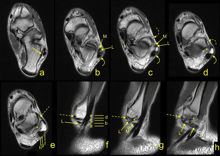
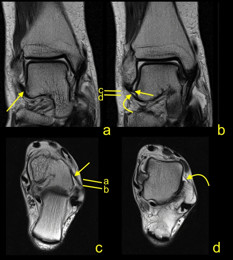
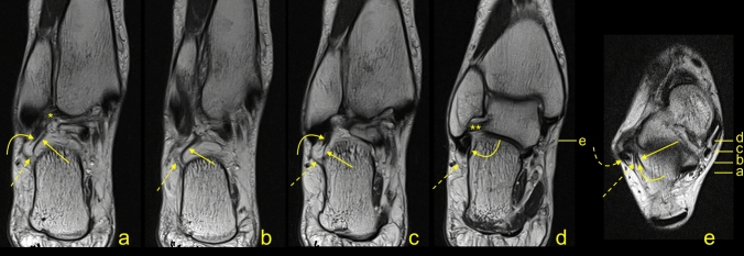
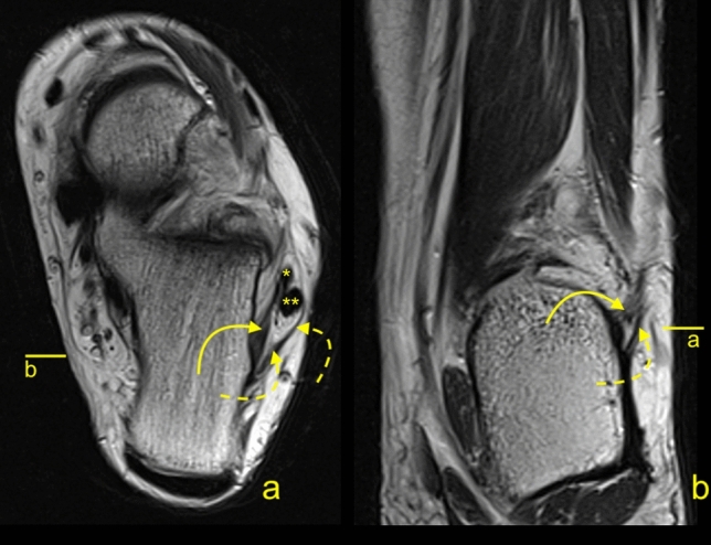
The anterior talofibular ligament and the calcaneofibular ligament are the most commonly injured ankle ligaments. This study aimed to investigate if the double fascicular anterior talofibular ligament and the calcaneofibular ligament are associated with the presence of interconnections between those two ligaments and connections with non-ligamentous structures. A retrospective re-evaluation of 198 magnetic resonance imaging examinations of the ankle joint was conducted. The correlation between the double fascicular anterior talofibular ligament and calcaneofibular ligament and connections with the superior peroneal retinaculum, the peroneal tendon sheath, the tibiofibular ligaments, and the inferior extensor retinaculum was studied. The relationships between the anterior talofibular ligament's and the calcaneofibular ligament's diameters with the presence of connections were investigated. Most of the connections were visible in a group of double fascicular ligaments. Most often, one was between the anterior talofibular ligament and calcaneofibular ligament (74.7%). Statistically significant differences between groups of single and double fascicular ligaments were visible in groups of connections between the anterior talofibular ligament and the peroneal tendon sheath (p < 0.001) as well as the calcaneofibular ligament and the posterior tibiofibular ligament (p < 0.05), superior peroneal retinaculum (p < 0.001), and peroneal tendon sheath (p < 0.001). Differences between the thickness of the anterior talofibular ligament and the calcaneofibular ligament (p < 0.001), the diameter of the fibular insertion of the anterior talofibular ligament (p < 0.001), the diameter of calcaneal attachment of the calcaneofibular ligament (p < 0.05), and tibiocalcaneal angle (p < 0.01) were statistically significant. The presence of the double fascicular anterior talofibular ligament and the calcaneofibular ligament fascicles correlate with connections to adjacent structures.


Conflict of interest statement

The authors declare no competing interests.

Figures

Figure 1
Figure 1
The protocol of the measurements a-f. Explanation in the section Material and Methods.
Figure 2
Figure 2
A 37-yeard-old patient with suspicion of a stress fracture of the navicular bone, which was not confirmed on the MRI. The arch-shaped communication between the ATFL (A), the L-CFL (L), the M-CFL (M), and the PTFL (curved dashed arrow) on the sagittal section.
Figure 3
Figure 3
A 33-yeard-old patient with suspicion of an osteochondral lesion of the talus, which was not confirmed on the MRI. The communications present in relation to the lateral malleolus. The CFL (straight arrow), the ATFL (dashed arrow), the SPR (curved arrow), the IER (curved dashed arrow), and the PTFL (*). On figure (b, c, f, g and h) two fascicles of the CFL are visible: L- the L-CFL and M- the M-CFL. Levels of cross-sections are labeled on figures (e) and (f).
Figure 4
Figure 4
A 45-yeard-old patient with suspicion of a soft tissue tumor, which was not confirmed on the MRI. The double fascicular ATFL, two fascicles with the same diameter. Sagittal section (a,b) and transverse section (c). The S-ATFL (straight arrow), the I-ATFL (curved arrow), the PTFL (dashed arrow), and the CFL (curved dashed arrow). Levels of cross-sections are labeled on figures (b,c).
Figure 5
Figure 5
A 20-yeard-old patient with suspicion of ganglion originating from the talonavicular joint, which was not confirmed on the MRI. The presence of the os subfibulare does not change the anatomical relations of the ligaments. ATFL (curved arrow) and CFL (straight arrow). The M-CFL (M) runs anterior to communicate with the ATFL, while the L-CFL (L) attaches to the fibula.
Figure 6
Figure 6
A 34-yeard-old patient with suspicion of the soft tissue tumor, which was not confirmed on the MRI. The double fascicular ATFL, two asymmetrical fascicles. Sagittal section (a,b) and transverse section (c). The S-ATFL (straight arrow), the I-ATFL (curved arrow), the PTFL (dashed arrow), and the CFL (curved dashed arrow). Levels of cross-sections are labeled on figures b and c.
Figure 7
Figure 7
A 43-yeard-old patient with suspicion of a tumor. The MRI did not reveal a tumor in the soft tissue. The oblique sections (ad) and the axial cross-section (e). Levels of cross-sections are labeled on figures (d,e). Connections of the CFL (curved arrow) with the FTCL (straight arrow), the PTS (dashed arrow), the superior peroneal retinaculum (dashed curved arrow), the PTFL (*), and the ATFL (**).
Figure 8
Figure 8
A 19-yeard-old patient with suspicion of the rheumatoid arthritis. No changes which correspond to this were found. The MRI did not reveal a tumor in the soft tissue. The communication between the CFL and the SPR. The axial section (a) and the corona section (b). Levels of cross-sections are labeled on both figures with lines and letters a and b. Connections of the CFL (curved arrow) with the superior peroneal retinaculum (dashed curved arrow), the peroneus brevis (*), and the peroneus longus (**).
Figure 9
Figure 9
A 41-yeard-old patient with suspicion of a stress fracture of the distal tibia. No fractures were revealed on the MRI. The variably occurring accessory fascicle of the CFL inserts on the talus (curved dashed arrow). The CFL (straight arrow) in the distal part is not divided, and the proximal part (P) attaches to the lateral malleolus. The PTS (dashed arrow) and the PTFL (*).

References

    1. Edama M, Kageyama I, Kikumoto T, Nakamura M, Ito W, Nakamura E, Hirabayashi R, Takabayashi T, Inai T, Onishi H. Morphological features of the anterior talofibular ligament by the number of fiber bundles. Ann. Anat. 2018;216:69–74. doi: 10.1016/j.aanat.2017.11.001. - DOI - PubMed
    1. Milner CE, Soames RW. Anatomical variations of the anterior talofibular ligament of the human ankle joint. J. Anat. 1997;191(Pt 3):457–458. doi: 10.1046/j.1469-7580.1997.19130457.x. - DOI - PMC - PubMed
    1. Raheem OA, O'Brien M. Anatomical review of the lateral collateral ligaments of the ankle: a cadaveric study. Anat. Sci. Int. 2011;86:189–193. doi: 10.1007/s12565-011-0109-7. - DOI - PubMed
    1. Siegler S, Block J, Schneck CD. The mechanical characteristics of the collateral ligaments of the human ankle joint. Foot Ankle. 1988;8:234–242. doi: 10.1177/107110078800800502. - DOI - PubMed
    1. Uğurlu M, Bozkurt M, Demirkale İ, Cömert A, Acar Hİ, Tekdemir İ. Anatomy of the lateral complex of the ankle joint in relation to peroneal tendons, distal fibula and talus: a cadaveric study. Joint Dis. Related Surg. 2010;21(3):153–158. - PubMed

Publication types