. 2021 Jun;93(6):3484-3495.
doi: 10.1002/jmv.26700. Epub 2020 Dec 17.

The clinically used serine protease inhibitor nafamostat reduces influenza virus replication and cytokine production in human airway epithelial cells and viral replication in mice

Affiliations

The clinically used serine protease inhibitor nafamostat reduces influenza virus replication and cytokine production in human airway epithelial cells and viral replication in mice

Mutsuo Yamaya et al. J Med Virol. 2021 Jun.

Abstract

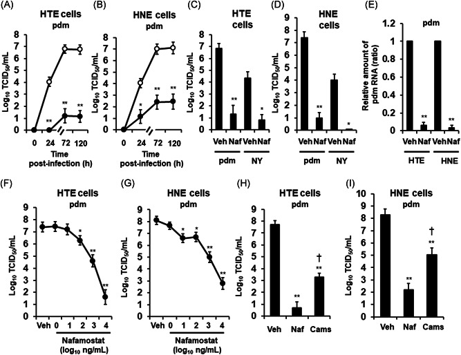
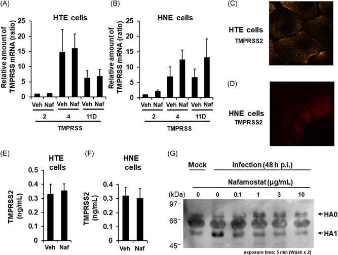
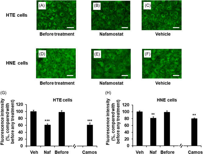
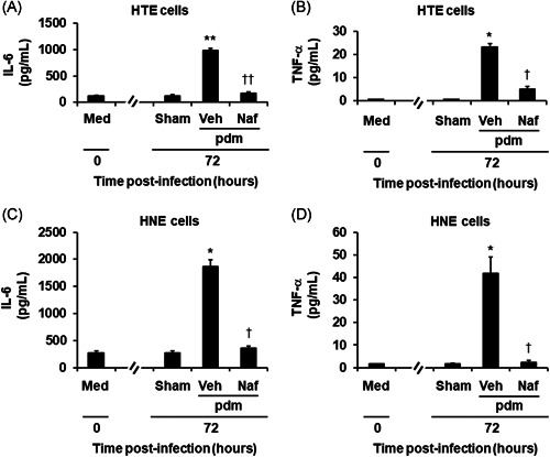
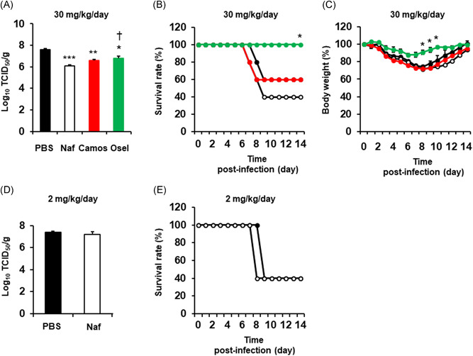
The effects of the clinically used protease inhibitor nafamostat on influenza virus replication have not been well studied. Primary human tracheal (HTE) and nasal (HNE) epithelial cells were pretreated with nafamostat and infected with the 2009 pandemic [A/Sendai-H/108/2009/(H1N1) pdm09] or seasonal [A/New York/55/2004(H3N2)] influenza virus. Pretreatment with nafamostat reduced the titers of the pandemic and seasonal influenza viruses and the secretion of inflammatory cytokines, including interleukin-6 and tumor necrosis factor-α, in the supernatants of the cells infected with the pandemic influenza virus. HTE and HNE cells exhibited mRNA and/or protein expression of transmembrane protease serine 2 (TMPRSS2), TMPRSS4, and TMPRSS11D. Pretreatment with nafamostat reduced cleavage of the precursor protein HA0 of the pandemic influenza virus into subunit HA1 in HTE cells and reduced the number of acidic endosomes in HTE and HNE cells where influenza virus RNA enters the cytoplasm. Additionally, nafamostat (30 mg/kg/day, intraperitoneal administration) reduced the levels of the pandemic influenza virus [A/Hyogo/YS/2011 (H1N1) pdm09] in mouse lung washes. These findings suggest that nafamostat may inhibit influenza virus replication in human airway epithelial cells and mouse lungs and reduce infection-induced airway inflammation by modulating cytokine production.

Keywords: antiviral agents; cell cultures; cytokines; influenza virus; protease inhibitor; respiratory tract.


Figures

Figure 1
Figure 1
A and B, Time course of viral release into supernatants of primary cultures of human tracheal (HTE) (A) or nasal (HNE) (B) epithelial cells showing levels at different times after exposure to the 2009 pandemic [A/Sendai‐H/108/2009/(H1N1) pdm09] influenza virus (pdm) in the presence of nafamostat (10 μg/ml) (closed circles) or vehicle (0.1% water) (control, open circles). C and D, The viral titers in supernatants collected between 24 h and 72 h after infection of HTE (C) or HNE (D) cells with the pdm or the seasonal [A/New York/55/2004 (H3N2)] (NY) influenza virus in the presence of nafamostat (Naf) or vehicle (0.1% water) (Veh). E, RNA levels of the pdm in HTE or HNE cells at 72 h after infection in the presence of nafamostat (Naf) or vehicle (Veh). The results are expressed as the relative amount of RNA (ratio) compared to the influenza virus RNA level in the vehicle‐pretreated cells. F and G, Concentration‐dependent effects of nafamostat on the release of the pandemic influenza virus (pdm) in the supernatants of HTE (F) or HNE (G) cells collected between 24 and 72 h after infection. H and I, Titers of the pandemic influenza virus (pdm) in supernatants collected between 24 and 72 h after infection of HTE (H) or HNE (I) cells pretreated with nafamostat (Naf, 10 μg/ml), camostat (Camos, 10 μg/ml) or vehicle (Veh). A‐I: Treatment with nafamostat (A–I) or camostat (H, I) was initiated 30 min before infection and continued during infection and after infection until the end of the experiments. Changes in the viral titers in supernatants are expressed as log10TCID50/ml (A–D, F–I). The results are expressed as the mean ± SEM of five (A–D, F, G) or four (E, H, I) different tracheal or nasal samples. Camos, camostat; HNE, human nasal; HTE, human tracheal; Naf, nafamostat; Veh, vehicle. Significant differences versus the vehicle alone group are indicated by *p < .05 and **p < .01. Significant differences versus the nafamostat group are indicated by p < .05
Figure 2
Figure 2
A and B, The mRNA expression levels of TMPRSS2, TMPRSS4, or TMPRSS11D in uninfected HTE or HNE cells treated with nafamostat (Naf, 10 μg/ml) or vehicle (Veh) for 72 h. The results for TMPRSS mRNA levels are expressed as the relative amount of mRNA (ratio) compared to the TMPRSS2 mRNA in the vehicle‐pretreated cells. The results are expressed as the mean ± SEM of five different tracheal or nasal samples. C and D, Indirect immunofluorescence staining of TMPRSS2 in HTE (C) and HNE (D) cells. TMPRSS2 was stained orange. Magnification; 630× (C) or 1000× (D). E and F, The TMPRSS2 protein concentration in the supernatants of HTE or HNE cells treated with nafamostat (Naf, 10 μg/ml) or vehicle (Veh) for 72 h. The results are expressed as the mean ± SEM of five different tracheal or nasal samples. G, Western blot analysis of proteins in the supernatants of primary cultures of HTE cells collected at 48 h post‐infection with the pandemic influenza virus in the presence of nafamostat (0.1, 1, 3, or 10 μg/ml) or vehicle (0) showing inhibition of HA0 cleavage. HA0, hemagglutinin precursor protein; HA1, hemagglutinin subunit; Mock, without infection
Figure 3
Figure 3
A–F, Changes in the distribution of acidic endosomes exhibiting green fluorescence in uninfected HTE (A–C) or HNE (D–F) cells at 72 h after treatment with Naf (10 μg/ml) (B and E) or Veh (C and F) or untreated cells cultured in medium alone (before treatment) (A and D) (scale bar = 100 μm). G and H, The effects of treatment with Naf (10 μg/ml), Veh, or Camos (10 μg/ml) on the fluorescence intensity of acidic endosomes in HTE cells (G) and HNE cells (H) at 72 h after treatment or in untreated cells (before treatment; before). The results are expressed as the relative intensity (%) compared to the mean intensity value of the vehicle‐treated cells. The results are expressed as the mean ± SEM for seven tracheal or nasal mucosa tissue samples. Camos, camostat; HNE, human nasal; HTE, human tracheal; Naf, nafamostat; Veh, vehicle. Significant differences versus values from the cells treated with Veh are indicated by **p < .01 and ***p < .001
Figure 4
Figure 4
The release of IL‐6 (A, C) and TNF‐α (B, D) into the supernatants of HTE (A, B) or HNE (C, D) cells treated with Naf (10 μg/ml) or Veh collected before (time 0) and between 24 and 72 h after infection with the pdm or after sham infection (Sham). The results are reported as the mean ± SEM for cells from five different subjects. IL‐6, interleukin‐6; pdm, pandemic influenza virus; TNF‐α, tumor necrosis factor‐α; Veh, vehicle. Significant differences versus values from the cells before infection (Med) are indicated by *p < .05 and **p < .01. Significant differences versus values from the cells infected with the pandemic influenza virus alone in the presence of vehicle (Veh) are indicated by p < .05 and †† p < .01
Figure 5
Figure 5
A, The titers of a pandemic [A/Hyogo/YS/2011 (H1N1) pdm09] influenza virus in lung samples collected from mice treated with 30 mg/kg/day nafamostat (Naf), camostat (Camos), oseltamivir (Osel), or vehicle (PBS) at 72 h after infection. The viral titers are expressed as TCID50/g of mouse lung. The results are expressed as the mean ± SEM of five mice. Significant differences versus the vehicle alone group are indicated by *p < .05, **p < .01 and ***p < .001. Significant differences versus the nafamostat group are indicated by p < .05. B and C, Time courses of the survival rate (B) or body weight (C) of mice treated with 30 mg/kg/day nafamostat (open circles with black line), camostat (closed red circles with red line), oseltamivir (closed green circles with green line), or PBS (closed circles with black line). Five mice were used in each group in the study. The survival rate and body weight at the time of infection were set to 100%. The results are expressed as the relative survival rate or body weight (%) compared to the values at the time of infection. The body weight results are expressed as the mean ± SEM. The body weight in the nafamostat group 9 days and later after infection was expressed as the mean value because the mouse number was reduced due to death. Significant differences versus the PBS alone group are indicated by *p < .05. D, The titers of a pandemic [A/Hyogo/YS/2011 (H1N1) pdm09] influenza virus in lung samples collected from mice treated with 2 mg/kg/day nafamostat (Naf) or PBS at 72 h after infection. The results are expressed as the mean ± SEM of five (Naf) or four (PBS) mice. E, Time course of the survival rate of mice treated with intraperitoneal injection of 2 mg/kg/day nafamostat (open circles with black line) or PBS (closed circles with black line). Five mice were used in the study. The survival rate at the time of infection was set to 100%

Similar articles

Cited by

References

    1. Treanor JJ, Hayden FG, Vrooman PS, et al. Efficacy and safety of the oral neuraminidase inhibitor oseltamivir in treating acute influenza: a randomized controlled trial. US Oral Neuraminidase Study Group. JAMA. 2000;283:1016‐1024. 10.1001/jama.283.8.1016 - DOI - PubMed
    1. Kumar A. Early versus late oseltamivir treatment in severely ill patients with 2009 pandemic influenza A (H1N1): speed is life. J Antimicrob Chemother. 2011;66:959‐963. 10.1093/jac/dkr090 - DOI - PubMed
    1. Hayden FG, Sugaya N, Hirotsu N, et al. Baloxavir marboxil for uncomplicated influenza in adults and adolescents. N Engl J Med. 2018;379:913‐923. 10.1056/NEJMoa1716197 - DOI - PubMed
    1. Perez‐Padilla R, de la Rosa‐Zamboni D, Ponce de Leon S, et al. INER Working Group on Influenza. Pneumonia and respiratory failure from swine‐origin influenza A (H1N1) in Mexico. N Engl J Med. 2009;361:680‐689. 10.1056/NEJMoa0904252 - DOI - PubMed
    1. Gooskens J, Jonges M, Claas EC, Meijer A, van den Broek PJ, Kroes AM. Morbidity and mortality associated with nosocomial transmission of oseltamivir‐resistant influenza A (H1N1) virus. JAMA. 2009;301:1042‐1046. 10.1001/jama.2009.297 - DOI - PubMed

MeSH terms