. 2021 Feb 24;223(4):589-599.
doi: 10.1093/infdis/jiaa737.

SARS-CoV-2-Specific Neutralizing Antibody Responses in Norwegian Health Care Workers After the First Wave of COVID-19 Pandemic: A Prospective Cohort Study

Collaborators, Affiliations

SARS-CoV-2-Specific Neutralizing Antibody Responses in Norwegian Health Care Workers After the First Wave of COVID-19 Pandemic: A Prospective Cohort Study

Mai-Chi Trieu et al. J Infect Dis. .

Abstract

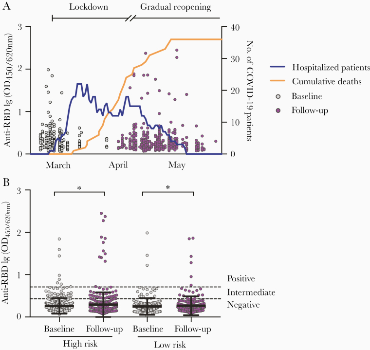
Background: During the coronavirus disease 2019 (COVID-19) pandemic, many countries experienced infection in health care workers (HCW) due to overburdened health care systems. Whether infected HCW acquire protective immunity against severe acute respiratory syndrome coronavirus 2 (SARS-CoV-2) is unclear.

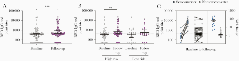
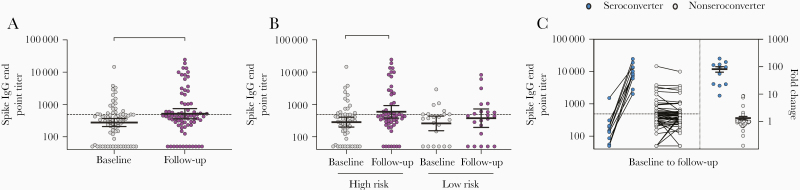
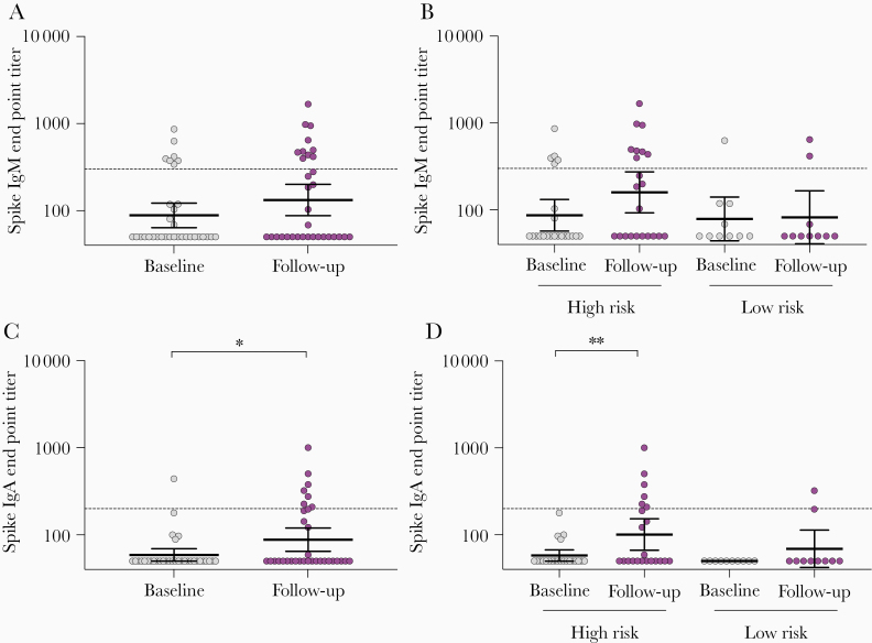
Methods: In a Norwegian prospective cohort study, we enrolled 607 HCW before and after the first COVID-19 wave. Exposure history, COVID-19-like symptoms, and serum samples were collected. SARS-CoV-2-specific antibodies were characterized by spike-protein IgG/IgM/IgA enzyme-linked immunosorbent and live-virus neutralization assays.

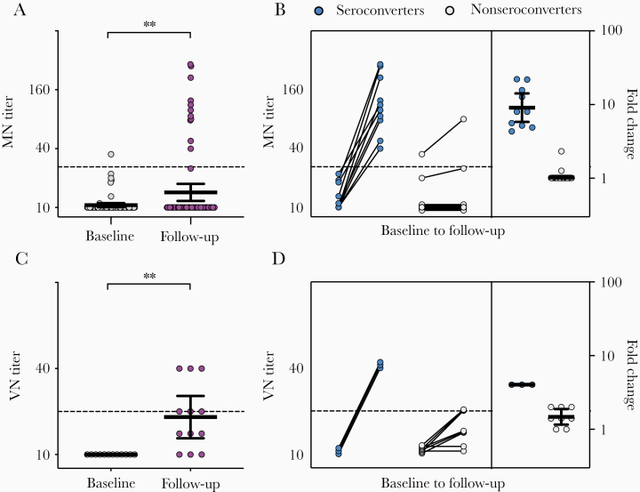
Results: Spike-specific IgG/IgM/IgA antibodies increased after the first wave in HCW with, but not in HCW without, COVID-19 patient exposure. Thirty-two HCW (5.3%) had spike-specific antibodies (11 seroconverted with ≥4-fold increase, 21 were seropositive at baseline). Neutralizing antibodies were found in 11 HCW that seroconverted, of whom 4 (36.4%) were asymptomatic. Ninety-seven HCW were tested by reverse transcriptase polymerase chain reaction (RT-PCR) during follow-up; 8 were positive (7 seroconverted, 1 had undetectable antibodies).

Conclusions: We found increases in SARS-CoV-2 neutralizing antibodies in infected HCW, especially after COVID-19 patient exposure. Our data show a low number of SARS-CoV-2-seropositive HCW in a low-prevalence setting; however, the proportion of seropositivity was higher than RT-PCR positivity, highlighting the importance of antibody testing.

Keywords: COVID-19; IgA; IgG; IgM; SARS-CoV-2; antibody characterization; health care workers; neutralizing antibody; seroconversion; spike protein.


Figures

Figure 1.
Figure 1.
Screening for SARS-CoV-2 RBD-specific antibodies in HCW before and after COVID-19 patient admissions. A, HCW (n = 607) were recruited between 6 March 2020 and 9 April 2020, and followed up after 6–10 weeks. Each circle represents 1 HCW and their anti-RBD antibodies measured in the screening ELISA as OD at 450/620 nm (left y-axis). The numbers of hospitalized COVID-19 patients (blue line) and cumulative deaths (orange line) in Bergen, Norway are plotted on the right y-axis. Lockdown was initiated in Norway on 12 March 2020 and a gradual reopening starting on 20 April 2020. B, HCW were grouped into high risk (testing facility, COVID-19–designated wards, and intensive care unit wards) and low risk (no known exposure to COVID-19 patients) of occupational exposure to SARS-CoV-2 according to their working department and information in their case report forms. Dotted lines are cutoffs for negative screening results (OD < 0.430) and positive screening results (OD ≥ 0.708) (see Supplementary Figure 1 for further information). Horizontal lines represent mean with standard deviation. OD values were log-transformed and compared between time points in mixed-effects models with adjustment for subject variation, age, sex, and other relevant demographic factors. *P < .05. Abbreviations: COVID-19, coronavirus disease 2019; ELISA, enzyme-linked immunosorbent assay; HCW, health care workers; OD, optical density; RBD, receptor-binding domain; SARS-CoV-2, severe acute respiratory syndrome coronavirus 2.
Figure 2.
Figure 2.
SARS-CoV-2 RBD-specific IgG antibodies in HCW before and after COVID-19 patient admissions. A, The RBD-specific IgG end point titers were measured for HCW with positive or intermediate RBD screening results (n = 76) by ELISA. B, RBD-specific IgG end point titers in high-risk and low-risk HCW groups. C, HCW were divided into seroconverters (blue circle) who were seropositive and had ≥4-fold increase in IgG titers at follow-up and nonseroconverters (gray circle) who were either seronegative or had <4-fold increase in IgG titers at follow-up. The fold changes are plotted on the right y-axis with horizontal lines representing the mean with standard error of the mean. Dotted lines represent cutoffs for positive results, calculated as 3 standard deviations above the mean of the prepandemic negative sera (RBD IgG end point titer ≥400). Individuals with undetectable antibodies were assigned an end point titer of 50 for plotting and calculation purposes. End point titers were log-transformed and compared between time points in mixed-effects models with adjustment for subject variation, age, sex, and other relevant demographic factors. **P < .01, ***P < .001. Abbreviations: COVID-19, coronavirus disease 2019; ELISA, enzyme-linked immunosorbent assay; HCW, health care workers; IgG, immunoglobulin G; RBD, receptor-binding domain; SARS-CoV-2, severe acute respiratory syndrome coronavirus 2.
Figure 3.
Figure 3.
SARS-CoV-2 spike-specific IgG antibodies in HCW before and after COVID-19 patient admissions. A, The RBD-specific IgG levels were measured for HCW with positive or intermediate RBD screening results (n = 76), which were confirmed in a confirmatory spike IgG ELISA. B, Spike-specific IgG end point titers in high-risk and low-risk HCW groups. C, HCW were divided into seroconverters (blue circle) who were seropositive and had ≥4-fold increase in IgG titers at follow-up and nonseroconverters (gray circle) who were either seronegative or had <4-fold increase in IgG titers at follow-up. The fold changes are plotted on the right y-axis with horizontal lines representing the mean with standard error of the mean. Dotted lines represent cutoffs for positive results, calculated as 3 standard deviations above the mean of the prepandemic negative sera (spike IgG end point titer ≥485). Individuals with undetectable antibodies were assigned an end point titer of 50 for plotting and calculation purposes. End point titers were log-transformed and compared between time points in mixed-effects models with adjustment for subject variation, age, sex, and other relevant demographic factors. *P < .05, **P < .01. Abbreviations: COVID-19, coronavirus disease 2019; ELISA, enzyme-linked immunosorbent assay; HCW, health care workers; IgG, immunoglobulin G; SARS-CoV-2, severe acute respiratory syndrome coronavirus 2.
Figure 4.
Figure 4.
SARS-CoV-2 spike-specific IgM and IgA antibodies in HCW before and after COVID-19 patient admissions. HCW with positive spike IgG results (n = 32) were further analyzed in spike IgM and IgA ELISA. A and C, Spike-specific IgM and IgA end point titers. B, and D, Spike-specific IgM and IgA end point titers in HCW in high-risk and low-risk groups. Each circle represents 1 HCW (gray baseline and purple follow-up). Horizontal lines represent geometric mean with 95% confidence interval. Dotted lines represent cutoffs for positive results, calculated as 3 standard deviations above the mean of the prepandemic negative sera (IgM end point titer ≥300, IgA end point tire ≥200). Individuals with undetectable antibodies were assigned an end point titer of 50 for plotting and calculation purposes. End point titers were log-transformed and compared between time points in mixed-effects models with adjustment for subject variation, age, sex, and other relevant demographic factors. *P < .05, **P < .01. Abbreviations: COVID-19, coronavirus disease 2019; ELISA, enzyme-linked immunosorbent assay; HCW, health care workers; IgG, immunoglobulin G; SARS-CoV-2, severe acute respiratory syndrome coronavirus 2.
Figure 5.
Figure 5.
SARS-CoV-2 neutralizing antibodies in HCW. A, MN titers of HCW with positive or intermediate SARS-CoV-2 RBD screening results (n = 76). C, Live VN titers of HCW positive with MN antibodies (n = 11). Each circle represents 1 HCW (gray baseline and purple follow-up). Horizontal lines represent geometric mean with 95% confidence interval. HCW were divided into seroconverters (blue circle) who had ≥4-fold increase in (B) MN and (D) VN titers and nonseroconverters (gray circle) who had <4-fold increase in titers. Their respective fold changes in MN and VN titers are plotted on the right y-axis with horizontal lines representing the mean with standard error of the mean. Dotted lines represent positive neutralizing antibody titers of 20. Individuals with undetectable antibodies were assigned a titer of 10 for plotting and calculation purposes. MN and VN titers were log-transformed and compared between time points in mixed-effects models with adjustment for subject variation, age, sex, and other relevant demographic factors. **P < .01. Abbreviations: HCW, health care workers; MN, microneutralization; RBD, receptor-binding domain; SARS-CoV-2, severe acute respiratory syndrome coronavirus 2; VN, virus neutralization.

Comment in

  • SARS-CoV-2 in health and care staff in Norway, 2020.
    Molvik M, Danielsen AS, Grøsland M, Telle KE, Kacelnik O, Eriksen-Volle HM. Molvik M, et al. Tidsskr Nor Laegeforen. 2021 Feb 9;141(3). doi: 10.4045/tidsskr.20.1048. Print 2021 Feb 23. Tidsskr Nor Laegeforen. 2021. PMID: 33624971 English, Norwegian.

References

    1. World Health Organization. Timeline of WHO’s response to COVID-19 https://www.who.int/news-room/detail/29-06-2020-covidtimeline. Accessed 30 July 2020.
    1. Livingston E, Bucher K. Coronavirus disease 2019 (COVID-19) in Italy. JAMA 2020; 323:1335. - PubMed
    1. Remuzzi A, Remuzzi G. COVID-19 and Italy: what next? Lancet 2020; 395:1225–8. - PMC - PubMed
    1. Lai X, Wang M, Qin C, et al. Coronavirus disease 2019 (COVID-2019) infection among health care workers and implications for prevention measures in a tertiary hospital in Wuhan, China. JAMA Netw Open 2020; 3:e209666. - PMC - PubMed
    1. Nguyen LH, Drew DA, Graham MS, et al. ; Coronavirus Pandemic Epidemiology Consortium Risk of COVID-19 among front-line health-care workers and the general community: a prospective cohort study. Lancet Public Health 2020; 5:e475–83. - PMC - PubMed

Publication types

Substances