

Anthropomorphic lung phantom based validation of in-room proton therapy 4D-CBCT image correction for dose calculation
- PMID: 33248812
- PMCID: PMC9948846
- DOI: 10.1016/j.zemedi.2020.09.004
Anthropomorphic lung phantom based validation of in-room proton therapy 4D-CBCT image correction for dose calculation
Abstract
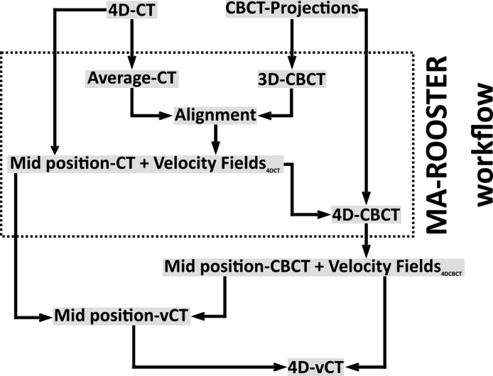
Purpose: Ventilation-induced tumour motion remains a challenge for the accuracy of proton therapy treatments in lung patients. We investigated the feasibility of using a 4D virtual CT (4D-vCT) approach based on deformable image registration (DIR) and motion-aware 4D CBCT reconstruction (MA-ROOSTER) to enable accurate daily proton dose calculation using a gantry-mounted CBCT scanner tailored to proton therapy.
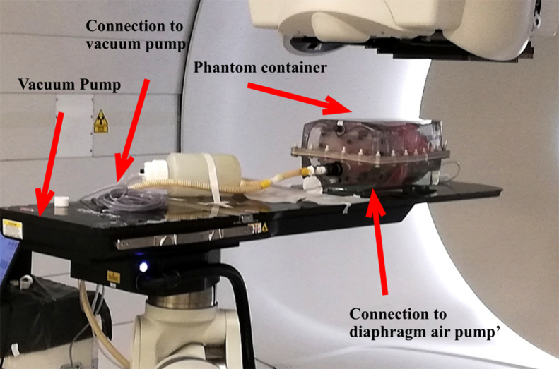
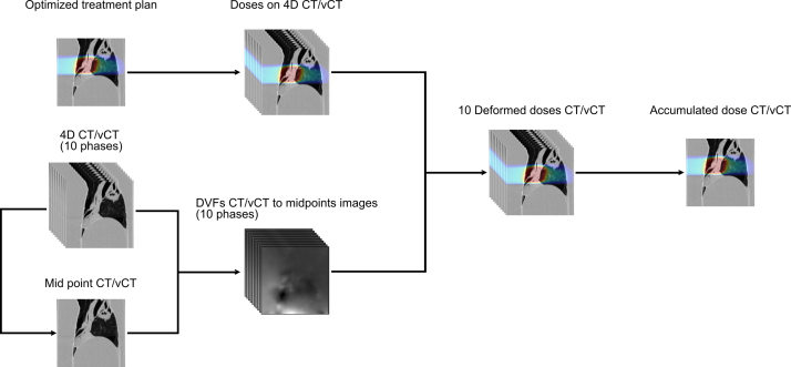
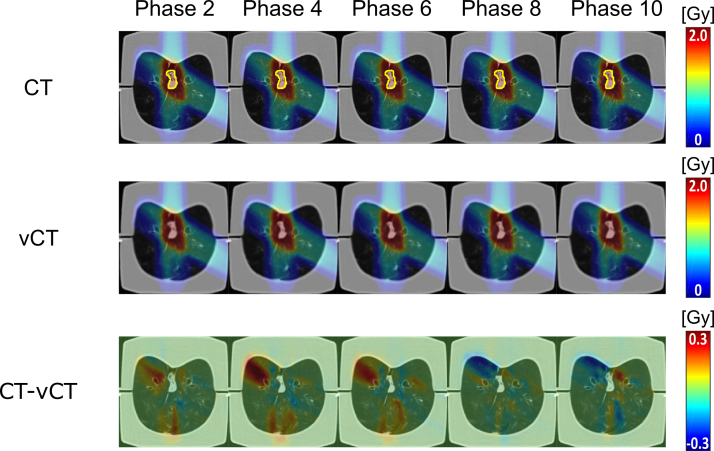
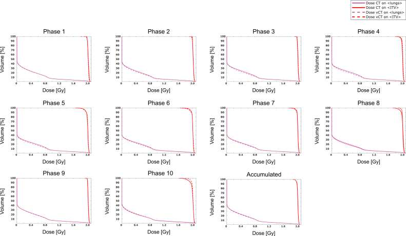
Methods: Ventilation correlated data of 10 breathing phases were acquired from a porcine ex-vivo functional lung phantom using CT and CBCT. 4D-vCTs were generated by (1) DIR of the mid-position 4D-CT to the mid-position 4D-CBCT (reconstructed with the MA-ROOSTER) using a diffeomorphic Morphons algorithm and (2) subsequent propagation of the obtained mid-position vCT to the individual 4D-CBCT phases. Proton therapy treatment planning was performed to evaluate dose calculation accuracy of the 4D-vCTs. A robust treatment plan delivering a nominal dose of 60Gy was generated on the average intensity image of the 4D-CT for an approximated internal target volume (ITV). Dose distributions were then recalculated on individual phases of the 4D-CT and the 4D-vCT based on the optimized plan. Dose accumulation was performed for 4D-vCT and 4D-CT using DIR of each phase to the mid position, which was chosen as reference. Dose based on the 4D-vCT was then evaluated against the dose calculated on 4D-CT both, phase-by-phase as well as accumulated, by comparing dose volume histogram (DVH) values (Dmean, D2%, D98%, D95%) for the ITV, and by a 3D-gamma index analysis (global, 3%/3mm, 5Gy, 20Gy and 30Gy dose thresholds).
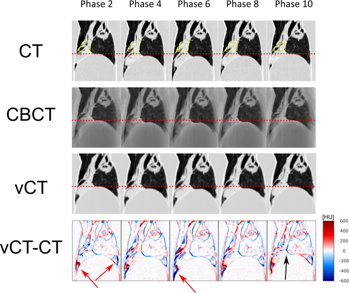
Results: Good agreement was found between the 4D-CT and 4D-vCT-based ITV-DVH curves. The relative differences ((CT-vCT)/CT) between accumulated values of ITV Dmean, D2%, D95% and D98% for the 4D-CT and 4D-vCT-based dose distributions were -0.2%, 0.0%, -0.1% and -0.1%, respectively. Phase specific values varied between -0.5% and 0.2%, -0.2% and 0.5%, -3.5% and 1.5%, and -5.7% and 2.3%. The relative difference of accumulated Dmean over the lungs was 2.3% and Dmean for the phases varied between -5.4% and 5.8%. The gamma pass-rates with 5Gy, 20Gy and 30Gy thresholds for the accumulated doses were 96.7%, 99.6% and 99.9%, respectively. Phase-by-phase comparison yielded pass-rates between 86% and 97%, 88% and 98%, and 94% and 100%.
Conclusions: Feasibility of the suggested 4D-vCT workflow using proton therapy specific imaging equipment was shown. Results indicate the potential of the method to be applied for daily 4D proton dose estimation.
Keywords: 4D-vCT; Cone-beam; Motion; Proton therapy; Thorax; Tomography.
Copyright © 2020. Published by Elsevier GmbH.
Figures
Similar articles
-
Investigating CT to CBCT image registration for head and neck proton therapy as a tool for daily dose recalculation.Med Phys. 2015 Mar;42(3):1354-66. doi: 10.1118/1.4908223. Med Phys. 2015. PMID: 25735290
-
Validation of proton dose calculation on scatter corrected 4D cone beam computed tomography using a porcine lung phantom.Phys Med Biol. 2021 Aug 30;66(17). doi: 10.1088/1361-6560/ac16e9. Phys Med Biol. 2021. PMID: 34293737
-
Feasibility of 4DCBCT-based proton dose calculation: An ex vivo porcine lung phantom study.Z Med Phys. 2019 Aug;29(3):249-261. doi: 10.1016/j.zemedi.2018.10.005. Epub 2018 Nov 14. Z Med Phys. 2019. PMID: 30448049
-
Quantitative use of cone-beam computed tomography in proton therapy: challenges and opportunities.Phys Med Biol. 2025 Apr 24;70(9). doi: 10.1088/1361-6560/adc86c. Phys Med Biol. 2025. PMID: 40269645 Review.
-
Adaptive proton therapy.Phys Med Biol. 2021 Nov 15;66(22):10.1088/1361-6560/ac344f. doi: 10.1088/1361-6560/ac344f. Phys Med Biol. 2021. PMID: 34710858 Free PMC article. Review.
Cited by
-
A systematic review of volumetric image guidance in proton therapy.Phys Eng Sci Med. 2023 Sep;46(3):963-975. doi: 10.1007/s13246-023-01294-9. Epub 2023 Jun 29. Phys Eng Sci Med. 2023. PMID: 37382744 Free PMC article.
-
ScatterNet for projection-based 4D cone-beam computed tomography intensity correction of lung cancer patients.Phys Imaging Radiat Oncol. 2023 Aug 18;27:100482. doi: 10.1016/j.phro.2023.100482. eCollection 2023 Jul. Phys Imaging Radiat Oncol. 2023. PMID: 37680905 Free PMC article.
-
Deep learning-based 4D-synthetic CTs from sparse-view CBCTs for dose calculations in adaptive proton therapy.Med Phys. 2022 Nov;49(11):6824-6839. doi: 10.1002/mp.15930. Epub 2022 Aug 27. Med Phys. 2022. PMID: 35982630 Free PMC article.
References
-
- Knopf A.-C., Lomax A. In vivo proton range verification: a review. Phys Med Biol. 2013;58:R131–R160. - PubMed
MeSH terms
LinkOut - more resources
Full Text Sources
Medical
